Image reconstruction method for differential phase contrast x-ray imaging

a phase contrast and image reconstruction technology, applied in the field of x-ray imaging techniques, can solve the problems of insufficient distinction between certain types of tissue structures in images, tumors and fluid-filled cysts may be difficult to distinguish on images, and the image reconstruction associated with such techniques may be subject to a variety of drawbacks

Active Publication Date: 2014-06-19
GENERAL ELECTRIC CO
View PDF3 Cites 34 Cited by
  • Summary
  • Abstract
  • Description
  • Claims
  • Application Information

AI Technical Summary

Problems solved by technology

However, absorption-based techniques may provide images with insufficient distinction between certain types of tissue structures.
For example, tumors and fluid-filled cysts may be difficult to distinguish on images generated by X-ray absorption of tissue.
However, image reconstruction associated with such techniques may be subject to a variety of drawbacks associated with factors such as image geometry, hardware constraints, and so forth.
Unfortunately, the noisy nature of the data may complicate the integration step, thereby resulting in blurred images having streak artifacts along the line of integration.

Method used


Image

  • Image reconstruction method for differential phase contrast x-ray imaging
  • Image reconstruction method for differential phase contrast x-ray imaging
  • Image reconstruction method for differential phase contrast x-ray imaging

Examples


Embodiment Construction

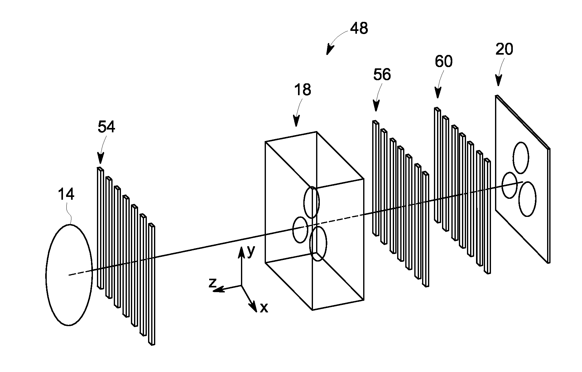
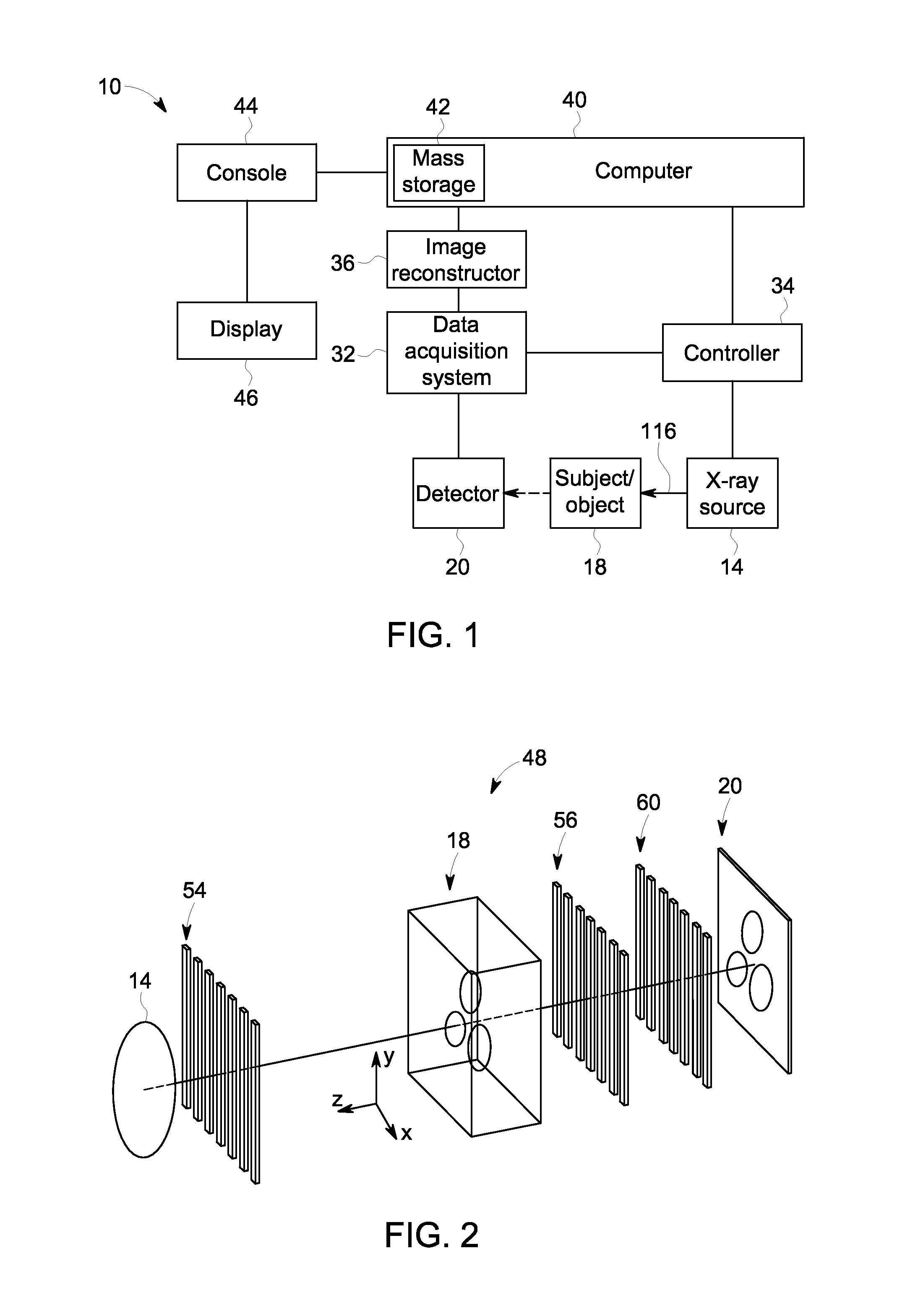
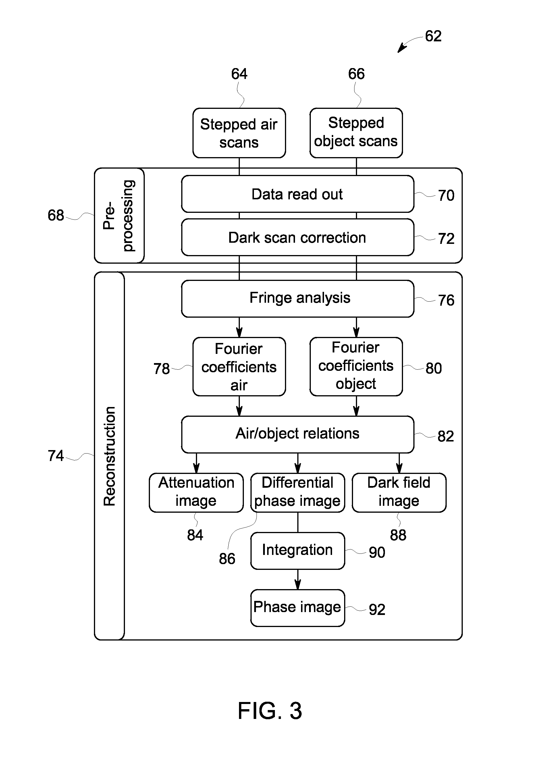
[0018]Provided herein are phase retrieval systems and methods for differential X-ray phase contrast (XPC) imaging that enable a phase image of a subject to be generated from a differential phase image of the subject. Since the measurement principle in differential XPC imaging is based on the refraction of an X-ray beam penetrating through matter, projections of the gradient of the cumulative phase shift due to the refractive index variability of the subject are generated in a direction orthogonal to the X-ray beam. The differential XPC projections have a differential nature because of this gradient, and thus, require integration to obtain a phase image of the subject. Presently disclosed embodiments may offer advantages over systems that perform a one dimensional integration step in the image domain to obtain the phase image from the differential phase image. For example, in certain embodiments, by applying an iterative reconstruction technique in the Fourier domain, the low frequen...


PUM

PropertyMeasurementUnit
phase retrieval methodaaaaaaaaaa
contrast imagingaaaaaaaaaa
differential phase imageaaaaaaaaaa

Abstract

A phase retrieval method for differential phase contrast imaging includes receiving data corresponding to a differential phase image generated from a measured signal. The measured signal corresponds to an X-ray signal detected by a detector after passing through a subject located with a grating arrangement between an X-ray source and the detector. The method further includes generating a phase image corresponding to the integration of the differential phase image. Generating the phase image includes performing an iterative total variation regularized integration in the Fourier domain.

Description

BACKGROUND[0001]The subject matter disclosed herein generally relates to X-ray imaging techniques and, in particular, to systems and methods for reconstructing images in X-ray phase contrast imaging.[0002]In non-invasive imaging systems, X-ray tubes are used in various X-ray systems and computed tomography (CT) systems as a source of X-ray radiation. The radiation is emitted in response to control signals during an examination or imaging sequence. Typically, the X-ray tube includes a cathode and an anode. An emitter within the cathode may emit a stream of electrons in response to heat resulting from an applied electrical current, and / or an electric field resulting from an applied voltage to a properly shaped metallic plate in front of the emitter. The anode may include a target that is impacted by the stream of electrons. The target may, as a result of impact by the electron beam, produce X-ray radiation to be emitted toward an imaged volume.[0003]Conventional X-ray imaging systems ...

Claims


Application Information

Patent Timeline
Patent Type & Authority Applications(United States)
IPC IPC(8): G06T5/00G01N23/04
CPCG01N23/04G06T5/001G01N23/046G01N2223/419G01N2223/612
Inventor SPERL, JONATHAN IMMANUELBEQUE, DIRK WIM JOS
Owner GENERAL ELECTRIC CO
Why Eureka
Try Eureka