Digital holographic microscopy apparatus and method for clinical diagnostic hematology

a holographic microscopy and clinical technology, applied in the field of digital holographic microscopy for clinical diagnostic hematology, can solve the problems of subjective manual counting, unreliable statistically, and inability to discriminate with respect to individual particle volume, and achieve the effect of cost-effectiveness and easy implementation

Inactive Publication Date: 2014-08-07
WET LABS
View PDF2 Cites 12 Cited by
  • Summary
  • Abstract
  • Description
  • Claims
  • Application Information

AI Technical Summary

Benefits of technology

[0015]The present invention enables adequate blood cell type differentials with

Problems solved by technology

CBC devices employing electroresistive methods can only discriminate with respect to individual particle volume.
Also, if results from an automated system are irregular, then typically a manual CBC is performed.
Manual counting can, however, be subjective, labor-intensive, and statistically unreliable (only 100-200 cells are counted as opposed to thousands with automated counters).
It takes experience to consistently make technically adequate smears and, even then, non-uniform distributions of white blood cells (WBCs) and RBCs over the smear create biases.
As mentioned, certain drawbacks of impedance

Method used


Image

  • Digital holographic microscopy apparatus and method for clinical diagnostic hematology
  • Digital holographic microscopy apparatus and method for clinical diagnostic hematology
  • Digital holographic microscopy apparatus and method for clinical diagnostic hematology

Examples


Embodiment Construction

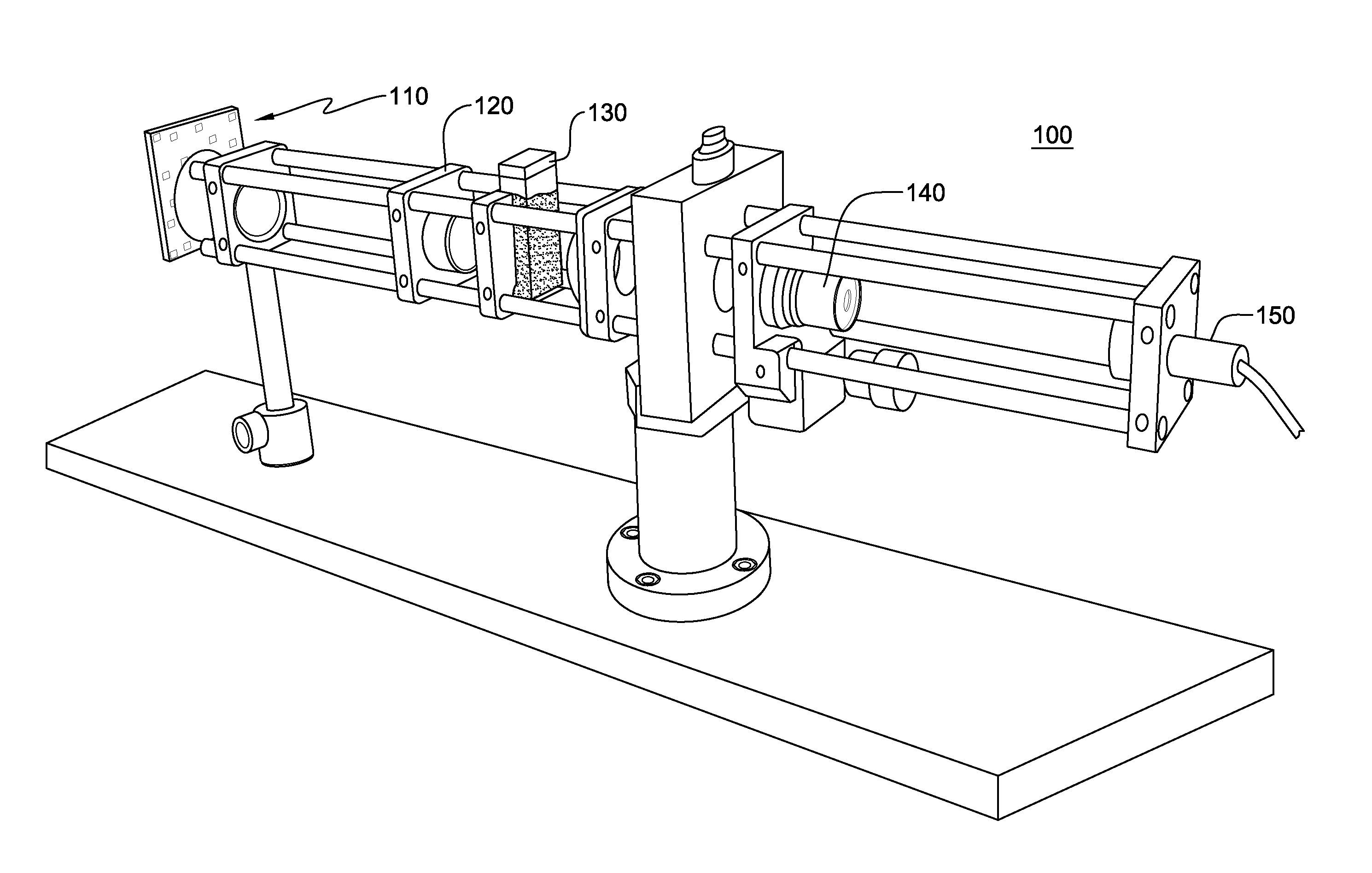
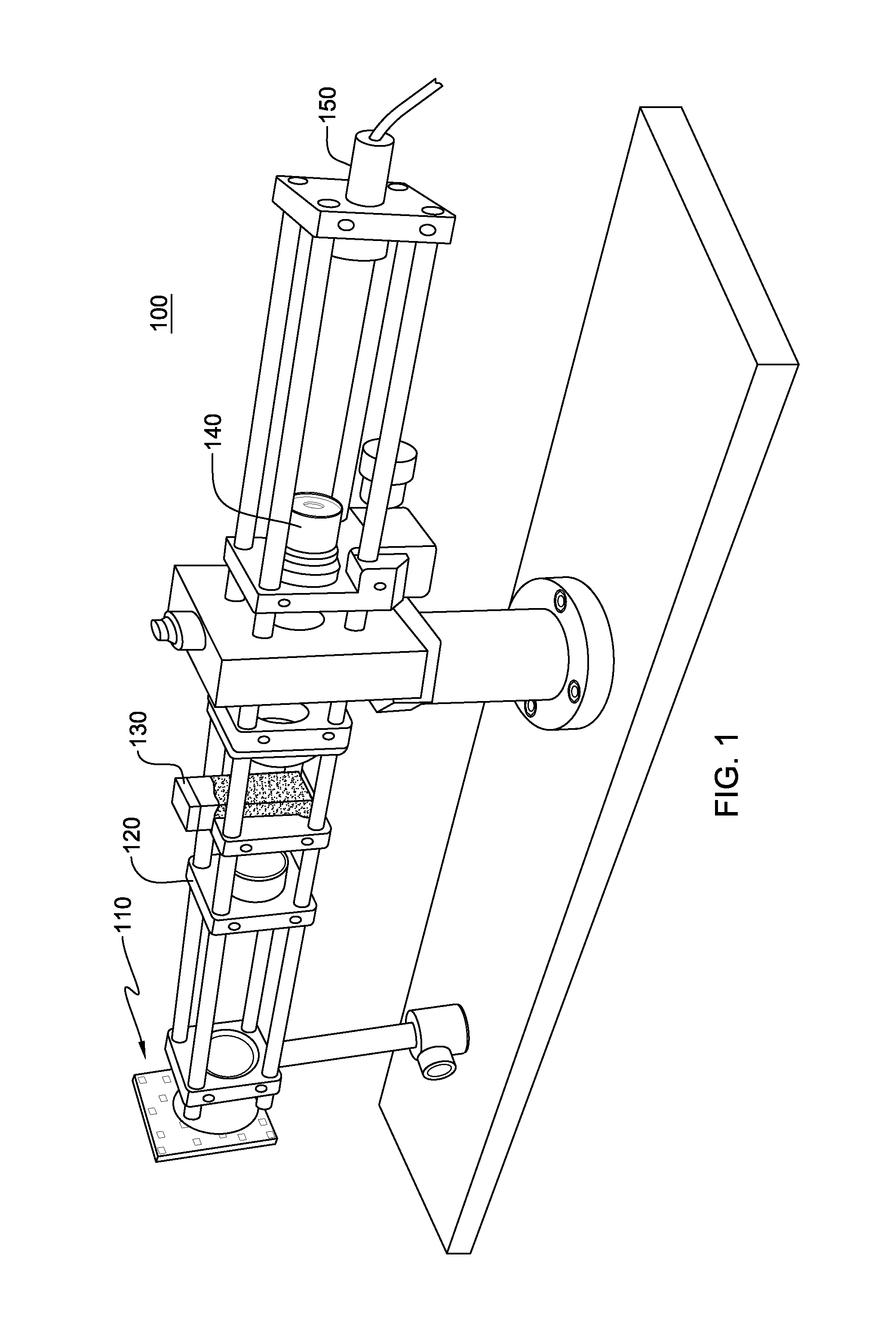
[0024]Transmission holographic imaging generically refers to recording the interference pattern of a reference beam with light that has been diffracted by particles in a suspension. When reconstructed, the result is a 3-D image of all the particles in the sample volume, all simultaneously in focus. There are a number of different optical arrangements for a holographic microscope, each having different advantages and disadvantages. The simplest optical setup, which is particularly suitable for characterizing small particles, is in-line holography. In this method, a collimated light source, typically from a laser, enters the sample. The diffraction pattern generated by the particle suspension is recorded along with the reference beam, which consists of that portion of the incident light that was not scattered. No separate optical path for the reference beam is needed. Placing a microscope objective in-line after the sample, a digital CCD camera can be used to record the magnified inte...


PUM


Abstract

An apparatus, method, and apparatus for hematology analysis comprising using a holographic microscope, in one embodiment a transmission-type holographic microscope. In one aspect, laser light is provided and split into first and second sample beams, the first sample beam for imaging with a first magnification, the second sample beam for imaging with a second magnification. The first and second sample beams are passed through a sample volume requiring hematology analysis. The first and second sample beams are combined with a reference beam and captured for digital analysis. The present invention enables adequate blood cell type differentials with a single, easily implemented, cost-effective holographic technique.

Description

CROSS-REFERENCE TO RELATED PATENT APPLICATION[0001]This application claims priority from U.S. provisional patent application Ser. No. 61 / 760,793, filed Feb. 5, 2013, and entitled “Digital Holographic Apparatus and Method for Clinical Diagnostic Hematology,” which is hereby incorporated herein by reference in its entirety.TECHNICAL FIELD[0002]This invention relates in general to digital holographic microscopy for clinical diagnostic hematology.BACKGROUND OF THE INVENTION[0003]Digital holographic microscopy (DHM) can provide non-intrusive, non-destructive, high-resolution, instantaneous 3-D imaging of particles at a resolution and sample volume size that few instruments can currently achieve. Recent advancements in lasers, CCD cameras and computing power have substantially reduced the cost, size and complexity of developing holographic analytical systems, rendering it an attractive option for enhanced particle characterization for a wide range of potential applications.[0004]Clinical ...

Claims


Application Information

Patent Timeline
IPC IPC(8): G01N21/45
CPCG01N21/453G01N15/0227G01N2015/0065G01N2015/0233G02B21/365
Inventor TWARDOWSKI, MICHAEL STEWARTSULLIVAN, JAMES MICHAEL
Owner WET LABS
Why Eureka
Try Eureka