
Device and method for displaying fetal positions and fetal biological signals using portable technology

a portable technology and positioning device technology, applied in the field of basic developmental biology, can solve the problems of reversing permanent poor health outcomes, debilitating, and affecting the quality of life of patients, and achieving the effect of reducing the number of patients

Inactive Publication Date: 2014-08-14
KIRALY MARIANNA
View PDF1 Cites 38 Cited by
  • Summary
  • Abstract
  • Description
  • Claims
  • Application Information

AI Technical Summary

Benefits of technology

This patent describes a new and cheap way to watch a baby during pregnancy without needing to put a ultrasound machine on its belly. The method and device use light to show different parts of the baby's body and how they are moving. This helps to create a 3D map of the baby's body and how it works, which can be useful for long-term monitoring of the baby's development.

Problems solved by technology

Despite major efforts, these prevalent, debilitating, life-long disorders remain biologically unexplained.
In most cases, it is already too late after birth to permanently reverse the poor health outcomes, as they are being developed early due to microenvironmental alterations.
It is a problem in the prior art to detect spontaneous brain activity in a developing fetus.
As a consequence, it is also a problem in the prior art to detect signs of epilepsy or other brain injuries or disorders in a developing fetus, as in most cases these are not correlated with responses to auditory stimuli.
It is a further problem and need in the prior art to provide a portable fetal-EEG recording device that is extremely sensitive, detecting potentials of even below 1-2 microvolts, capable of detecting and recording signals over an extended period of time, and perform the steps of analyzing the recorded signals for signs of developmental brain disorders in the developing fetus.
It is also a problem in the prior art to provide visual control of the fetus, in order to determine the movement of the fetus between the time of application of the electrodes to the time of later measurements.
It is a problem in the prior art to determine the position of a fetus accurately without ultrasound scanning, which is a crucial criterion for signal interpretation.

Method used


Image

  • Device and method for displaying fetal positions and fetal biological signals using portable technology
  • Device and method for displaying fetal positions and fetal biological signals using portable technology
  • Device and method for displaying fetal positions and fetal biological signals using portable technology

Examples


Embodiment Construction

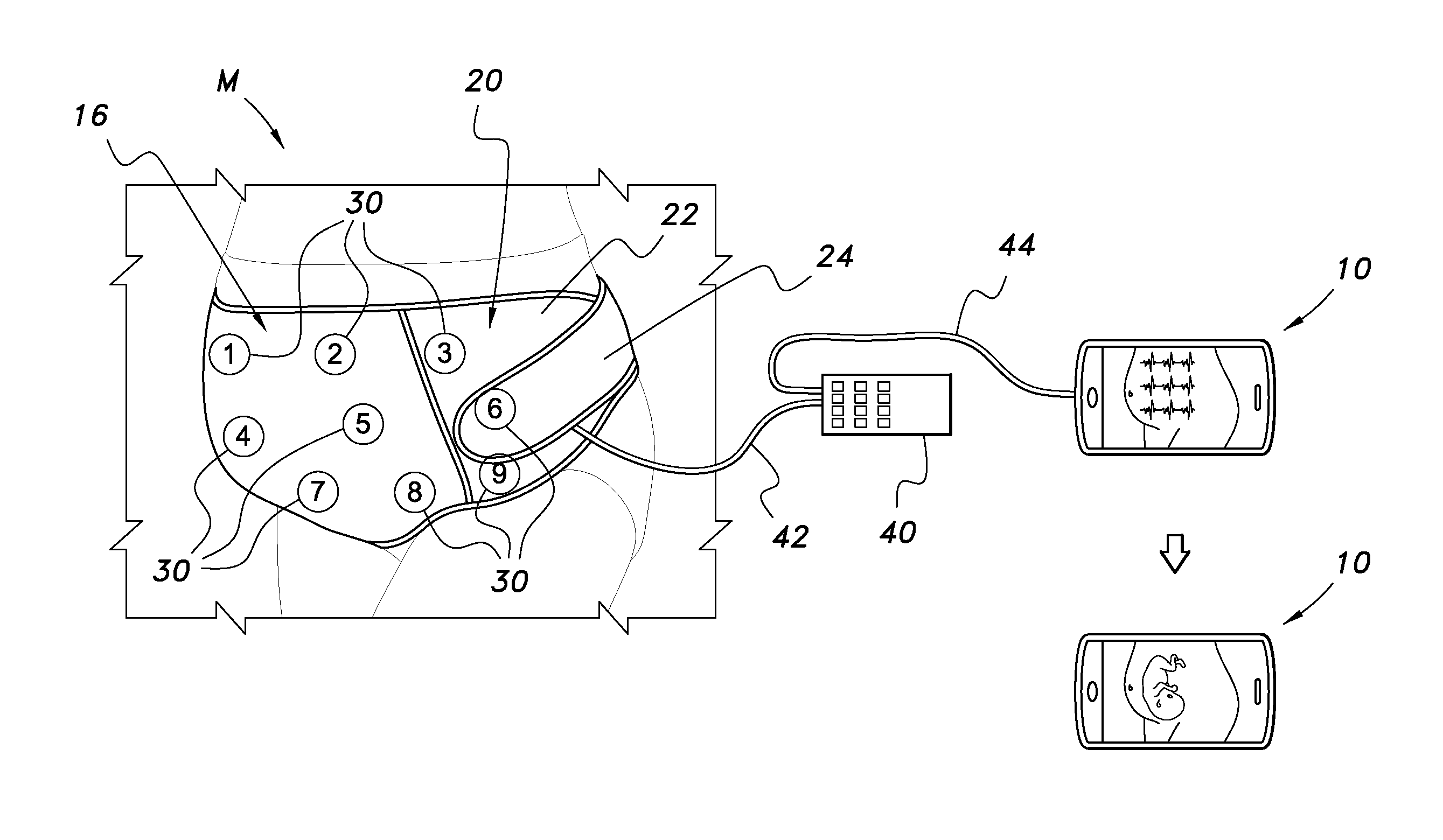
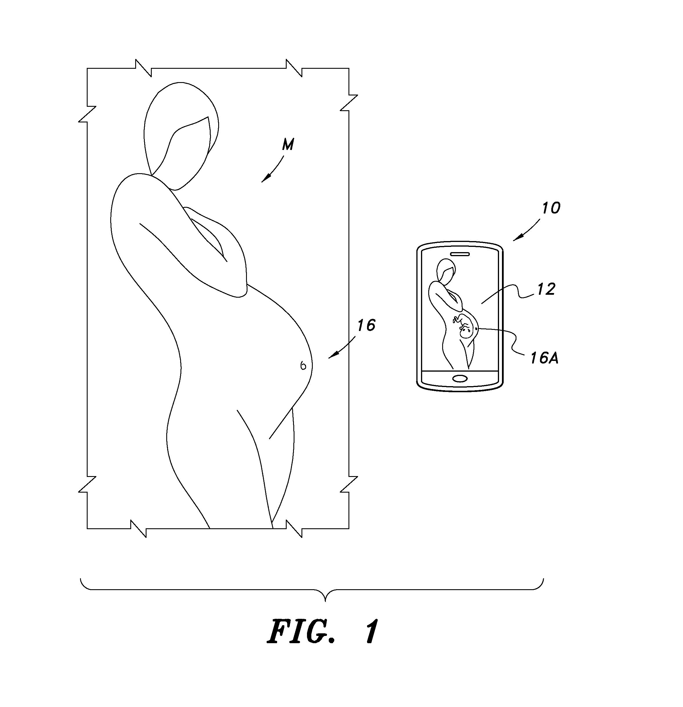
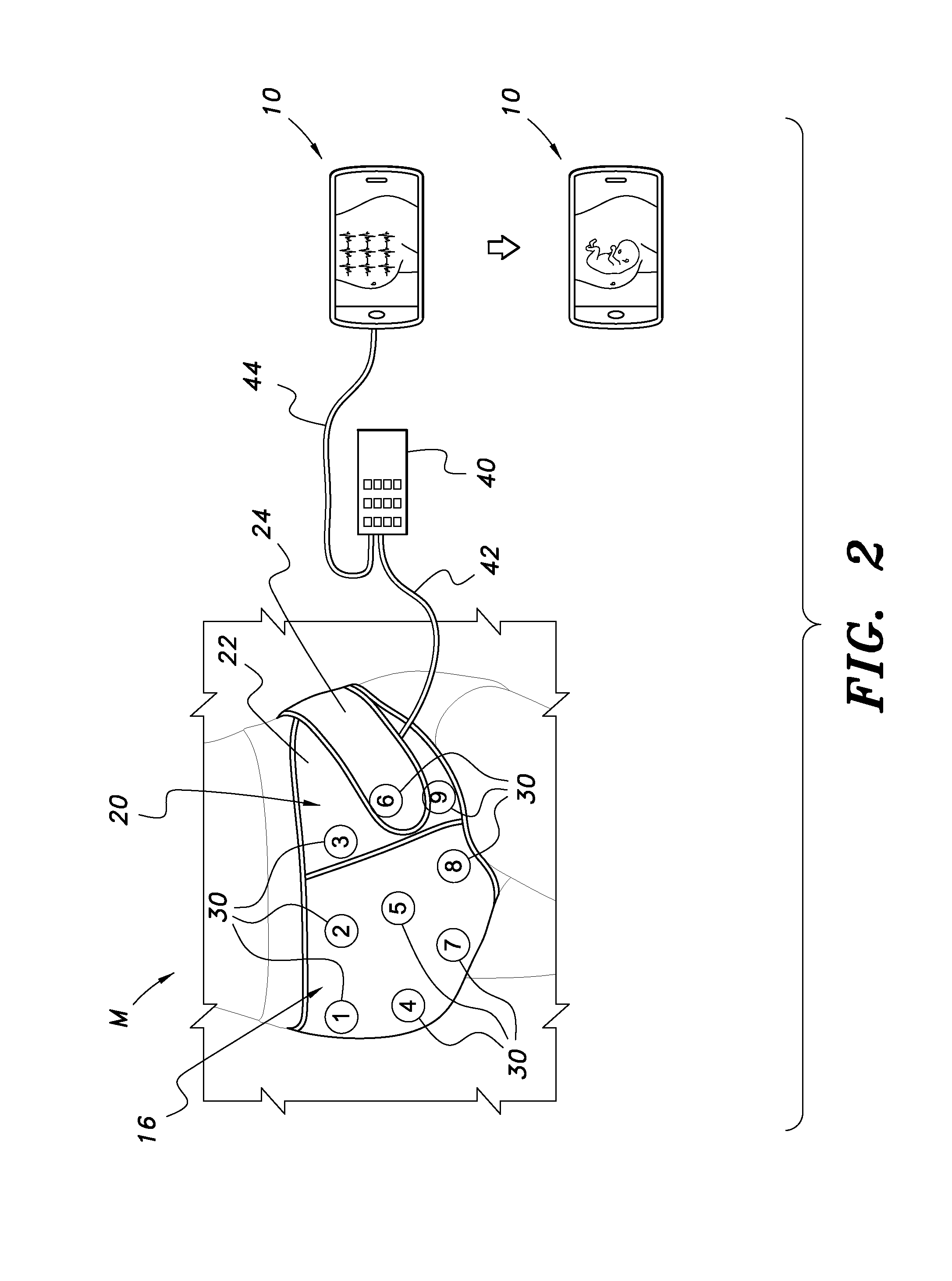
[0021]The present invention, discussed in detail hereunder, relates to a portable device and a method for using the portable device to detect fetal heart rate and EEG signals, and to detect signs of normal and abnormal fetal development. The device of the present invention provides an Internet connection, and it serves as an apparatus for performing and analyzing fetal-EEG and ECG recordings in parallel with fetal visualization. Furthermore it is also capable of associating 3D maternal abdominal locations to fetal tissue-specific optical features (light absorption, reflection), and matches fetal body anatomy with functional organ-specific electric activity patterns.

[0022]In many countries, practically all pregnant women undergo routine obstetric ultrasound (US) examinations, once or several times during pregnancy. Usually, one of the examinations is done at 16 to 22 gestational weeks for detection of fetal anomalies. At this time of pregnancy, the migration of neurons into the fetal...


PUM


Abstract

A new, inexpensive and non-invasive fetal visualization process helps to locate fetal body parts and identify fetal positions without exposing the fetus to prolonged ultrasound irradiation. Associating location data with biological electrophysiology signal patterns and / or light absorption / reflection-related tissue-specific local fetal body composition data enables the generation of a 3D anatomical and functional map of the fetal body through the expecting mother's womb, which will be essential for long-term home monitoring of a fetus during pregnancy.

Description

CLAIM FOR PRIORITY AND CROSS-REFERENCE TO RELATED APPLICATIONS[0001]This application is a continuation-in-part of U.S. Ser. No. 14 / 068,951 filed on Oct. 31, 2013, now pending, which in turn is a continuation-in-part of U.S. Ser. No. 13 / 396,233 in the name of the inventor Marianna Kiraly filed on Feb. 14, 2012, now pending, which in turn claims the priority of Provisional Patent Application Ser. No. 61 / 627,626, filed on Oct. 14, 2011, in the name of the inventor Marianna Kiraly. These applications are hereby expressly incorporated herein in their entirety by reference thereto.FIELD OF THE INVENTION[0002]The present invention relates to basic developmental biology, portable medical obstetric procedures and devices, and more particularly to the non-invasive monitoring of the positions and certain body parts of a developing human fetus inside the mother's womb.BACKGROUND OF THE INVENTION[0003]Many of the developmental disorders of childhood—cerebral palsy, epilepsy, cognitive impairment...

Claims


Application Information

Patent Timeline
IPC IPC(8): A61B8/00A61B8/14A61B8/08A61B19/00A61B5/0478A61B5/0408A61B5/04A61B5/044A61B5/00A61B5/0205
CPCA61B8/02A61B8/4227A61B5/02411A61B5/0448A61B5/0476A61B2562/046A61B5/0077A61B5/4362A61B5/6823A61B8/0866A61B5/0205A61B5/0478A61B5/04017A61B5/044A61B19/54A61B2019/5437A61B5/0011A61B5/04085A61B8/488A61B90/39A61B2090/3937A61B5/282A61B5/288A61B5/291A61B5/316A61B5/339A61B5/369
Inventor KIRALY, MARIANNA
Owner KIRALY MARIANNA
Learn More