
Devices and methods for forming an anastomosis

a technology of anastomosis and device, applied in the field of medical methods and equipment, can solve the problems of presenting a substantial risk to the patient, affecting the patient's recovery, so as to achieve the effect of reducing the risk of recurren

Active Publication Date: 2014-08-21
BOSTON SCI SCIMED INC
View PDF5 Cites 75 Cited by
  • Summary
  • Abstract
  • Description
  • Claims
  • Application Information

AI Technical Summary

Benefits of technology

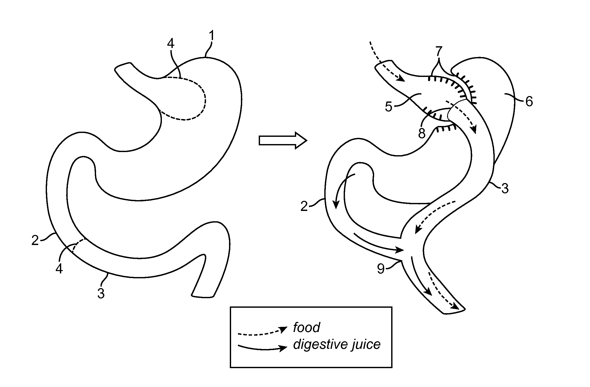
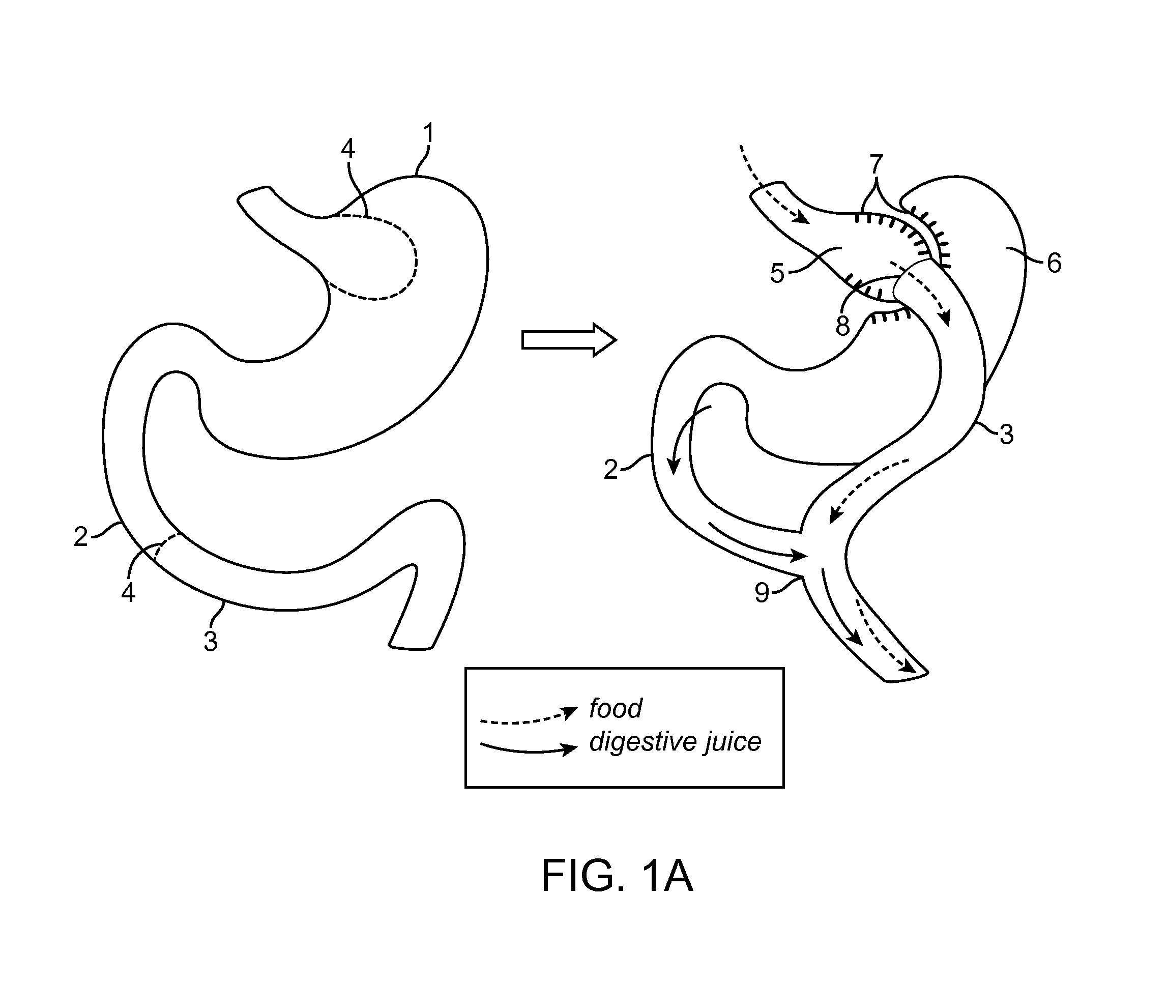
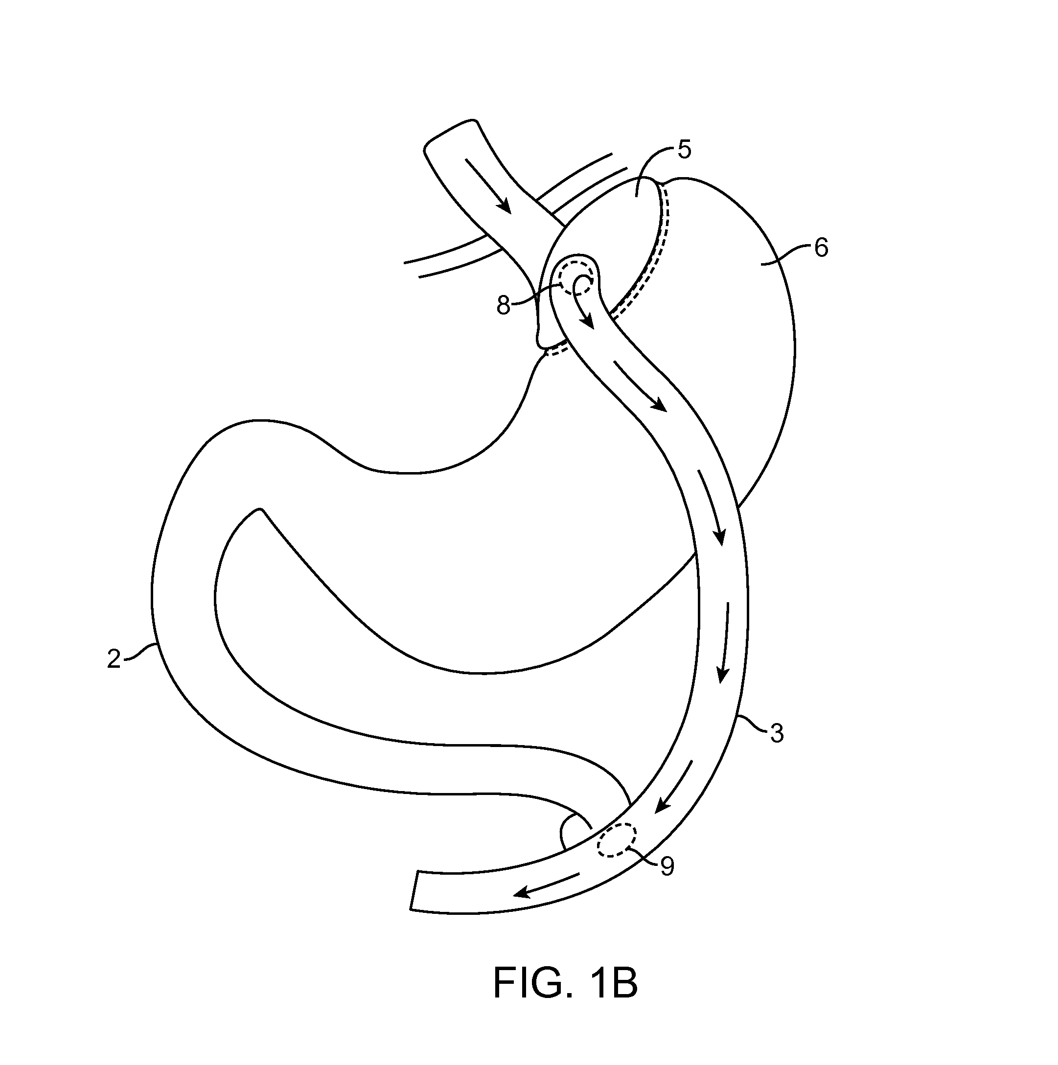
The present invention relates to improved stents for forming anastomosis within the digestive tract, laparoscopic and endoscopic tools for delivering stents, and methods for using them. The stents are designed to be easily delivered and retrievable, with a covered cylindrical region that allows for the flow of fluid, digested food, and partially digested food therethrough. The stents have a proximal flange and distal flange that can atraumatically engage bodily tissue. The technical effects of the invention include improved stent design, improved tool design, and improved methods for forming anastomosis.

Problems solved by technology

When initially forming such access penetrations, there is a significant risk of leakage from either or both of the access body lumen and the target body lumen into the surrounding space including, but not limited to the peritoneal cavity.
In some procedures, such as those involving bariatric, transgastric, or transduodenal bile duct access, loss of body fluid into surrounding tissues and body cavities can present a substantial risk to the patient.
The risk can be exacerbated when it is necessary to not only penetrate the luminal walls to gain initial access, usually with a needle, but to subsequently enlarge or dilate the initial penetration.
If the patient eats too much they will throw up.
It is possible to stretch the new gastric pouch but it is difficult.
Leakage can cause severe complications in the patient after surgery.
However, the stapling process can be lengthy using laparoscopic procedures and the diameter of the anastomoses formed from stapling vary between patients and doctors.
Other complications can also occur, such as the formation of a stricture at the anastomosis site.

Method used


Image

  • Devices and methods for forming an anastomosis
  • Devices and methods for forming an anastomosis
  • Devices and methods for forming an anastomosis

Examples


Embodiment Construction

[0103]Methods and devices are disclosed herein for forming an anastomosis. The devices and methods disclosed herein can be used to form multiple anastomoses. Tissue anchors and stents can be used to form the anastomosis. The anastomosis can be made using an endoscope, catheter, laparoscopic surgical instrument, laparoscopic, general surgery device, or a combination of one or more of these devices. The stents disclosed herein can be delivered using catheter based systems. In some embodiments the stents disclosed herein can be delivered using a laparoscopic device. In some embodiments the stents disclosed herein can be delivered using a rigid catheterless system. In some embodiments the stents can be delivered using a combination of a catheter and laparoscopic tools, for example the stent can be deployed with the catheter device and navigation and visualization assistance can be provided by the laparoscopic tools.

[0104]Improved access tools, improved stent designs to form consistent l...


PUM


Abstract

Devices and methods for deploying an anastomotic stent between portions of the gastro-intestinal (GI) tract are disclosed. The anastomotic stents are configured to atraumatically engage the tissue walls and to permit the flow of fluid, partially digested food, and food. The stents can be deployed using endoscopic catheter devices, laparoscopic tools, and combinations of both endoscopic tools and laparoscopic tools. Examples of anastomoses include anastomoses between the stomach and a portion of the intestines such as the jejunum. Anastomoses can also be formed between two closed ends of the intestines, such as two closed ends of the colon formed during a colon resection procedure. Anastomoses can also be formed between a fundal pouch formed during a gastric bypass procedure and the jejunum. Laparoscopic tools are disclosed to deploy a stent by selectively removing a radial restraint on a self expanding stent with the restraint removed through the laparoscopic access points.

Description

CROSS REFERENCE TO RELATED APPLICATIONS[0001]This application claims priority to U.S. Provisional Application No. 61 / 767,577 filed on Feb. 21, 2013 titled “Devices and Methods for Forming an Anastomosis”, the disclosure of which is incorporated by reference herein in its entirety.[0002]This application is related to U.S. Provisional Application No. 61 / 648,544 filed on May 17, 2012 and U.S. Provisional Application No. 61 / 727,629 filed on Nov. 16, 2012, the disclosures of which are incorporated herein by reference.INCORPORATION BY REFERENCE[0003]All publications and patent applications mentioned in this specification are herein incorporated by reference to the same extent as if each individual publication or patent application was specifically and individually indicated to be incorporated by reference.FIELD[0004]The present disclosure relates generally to medical methods and apparatus. More particularly, the present disclosure relates to methods and apparatus for forming an anastomosi...

Claims


Application Information

Patent Timeline
Patent Type & Authority Applications(United States)
IPC IPC(8): A61M27/00A61B17/11A61F5/00
CPCA61M27/002A61B17/1114A61F5/0076A61B2017/0034A61B2017/00477A61B2017/00876A61B2017/1132A61B2017/1135A61B2017/1139A61B2017/00278A61B2090/306A61B2090/378A61B2090/3925A61B2017/00004A61B2017/00867
Inventor BINMOELLER, KENNETH F.BROWN, PETERLEPULU, KEKEDONOVAN, RYAN
Owner BOSTON SCI SCIMED INC
Learn More