
Low intensity ultrasound therapy

a low-intensity ultrasound and therapy technology, applied in the field of hyperproliferative diseases and disorders, can solve the problems of necrosis, tissue damage, various kinds of tissue damage, etc., and achieve the effect of effective treatment and short period of tim

Active Publication Date: 2014-09-11
B G NEGEV TECH & APPL LTD
View PDF9 Cites 8 Cited by
  • Summary
  • Abstract
  • Description
  • Claims
  • Application Information

AI Technical Summary

Benefits of technology

This patent describes a method and device that uses ultrasound to treat cancerous and non-cancerous cells. The invention has the advantage of being able to work at any ultrasound frequency, making it more versatile than other methods. It uses low intensity ultrasound that is selective towards cancerous cells, minimizing damage to healthy cells. The invention can be administered continuously or intermittently, with short treatment periods. The duty cycle refers to the percentage of time the ultrasound is on and off, with values ranging from 10% to 90%. Overall, this technology offers a promising approach to treating cancer.

Problems solved by technology

All of these methods require low intensity ultrasound, so that to avoid damaging the skin more than needed to increase its permeability, and have a short range of impact.
Heating a tissue beyond a critical temperature for a period of time causes necrosis, the destruction of tissue.
At high enough acoustic intensities both cavitation (abrupt microbubble formation as a result of liquid medium exposure to ultrasound) and temperature elevation can occur, which in turn, cause various kinds of tissue damage.
Furthermore, a disadvantage of methods relying on ablation and / or thermal elevation is that the tissue damage is not limited to the cancerous tissue, but unselectively affects all the neighboring healthy tissue within the treated area.
However, this method is “blind” to cancerous cells having the same size of normal cells, and furthermore the frequencies taught in this application are unconventionally high for therapeutic uses, and reach 16 MHz and higher.
However, none of the studies conducted so far have been able to provide a therapeutically non-invasive selective treatment method of cancer and / or other proliferative diseases, which will avoid causing damage to a healthy tissue, cells or organs, while being solely based on ultrasound at low intensities, as opposed to HIFU, which is also non-invasive, but uses high intensities of ultrasound, and avoided damage to tissues only by its focusing mechanism.

Method used


Image

  • Low intensity ultrasound therapy
  • Low intensity ultrasound therapy
  • Low intensity ultrasound therapy

Examples


example 1

Effect of Ultrasound at Low Intensities on Cancerous Cell and Normal Cells

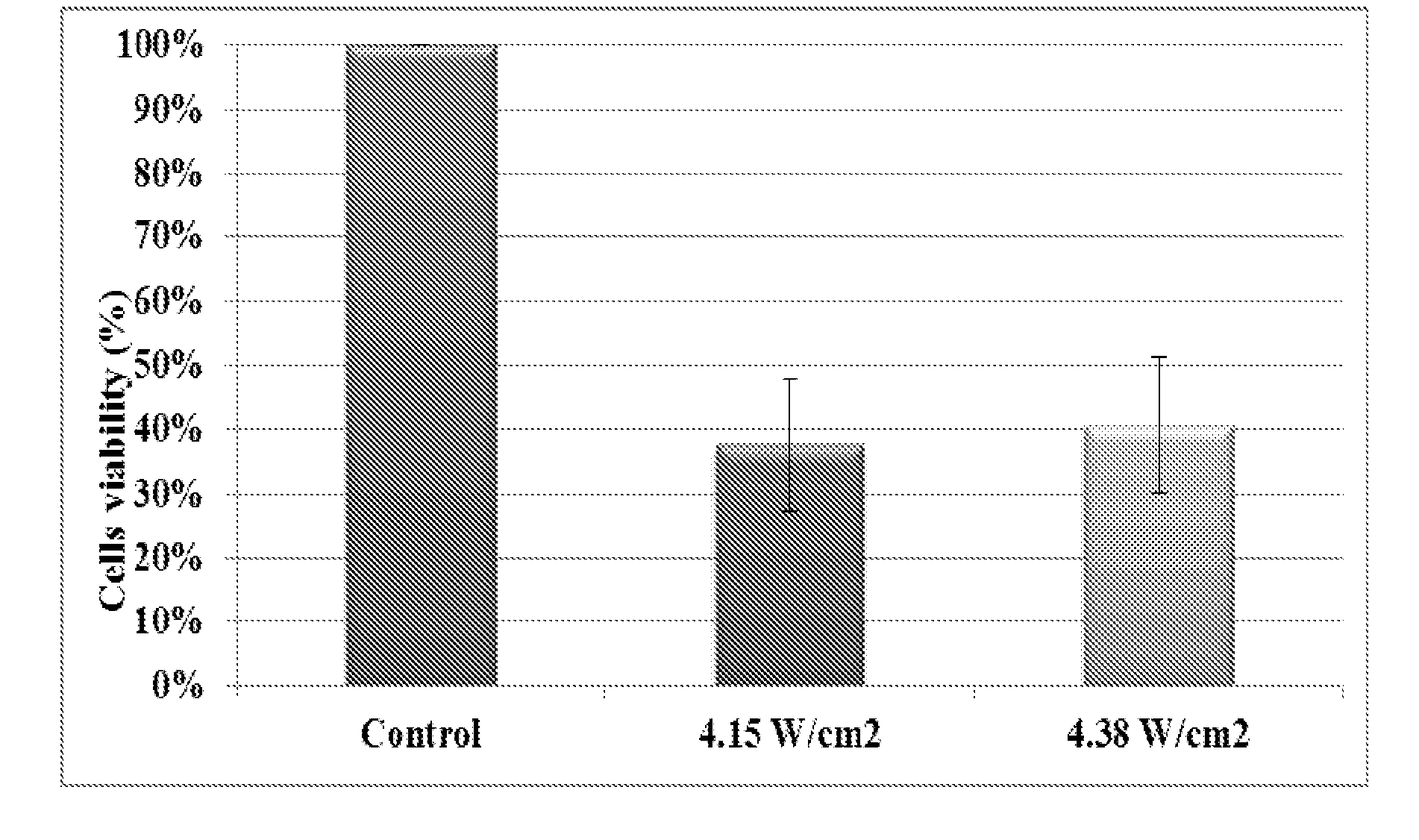
[0117]The effect of ultrasound at low intensities was tested on three cell lines: HeLa cells which are cancerous cells of ovarian cancer, NCI / ADR-RES (NAR) cells which are cancerous cells of ovarian cancer resistant to chemotherapy and MCF10A cells which are non-cancerous cells. The effects of intensity, duration and duty cycle of ultrasound on cells' mortality were evaluated.

[0118]Ultrasound transducer used for all of the experiments was with a frequency of 20 kHz (MISONIX, Model S-4000-010). Cell mortality was determined using a standard viability test using 3-(4,5-Dimethylthiazol-2-yl)-2,5-diphenyltetrazolium bromide (MTT) (available from Sigma, catalogue number M2128, CAS number: 298-93-1) that stains live cells only.

Cell Cultures

[0119]NAR cells were cultured in RPMI (Roswell Park Memorial Institute medium, Biological industries, 500 mL, Catalog: 01-109-1A) growth media containing 1% L-glutamine (Biologica...

example 2

Effect of Ultrasound Duty Cycle (DC) on Viability of Cancerous Cell and of Normal Cells

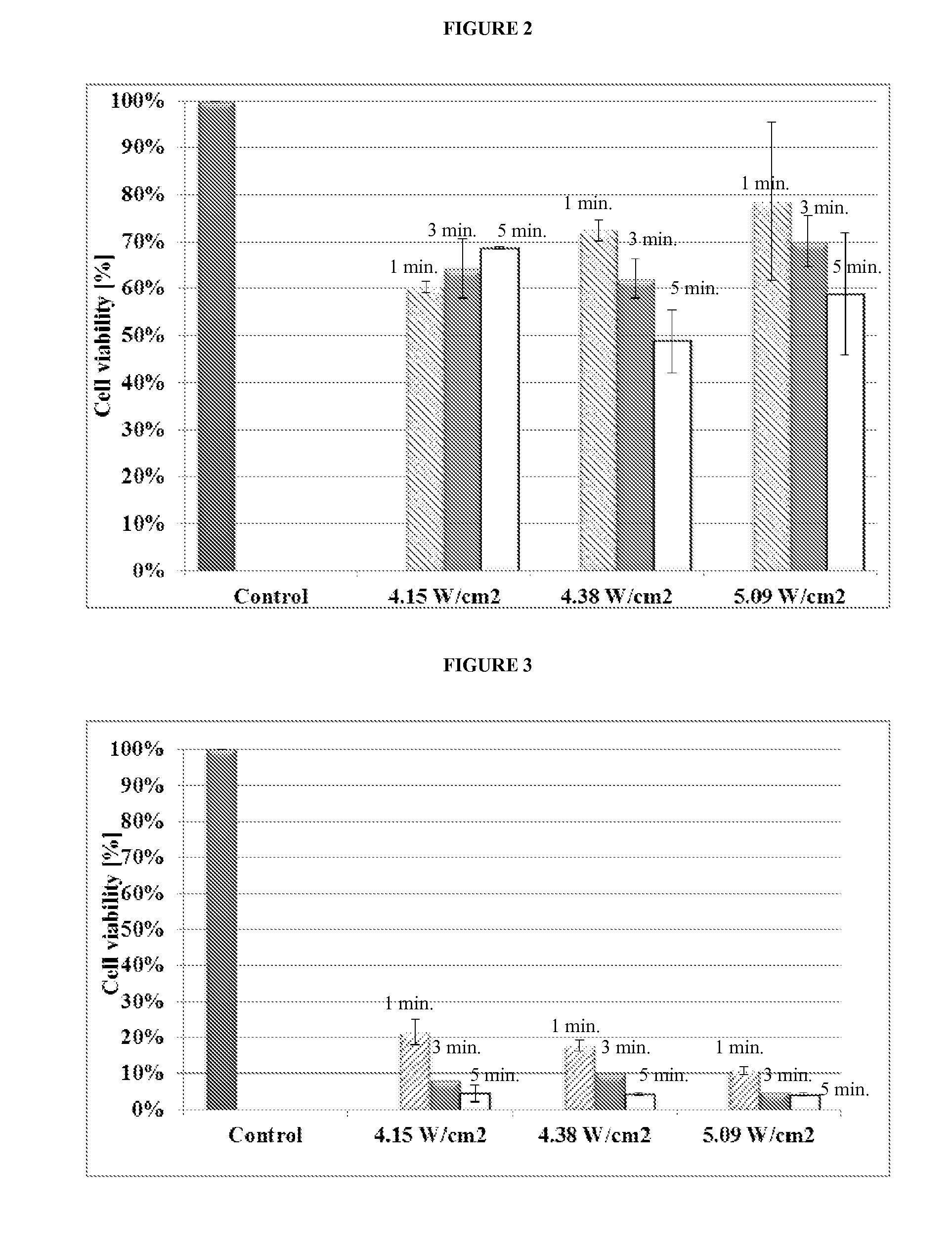
[0130]This study was conducted utilizing the most extreme ultrasound conditions of 5 minutes exposure at 5% amplitude (equals 5.09 W / cm2). This intensity was below the cavitational threshold intensity. Ultrasound frequency in this study was set to 20 kHz. In all previous experiments, duty cycle (DC) was set at 50% to avoid thermal effect. In this study 10% and 90% DC were also evaluated for their effect on NAR and MCF10A cell types. Cell cultures were prepared as described in Example 1.

[0131]The effect of DC on cells viability can be observed in FIGS. 4 and 5.

[0132]FIG. 4 shows MCF10A cell type average viability after 5 minutes exposure to ultrasound (20 kHz) at 5% amplitude for different US duty cycles (10%, 50% and 90%). (P value<0.1 compared to control experiment.)

[0133]FIG. 5 shows NAR cell type average viability after 5 minutes exposure to ultrasound (20 kHz) at 5% amplitude for different US ...

example 3

In-Vivo Study in BALB C / J Mice Bearing KHJJ Mouse Breast Adenocarcinoma

[0136]This study was carried out in BALB C / J white albino mice bearing KHJJ mouse breast adenocarcinoma. BALB C / J mice are infected by implantation under the skin of one of the mouse's thighs. A total of 18 mice were used, divided into three groups (6 mice per group, mixed genders). The three groups consisted of control group (group 1) and two groups assessing the effect of two different low intensity ultrasound protocols (groups 2 and 3). The second thigh (uninfected with cancer) in groups 2 and 3 serves as the reference organ. Group 1 was not being exposed to ultrasound at any stage. Group 2 and 3 were exposed to two different low intensity ultrasound protocols. Mice were anesthetized prior to ultrasound exposure with a formula containing Ketamine and Xylazine, administered in an amount depending on animal weight (Ketamine 75 mg / kg and Xylazine 5 mg / kg animal body weight).

[0137]The low intensity ultrasound prot...


PUM


Abstract

Methods of treating a subject suffering from a disease or a disorder associated with hyperproliferating cells. The method includes non-invasively administering to the subject ultrasound at a low intensity in a dose effective to selectively prevent the growth of the hyperproliferating cells or to eliminate the hyperproliferating cells in the subject, while substantially not affecting the cell viability of normal cells of the subject. The low intensity is pre-determined to be lower than a cavitational threshold intensity for a selected frequency.

Description

CROSS-REFERENCE TO RELATED APPLICATION(S)[0001]This application is based upon and claims the benefit of U.S. Provisional Patent Application No. 61 / 773,169, filed on Mar. 6, 2013. The entire contents of which are incorporated herein by reference.FIELD OF THE INVENTION[0002]The present disclosure relates generally to the field of treatment of hyperproliferative diseases and disorders. More specifically, methods of treating a subject afflicted with cancer, neoplasms or metastatic cancer using ultrasound are disclosed.BACKGROUND OF THE INVENTION[0003]Ultrasound has been used for several decades in the medical field, mostly as a diagnostic tool, and to a lesser extent as a therapeutic device.[0004]In most therapeutic methods ultrasound has been applied topically, either externally (on the skin surface) or internally (tympanal, buccal, vaginal or nasal applications). The common feature in this type of application is that the therapeutic effect results from increasing the penetration of th...

Claims


Application Information

Patent Timeline
Patent Type & Authority Applications(United States)
IPC IPC(8): A61N7/00
CPCA61N2007/0004A61N7/00
Inventor KOST, JOSEPHAZAGURI, AHARONYUDILEVICH, YANA
Owner B G NEGEV TECH & APPL LTD
Learn More