Enhanced expression of picornavirus proteins

a technology of picornavirus and protein, applied in the field of enhanced expression of picornavirus proteins, can solve the problems of disease and disorder, the efficiency of vaccine production can also be a barrier to commercial viability, and the ineffectiveness of killing virus, so as to enhance the production of polypeptides and induce immune responses

Inactive Publication Date: 2014-09-18
NOVAVAX
View PDF3 Cites 7 Cited by
  • Summary
  • Abstract
  • Description
  • Claims
  • Application Information

AI Technical Summary

Benefits of technology

This patent is about creating proteins that have a specific part at the beginning called a signal peptide. These signal peptides can be attached to other proteins to help them make their way through the body. This can be useful for making proteins that are not usually secreted or transmembrane proteins. These fusion proteins can be used to diagnose disease and to help induce immune responses. The polypeptides used can be from viruses, bacteria, or fungi.

Problems solved by technology

Killed viruses and live-attentuated viruses can be associated with deleterious effects, however, in some cases, the killed virus is not effective or indeed can exacerbate disease.
Efficiency of vaccine production can also be a barrier to commercial viability.
In some cases, a long lead time as required before production of the vaccine, and in other vases preparing the live virus before inactivation can result in disease and disorder.
The current vaccine is a killed whole virus vaccine with recognized limitations, such as those posed by growing the live virus prior to inactivation, poor growth of some strains, and reduced immunogenicity on storage.
However, while recombinant approaches overcome issues with production of live virus and offer the potential for rapid synthesis, producing suitable amounts of immunogens can be challenging for a variety of reasons.

Method used


Image

  • Enhanced expression of picornavirus proteins
  • Enhanced expression of picornavirus proteins
  • Enhanced expression of picornavirus proteins

Examples


example 1

Preparation and Expression of FMDV Polyprotein

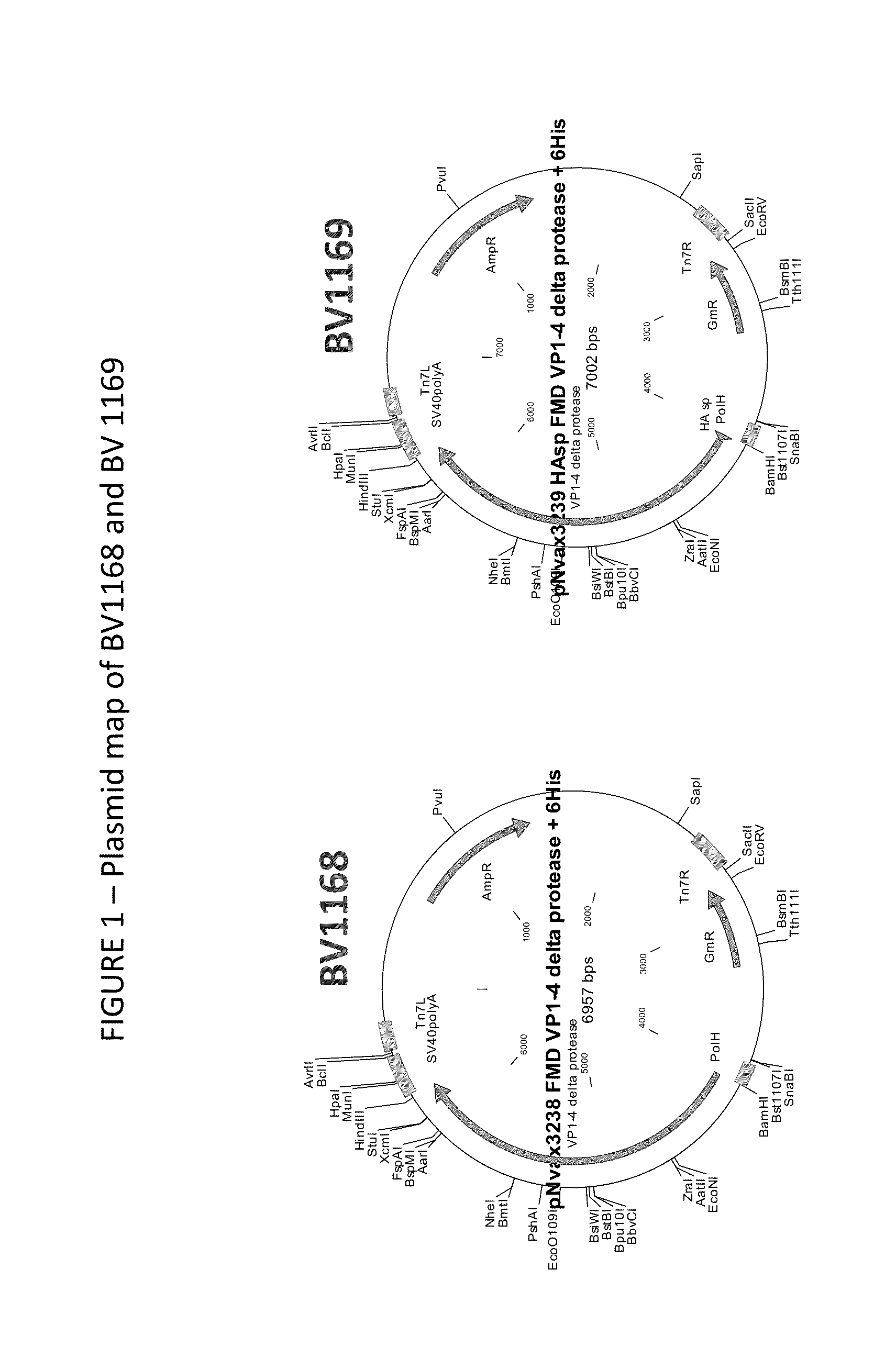
[0077]The FMDV P1 polyprotein was cloned into two vectors, BV1168 and BV1169 (FIG. 1). The vectors are identical except that BV1169 contained the A / Indonesia / 5 / 05 signal peptide sequence at the N-terminus (SEQ ID NO:3; MEKIVLLLAIVSLVK). In both cases, the P1 polyprotein contained an N-terminal His6 tag. The protein and nucleotide sequence encoded by BV1168 are shown in FIGS. 4C and 4D, respectively. The protein and nucleotide sequence encoded by BV1168 are shown in FIGS. 4A and 4B, respectively.

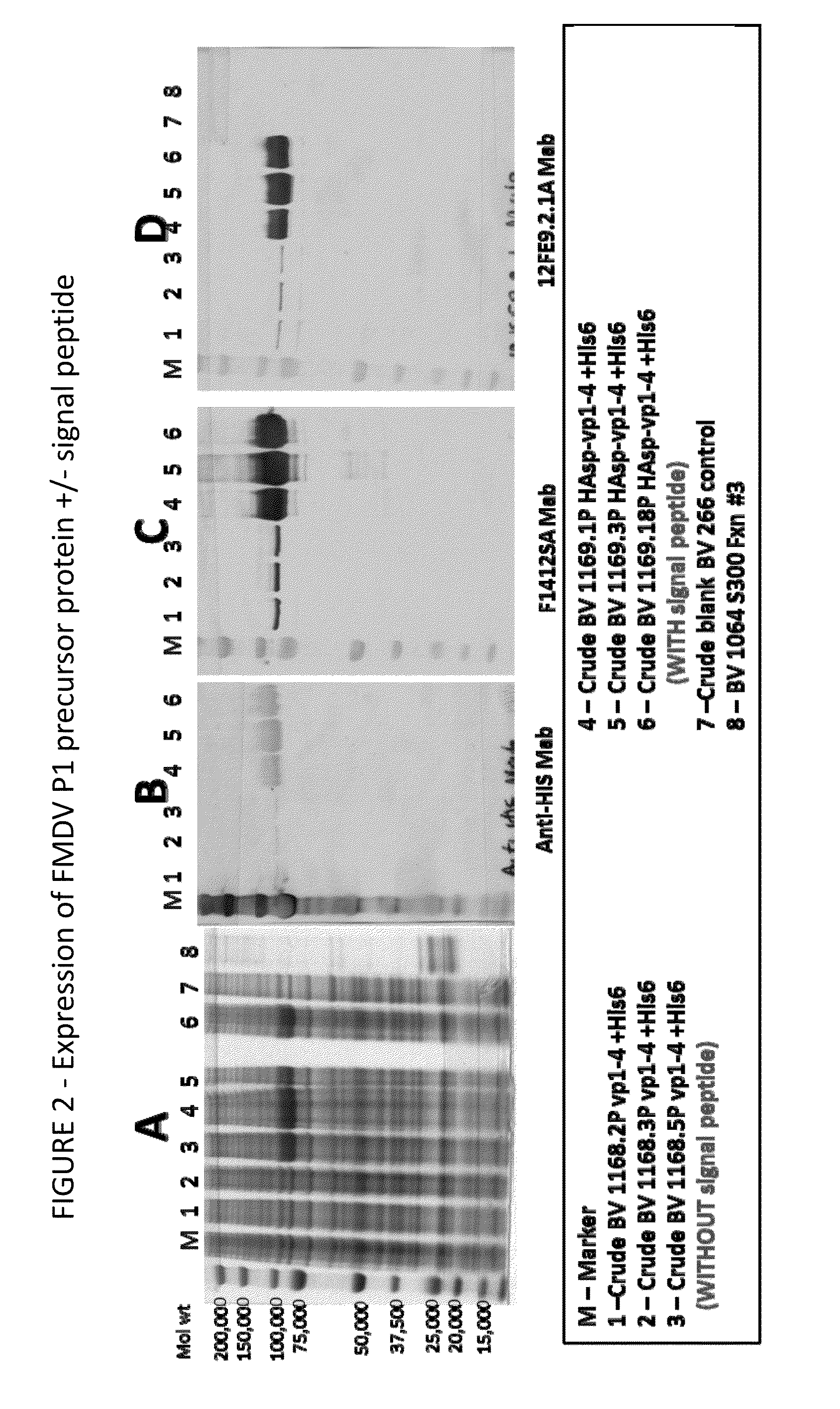
[0078]Sf9 cells were infected with three clones of BV1168 and BV1169 at an MOI of 0.5 ffu / cell and incubated at 27° C., 150 rpm for ˜70 hours. Crude harvest (cells and media) were analyzed by SDS-PAGE and western blot as shown in FIG. 2. The lanes are as follows: M-Marker. Lanes 1-3 show FMDV P1 protein with a hexa-histidine tag with out the signal peptide expressed from BV1168. Lanes 4-6 show FMDV P1 protein with a hexa-histidine tag and the sign...

example 2

Enhanced Expression of Single FMDV Polypeptide

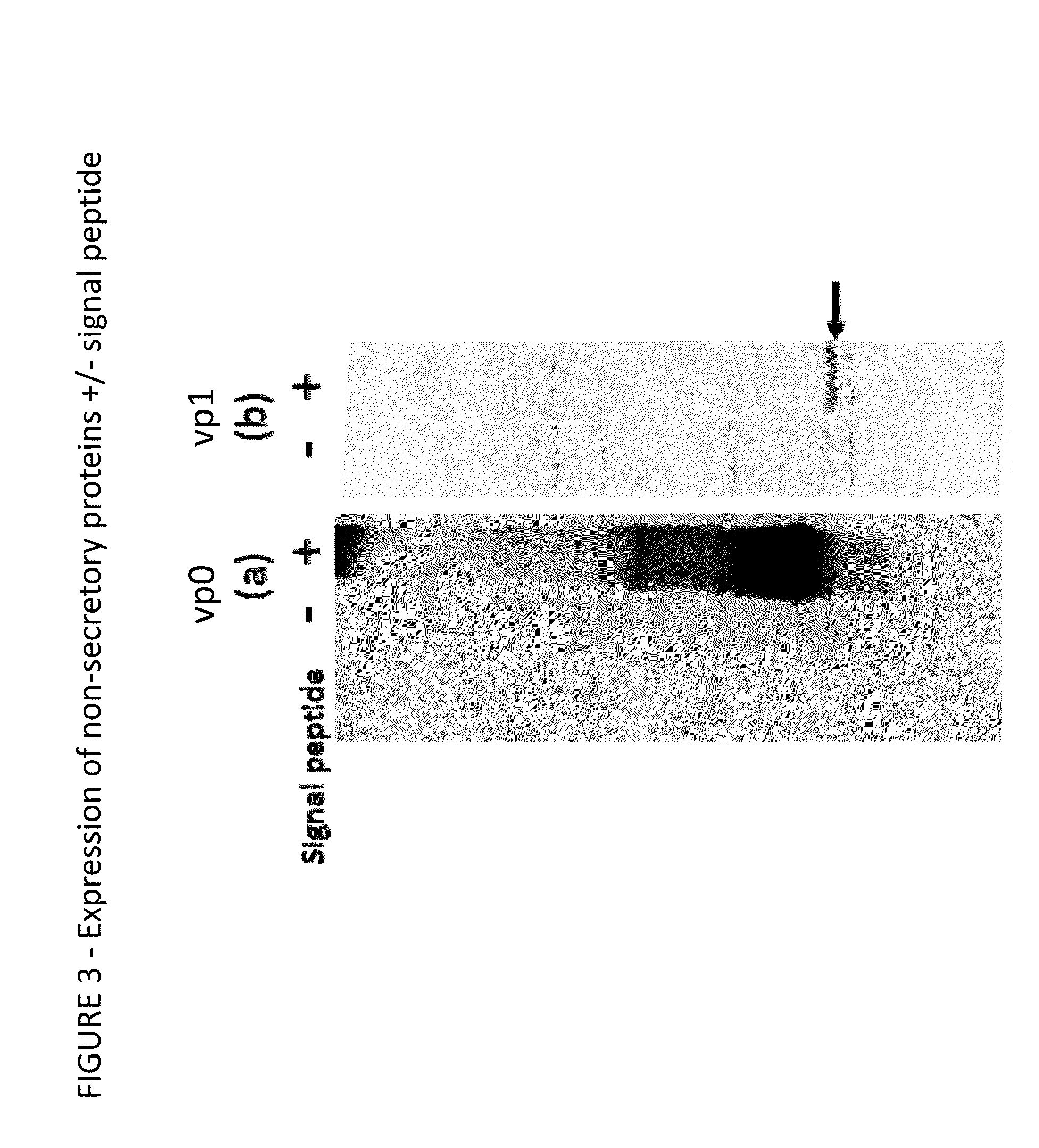
[0080]Expression of individual non-secretory proteins is enhanced when fused to a signal peptide. Sf9 cells were infected with Baculovirus expressing FMDV vp0 or FMDV vp1 proteins each with and without a signal peptide. Cells were harvested 70 hours post-infection. The harvested crude material (i.e. cells and medium) was analyzed by SDS-PAGE and by western blot using monoclonal antibody specific to the recombinant protein. FIG. 3, panel (a), the left panel, shows expression of FMDV vp0. FIG. 3, panel (b), the right panel, shows expression of FMDV vp1 protein. In each case, “+” recombinant protein contained a signal peptide, “−” recombinant protein did not have a signal peptide. The arrow indicates the expressed vp1 protein.

example 3

Expression of FMDV Polypeptide Containing Single and Multiple proteins

[0081]The signal peptide increases expression of expression of single proteins and proteins in tandem. FIG. 5 shows elevated expression with a variety of different constructs. Sf9 cells were infected with recombinant baculovirus (BV) expressing proteins 1, 2, 3, and / or 4 with (+) or without (−) a signal peptide and harvested ˜65 hrs post-infection. Crude samples (i.e. cells and medium) were harvested and analyzed by SDS-PAGE and western blots. The expressed recombinant proteins are indicated by the dot. BV1 expresses FMDV vp1. BV2 expresses FMDV vp0 vp3. BV3 expresses FMDV vp1, vp0 and vp3. BV4 expresses the FMDV P1 polyprotein (i.e., vp0-vp3-vp1). FIG. 5A shows a total protein stain. FIGS. 5B and 5C show binding of vp1 and vp2 antibodies, respectively. Note that the vp0 protein contains the vp2 protein. In each case, elevated expression of the proteins was obtained.


PUM

PropertyMeasurementUnit
Compositionaaaaaaaaaa
Pharmaceutically acceptableaaaaaaaaaa
Immunogenicityaaaaaaaaaa

Abstract

The disclosure provides fusion proteins containing N-terminal signal peptides fused to immunogenic polypeptides. The immunogenic polypeptides may be from viruses, bacteria, or fungi. The disclosure also provides elevated expression of the immunogenic polypeptides using the N-terminal signal peptide. The N-terminal signal peptides enhance synthesis of the protein, particularly where the protein is neither a secretory nor a transmembrane peptide. The fusion proteins may be used to diagnose disease and to induce immune responses.

Description

CROSS REFERENCE TO RELATED APPLICATIONS[0001]This application claims priority to U.S. Provisional Application Ser. No. 61 / 790,114, filed Mar. 15, 2013. The contents of which is hereby incorporated in full for all purposes.CROSS REFERENCE TO SEQUENCE LISTING[0002]This application incorporates by reference the contents of the 24 kb Sequence Listing titled “NOVV—52_SeqList.txt” created on Mar. 14, 2013.BACKGROUND[0003]Humans and animals suffer from diseases and disorders caused by pathogens, such as bacteria, fungi, and viruses. The use of vaccines to induce immunity against infection by a pathogen is well known in the art. Vaccines have historically been prepared using a variety of approaches, including killed viruses and live attenuated vaccines. Killed viruses and live-attentuated viruses can be associated with deleterious effects, however, in some cases, the killed virus is not effective or indeed can exacerbate disease.[0004]Efficiency of vaccine production can also be a barrier t...

Claims


Application Information

Patent Timeline
IPC IPC(8): C07K14/005A61K47/48A61K39/135
CPCC07K14/005A61K47/4833A61K39/135C07K2319/02C07K2319/21C07K2319/40A61K39/12C12N7/00C12N2760/16122C12N2770/32122C12N2770/32134C12N2770/32151A61K47/646
Inventor MASSARE, MICHAEL J.
Owner NOVAVAX
Why Eureka
Try Eureka