Zap-70 detection in chronic lymphocytic leukemia

a lymphocytic leukemia and detection method technology, applied in the field of methods for diagnosing disease, can solve the problems of difficult to standardize the protocol of flow cytometry for detection of zap-70, difficult to establish a clear cutoff value for flow cytometry, and difficult to implement a zap-70 approach in clinical flow cytometry laboratories, etc., to achieve the elimination of multiple steps in sample pretreatment, rapid and sensitive assay, and reliable and reproducible

Inactive Publication Date: 2014-10-09
CREATV MICROTECH
View PDF3 Cites 1 Cited by
  • Summary
  • Abstract
  • Description
  • Claims
  • Application Information

AI Technical Summary

Benefits of technology

[0010]In a second embodiment, the present invention is directed to immunoassays that can be used in the detection of selected proteins in a test sample. The immunoassays can be used in separately or in conjunction with the diagnostic methods of the present invention. In one aspect of this embodiment, the immunoassays can be used for the detection of ZAP-70 in a test sample, such as a population of B cells. The B cells may be from patients with chronic lymphocytic leukemia (CLL), for example (e.g., leukemic B cells). In conjunction with the detection of ZAP-70 in a test sample, the immunoassay is termed the VeriZAP™ assay system. The novel immunoassay system has the advantage of not including a step of conventional flow cytometry. The immunoassay system comprises a method for detection of ZAP-70 protein in a liquid-phase from a lysed population of cells (e.g., leukemic B cells) rather than counting whole cells expressing the detectable ZAP-70 marker.
[0011]Another aspect of the invention includes reagents, protocols and instrumentation useful in performing the immunoassays, including preparation of populations of B cells from a blood sample, extraction of intracellular ZAP-70 from the B cells, a liquid-phase fluorescence antibody assay, and measurement of ZAP-70 signal. In particular, populations of cells (e.g., leukemic B cells) that can be subjected to the immunoassays of the invention can be isolated from patient blood by a variety of methods. The purified cells are lysed to release all of the intracellular ZAP-70 proteins into a cell lysate. The released ZAP-70 proteins are extracted by immunomagnetic separation followed by detection with the fluorescence immunoassay. The invention provides a rapid and sensitive assay system for quantitative detection of ZAP-70 expression in the cells without use of the conventional flow cytometry. Further, the immunoassay system is simple, reliable and reproducible with the added advantage of eliminating multiple steps in sample pre-treatment, such as red blood cell lysis, cell fixation or permeabilization. The fluorescent ZAP-70 signal achieved using the immunoassay system of the invention is linear over a wide dynamic range, which enables quantitative assessment of small changes in ZAP-70 expression over the course of the disease and in response to therapeutic intervention.

Problems solved by technology

However, attempts to implement a ZAP-70 approach in clinical flow cytometry laboratories have been problematic since many commercially available antibodies give unreliable results (Admirand et al., 2010, Mod Pathol.
Due to these variations, it is difficult to standardize the protocols of flow cytometry for detection of ZAP-70.
Further, flow cytometry also suffers from lack of establishment of a clear cutoff value.
Therefore, a clear cutoff cannot be established for distinguishing the aggressive type from the indolent type of CLL (U.S. Pat. No. 7,759,076).
These methods are designed for qualification of total human ZAP-70 or for assaying kinase activity, and they are typically expensive and require assay times of from 4 hours to overnight.
Translating these methods into the clinic laboratory as routine diagnostic tools has not been achieved because they have not been standardized and validated, resulting in the lack reproducibility and multiple issues related to assay time, cost, complexity and data analysis.
Currently, a reliable and quantitative method for assessing ZAP-70 in leukemic cells is lacking.

Method used


Image

  • Zap-70 detection in chronic lymphocytic leukemia
  • Zap-70 detection in chronic lymphocytic leukemia
  • Zap-70 detection in chronic lymphocytic leukemia

Examples


example i

Preparations of Assay Reagents, Controls and Cell Lysates

[0104]Preparations of Positive Control, Antibodies and Reagents.

[0105]The immunoassay is based on quantitative, rather than qualitative, analysis. Improvements include use of controls of ZAP-70-positive cells (Jurkat cells) and ZAP-70-negative cells (normal B cells). In particular, purified recombinant ZAP-70 protein is included as target molecule control for validation and construction of standard curve. Human lymphocyte-derived cell lines can be obtained from the American Type Culture Collection (ATCC). One cell line, commonly used as ZAP-70 positive control, is the E6-1 clone of wild-type Jurkat cells (ATCC No. TIB-152). This cell line expresses ZAP-70 protein and serves as a positive control in the methods of the invention. The cell line was grown in the complete growth medium of RPMI-1640 according to ATCC protocols. All antibodies and reagents useful in the methods of invention are listed in Table 2. The biotinylated ant...

example ii

Selection of Antibodies and Fluorescence Dyes

[0112]Selection of Anti-ZAP-70 Antibodies.

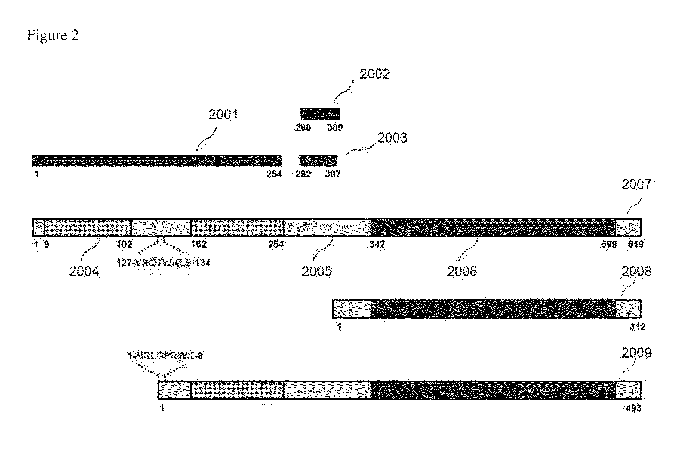
[0113]In this experiment, three different anti-ZAP-70 monoclonal antibodies were evaluated for detection of ZAP-70 in this study (clones 2F3.2, SBZAP and 1E7.2). The locations of the immunogens used to generate these antibodies are shown in FIG. 2. The clones 2F3.2 and SBZAP were used as a capture antibody, whereas 1E7.2 was used as a detector antibody, forming two combinations of antibody pairs: 2F3.2 / 1E7.2 and SBZAP / 1E7.2. The capture antibody 2F3.2 was immobilized on the surface of magnetic beads through biotin / avidin interaction. Phycoerythrin-Cy5.5 (PE-Cy5.5)-conjugated 1E7.2 (eBioscience) was used as the detector antibody. Two ZAP-70-positive controls, Jurkat cell lysate and CLL cell lysate (P8) were tested with these antibody pairs. The results showed that antibody pair 2F3.2 / 1E7.2 produced higher fluorescence signal than antibody pair SBZAP / 1E7.2 for Jurkat cell lysate (1.6-fold) and for C...

example iii

Detection of ZAP-70 in Jurkat Cell Lysates

[0116]Detection of ZAP-70 in Jurkat Cell Lysate.

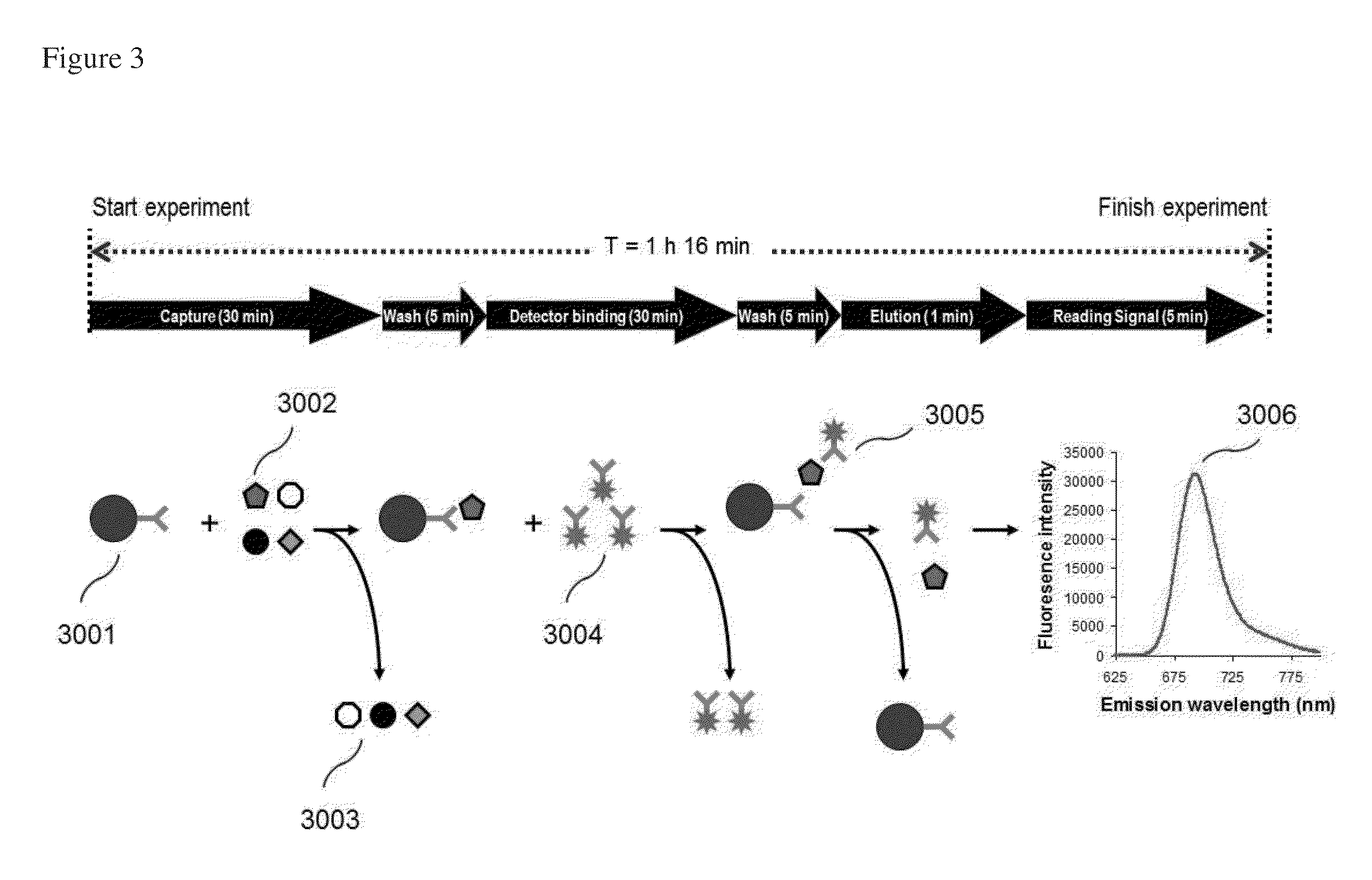
[0117]Jurkat cell lysate, the most common reference for ZAP-70, was used for evaluation of the method of the invention. To determine the detection sensitivity, two-fold serial dilutions of Jurkat cell lysate were prepared with a dilution solution (PBS containing 2% lysis buffer) to obtain final concentrations ranging from 63-1,000 cells / reaction. These serial dilutions were tested using PE-Cy5.5-conjugated detector antibody. Fluorescence signal was measured with excitation / emission wavelengths (Ex / Em) at 635 / 692 nm. Fluorescence intensity (FI) of each sample was measured with Signalyte™-II. In order to obtain a very low limit of detection, it was necessary to normalize the FI signal by subtracting the FI of a buffer control from the FI of each sample. This resulted in the normalized fluorescence intensity (NFI) of each sample. The relationship between NFI and Jurkat cell concentration is plotte...


PUM

PropertyMeasurementUnit
excitation angleaaaaaaaaaa
emission wavelengthsaaaaaaaaaa

Abstract

Detection of ZAP-70 expression provides important information about disease progression and overall survival in patients with chronic lymphocytic leukemia (CLL). The invention provides methods for diagnosing CLL in a subject, as well as methods for clearly distinguishing CLL patients with aggressive form of the disease. A consistent number of B cells from patient blood is isolated and lysed to release all of the intracellular ZAP-70 protein. The released ZAP-70 protein is subsequently extracted by immunomagnetic separation followed by detection with fluorescence immunosandwich assay. The ZAP-70 fluorescence signal is measured with Signalyte™-II spectrofluorometer. The VeriZAP™ assay is a simple, reliable, and reproducible method for quantitative detection of ZAP-70 in patient leukemic cells, and can be used as a prognostic test to distinguish indolent versus aggressive CLL patients.

Description

CROSS-REFERENCE TO RELATED APPLICATION[0001]This application claims benefit under 35 U.S.C. §119(e) of U.S. provisional application Ser. No. 61 / 555,199, filed Nov. 3, 2011, which is incorporated herein by reference in its entirety.FIELD OF THE INVENTION[0002]The present invention provides methods for diagnosing disease in a subject based by determining the amounts of one or more selected proteins in a test sample. In one aspect, the methods of the present invention can be used to discern between different forms of the same disease based on the relative amounts of the one or more selected proteins in the test sample. For example, aggressive disease can be distinguished from an indolent form of the disease. The diagnostic methods of the present invention are based on a novel immunoassay system that permits the quantitative detection of a selected protein in a test sample, and materials and equipment used therein. In one aspect of the invention, a fluorescence immunoassay, reagents and...

Claims


Application Information

Patent Timeline
Patent Type & Authority Applications(United States)
IPC IPC(8): G01N33/574
CPCG01N33/57426G01N33/57407G01N2800/56G01N33/582
Inventor TANG, CHA-MEIZHU, PEIXUAN
Owner CREATV MICROTECH
Why Eureka
Try Eureka