Coupling an Ultrasound Probe to the Skin

an ultrasound probe and skin technology, applied in the field of coupling ultrasound probes to the skin, can solve the problems of disadvantage in adhesion and achieve the effects of convenient removal, good acoustic coupling, and optimal adhesion of the prob

Active Publication Date: 2015-01-15
RESPINOR
View PDF2 Cites 5 Cited by
  • Summary
  • Abstract
  • Description
  • Claims
  • Application Information

AI Technical Summary

Benefits of technology

[0040]The combination of the tape with the roughened surface has been found to provide optimal adhesion of the probe to the skin, which allows ultrasound measurements such as those discussed above in relation to detecting extravasation and determining lung inspiration to be made effectively. The tape and probe may include optional features as discussed above.
[0041]In a preferred embodiment, the tape is sized to fit over the probe surface with a slight overlap. This ensures a good acoustic coupling between the transducers and the skin, and also enables the tape to be more eas

Problems solved by technology

Traditional ultrasound gels may not have the necessary properties for adhesion to an ultrasound probe, but instead are intended to be used wi

Method used


Image

  • Coupling an Ultrasound Probe to the Skin
  • Coupling an Ultrasound Probe to the Skin
  • Coupling an Ultrasound Probe to the Skin

Examples


Embodiment Construction

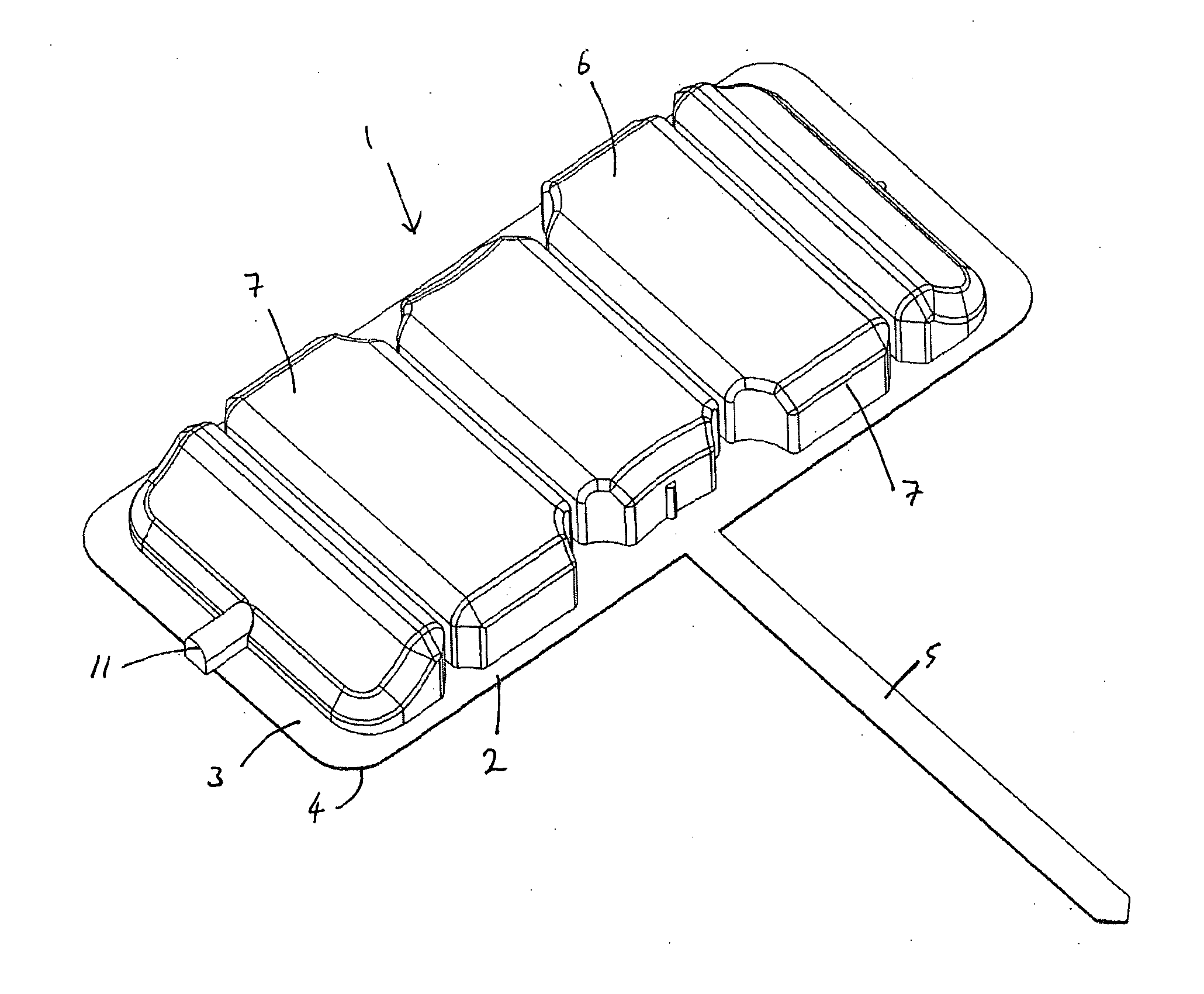
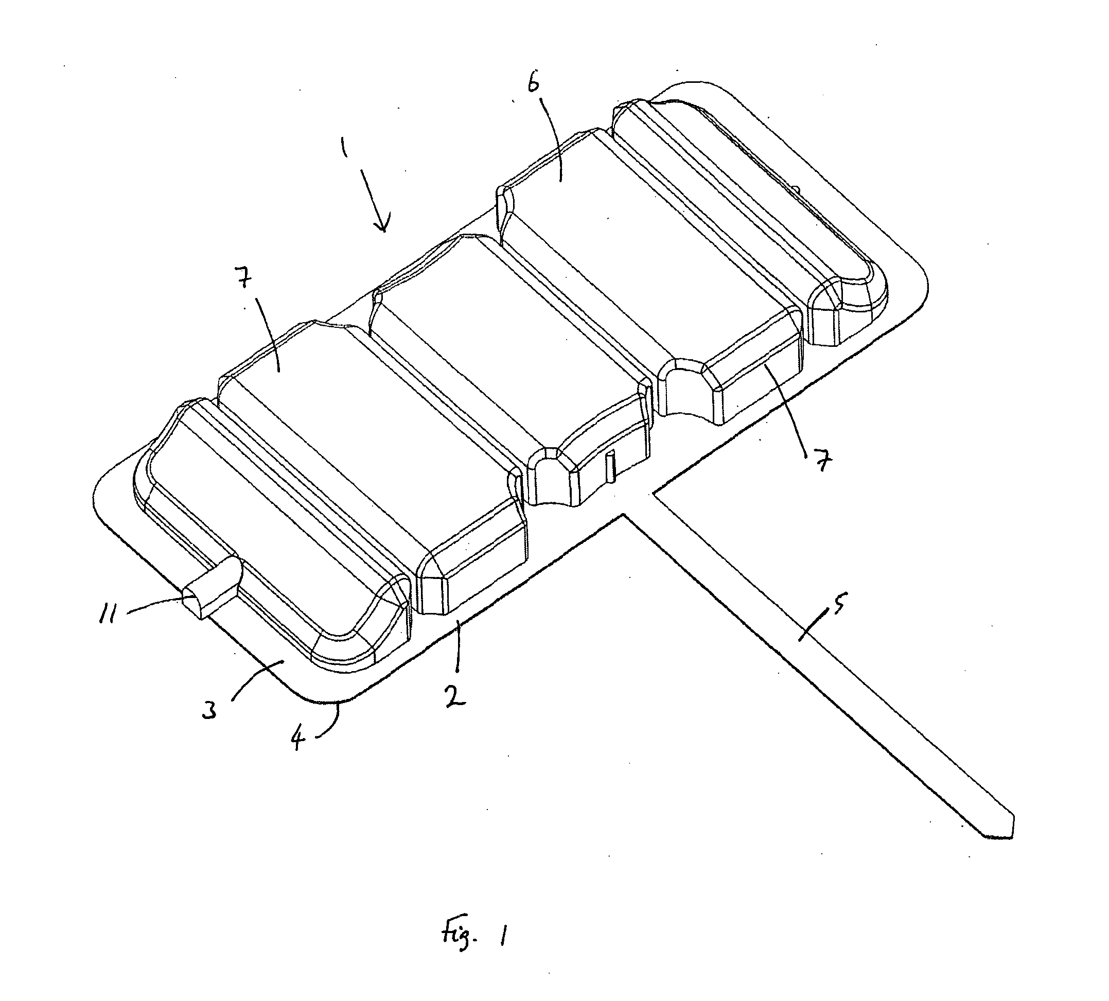
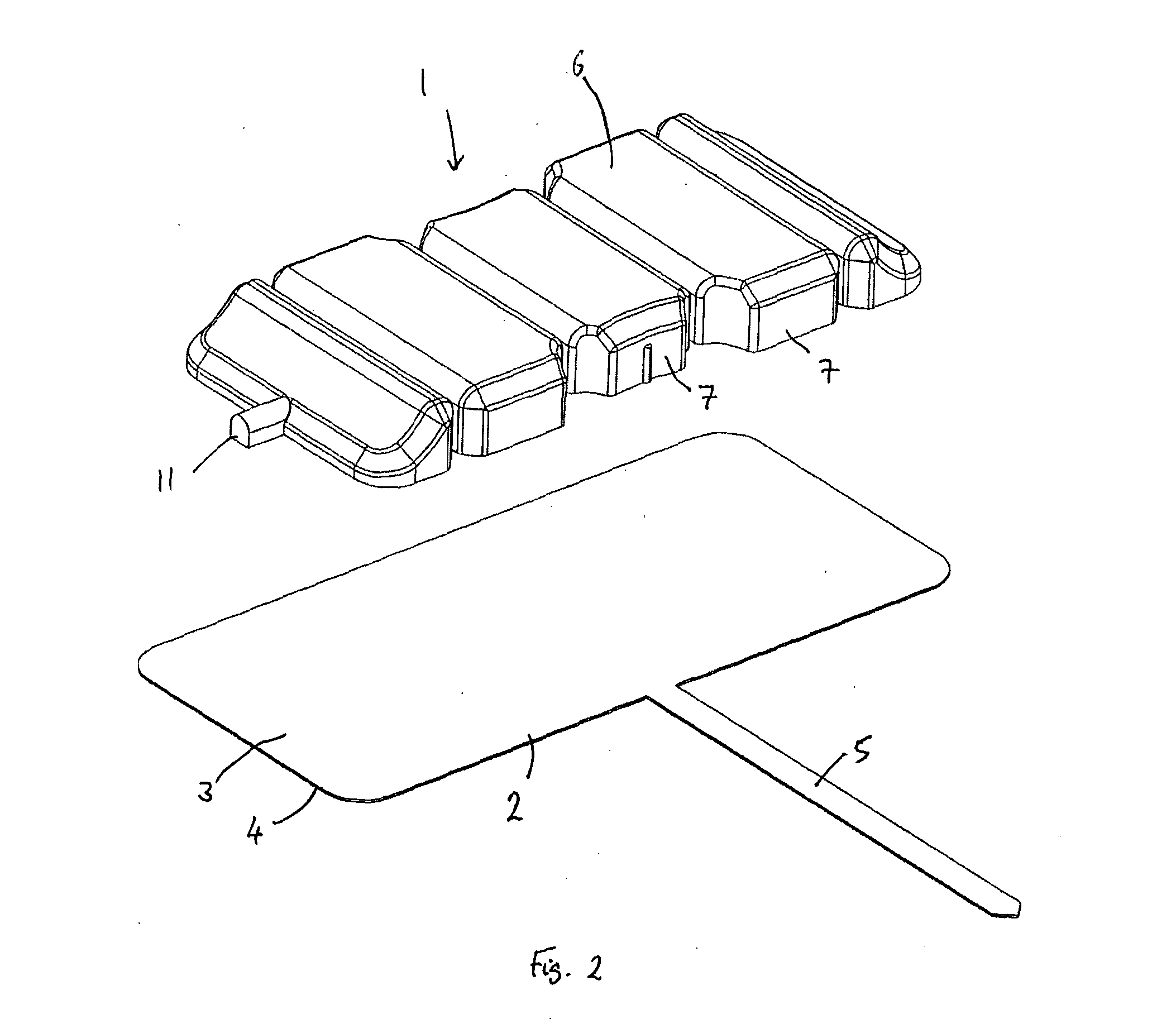
[0057]FIGS. 1 and 2 show an ultrasound probe 1 and tape 2 in perspective view, from above. The surface of the tape 2 that adheres to the skin is facing downward. The tape 2 is relatively thin, about 400 μm in the preferred embodiment. Tape thicknesses of between 200 to 500 μm may be used. The tape 2 comprises a first sonolucent gel layer 4, facing downward in FIGS. 1 and 2, and a second sonolucent gel layer 3. In the preferred embodiment, these layers are silicone adhesive adhered to a central sonolucent structural layer, which is preferably thin layer (e.g. 26 g / m2) of a non-woven material such as Reemay™. In the embodiment of FIGS. 1 and 2, the tape 2 includes an optional spacer 5, which extends outward from the probe 1 by a set distance. The spacer 5 can be used to ensure correct placement of the probe and tape combination relative to a desired location on the body.

[0058]The preferred silicone based adhesive tape is manufactured using a Silbione™ skin compatible silicone adhesive...


PUM

PropertyMeasurementUnit
Surface energyaaaaaaaaaa
Adhesion strengthaaaaaaaaaa
Transmissionaaaaaaaaaa

Abstract

A tape for securing an ultrasound probe to the skin may include a sonolucent silicone gel for transmitting ultrasound from an ultrasound transducer to the body. A method of manufacturing an adhesive silicone product may include a step of treating the adhesive composition or components of the composition to remove air or prevent the formation of air bubbles, in order to provide a sonolucent adhesive product.

Description

RELATED APPLICATIONS[0001]The present application is a continuation of U.S. patent application Ser. No. 13 / 642,865, filed on Nov. 16, 2012, now pending. U.S. patent application Ser. No. 13 / 642,865 is incorporated herein by reference in full.TECHNICAL FIELD The invention concerns devices and products for securing an ultrasound probe to the skin, and to related methods of manufacture.BACKGROUND OF THE INVENTION[0002]Ultrasound imaging or ultrasonography is an ultrasound-based diagnostic imaging technique used to visualize subcutaneous body structures including tendons, muscles, joints, vessels and internal organs for possible pathology or lesions. Obstetric sonography is commonly used during pregnancy and is widely recognized by the public. Various other diagnostic and therapeutic applications are practiced in medicine.[0003]In order to obtain a good quality image of the body structures of interest, it is necessary for the pulses of sound emitted by an ultrasound transducer to be pass...

Claims


Application Information

Patent Timeline
IPC IPC(8): A61B8/00C08J5/00C09J7/04C09J183/00C09J7/21C09J7/30
CPCA61B8/4236A61B8/4444C09J7/041Y10T428/2852C09J183/00A61B8/4483C08J2383/00C08J5/00A61B8/4281C08J5/128C09J2483/00C09J7/30C09J7/21Y10T442/674C09J183/04
Inventor BERARD-ANDERSEN, NICOLAYOLSEN, GJERMUND FJELD
Owner RESPINOR
Why Eureka
Try Eureka