Devices and methods for processing a biological sample

a biological sample and device technology, applied in the field of biological sample devices and methods, can solve the problems of difficult and inefficient disaggregation of complex biological samples, high probability of cells forming aggregates, and aggregation of cells

Inactive Publication Date: 2015-06-25
BECTON DICKINSON & CO
View PDF5 Cites 16 Cited by
  • Summary
  • Abstract
  • Description
  • Claims
  • Application Information

AI Technical Summary

Benefits of technology

[0004]Aspects of the present disclosure include methods for processing a biological sample. Methods according to certain embodiments include disrupting a biological sample to produce a disrupted biological sample and acoustically separating larger components from smaller components in the disrupted biological sample. In certain embodiments, methods may include monitoring aggregation while the biological sample is being proce

Problems solved by technology

This generally entails a manual step involving centrifugation, which may be time consuming, damage the cells, or lead to aggregation of the cells in the sample.
Further, because the cells often sit for extended periods of time, the likelihood of cells forming aggregates is high.
Tissue disaggregation and breaking up clumps of cells can often requires laborious purification and separation protocols in order to separate disaggregated cells from tissue and other biol

Method used


Image

  • Devices and methods for processing a biological sample
  • Devices and methods for processing a biological sample
  • Devices and methods for processing a biological sample

Examples


example 1

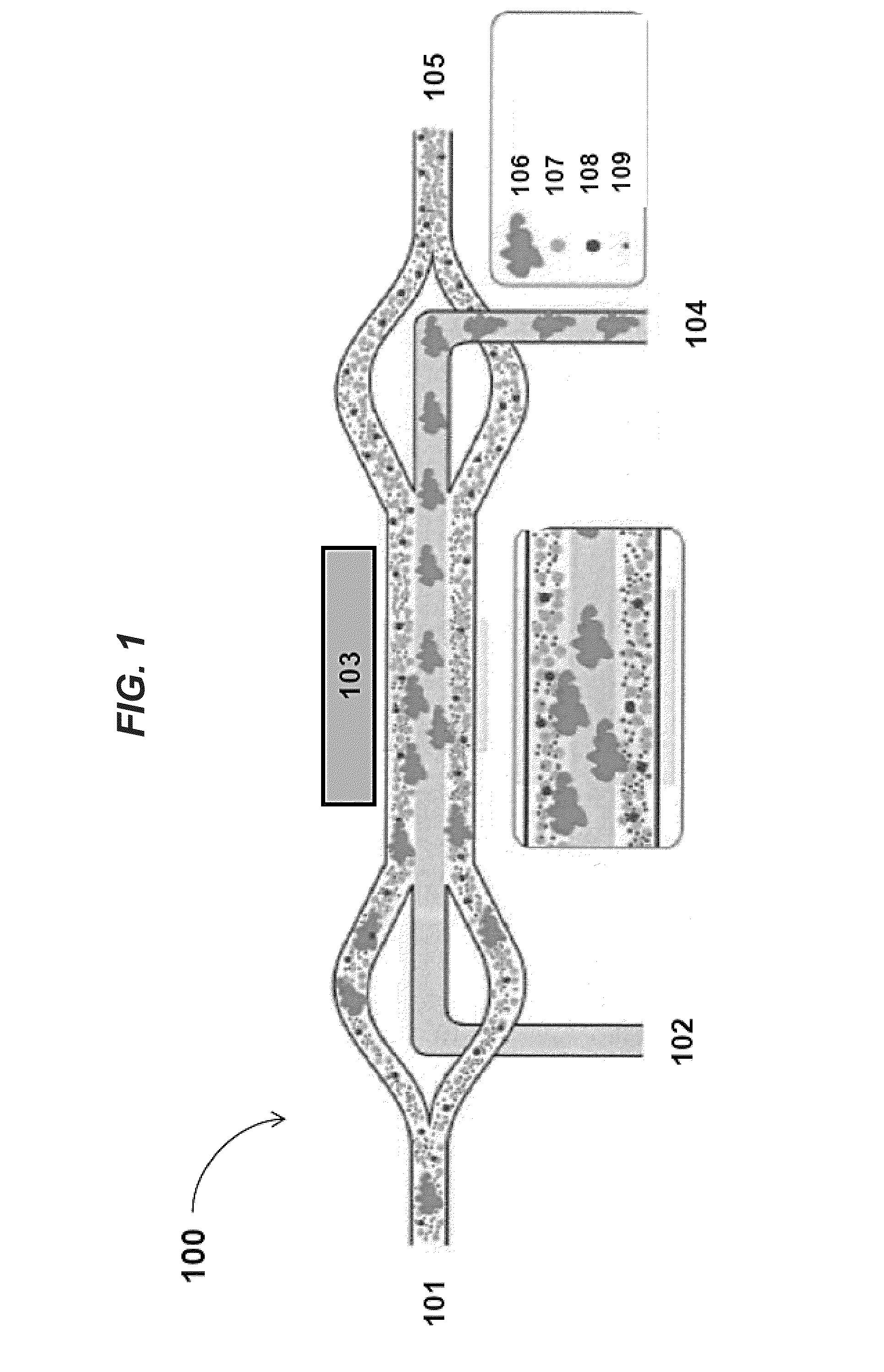
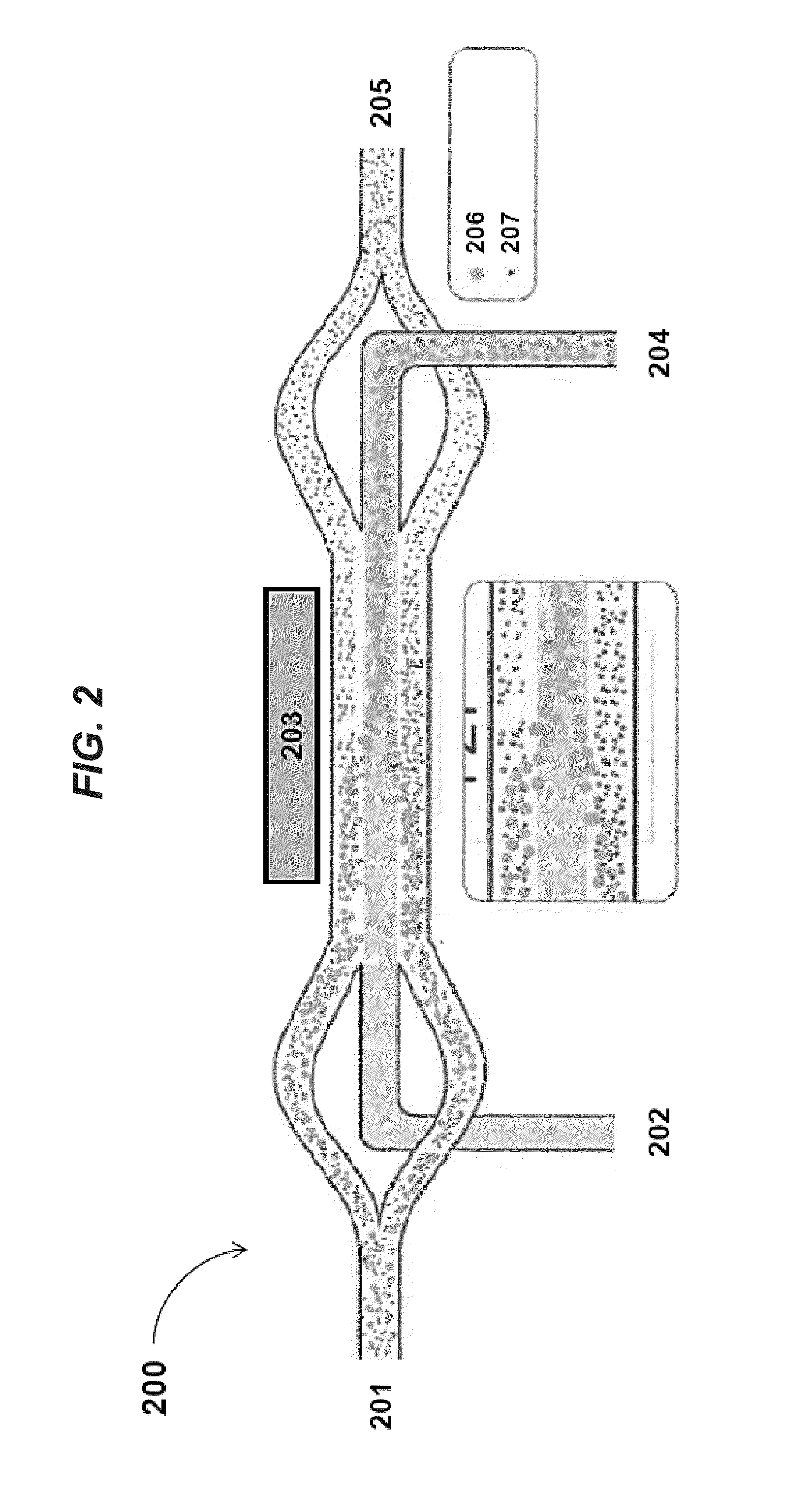
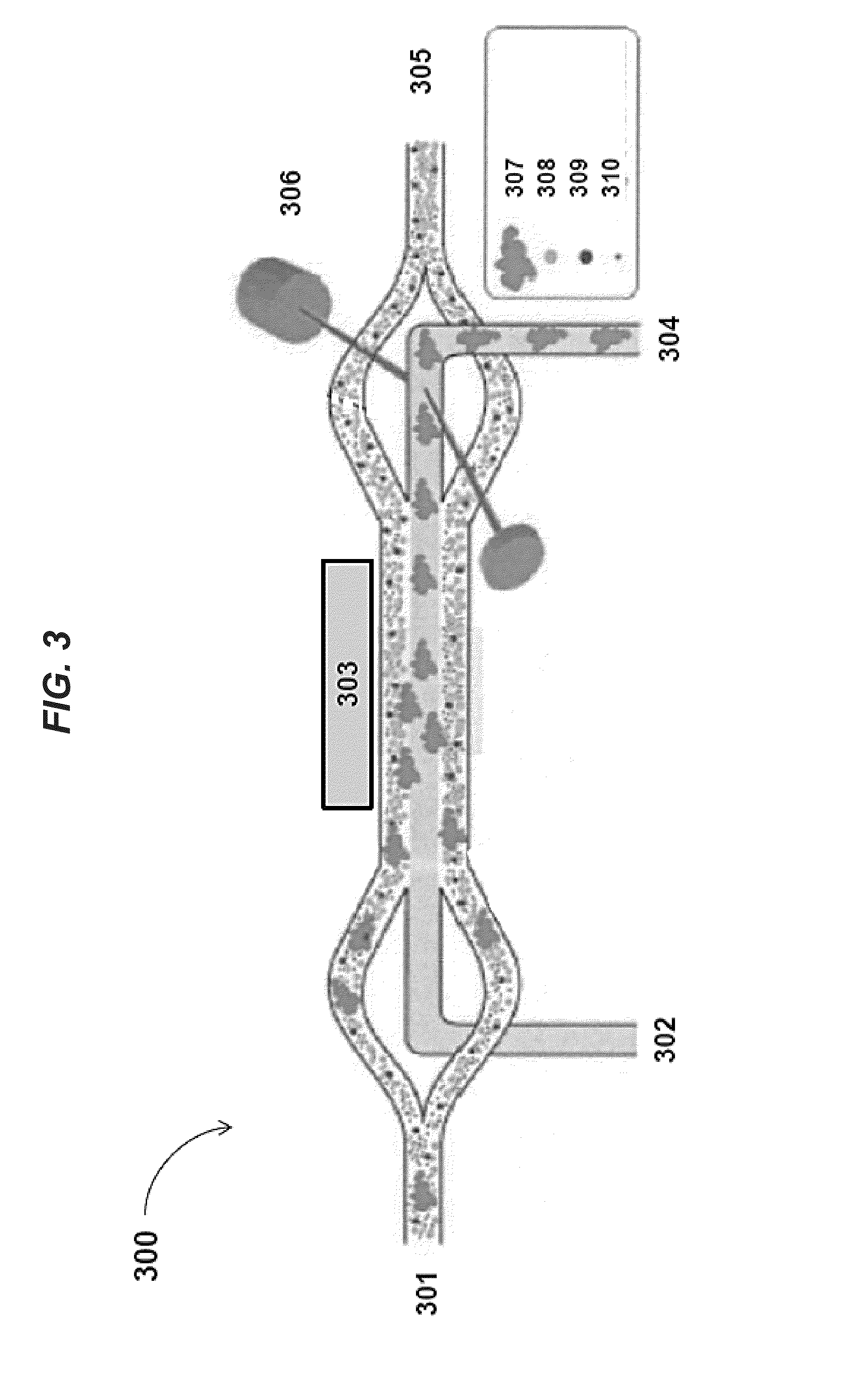
[0198]FIGS. 9a-b shows images of separating particles of different sizes in an acoustic concentrator device at low flow rates (e.g., 4 μL / minute). In FIG. 9a, when the piezoelectric transducer is not activated and no acoustic radiation force is exerted on the flowing sample, inertial forces cause only a few large particles (diameter about 50 μm) to move to the center stream within the channel of the acoustic concentrator device. In contrast, in FIG. 9b, when the piezoelectric transducer is operated at a power level of about 200 mV, the applied acoustic standing wave exerts radiation pressure to focus larger components (e.g., particles having diameters of about 50 μm) to the center stream while smaller particles (diameters less than 50 μm) remain in the laminating flow along the sides of the acoustic concentrator device channel.

example 2

[0199]FIGS. 10a-b shows that degree of hydrodynamic focusing in the acoustic concentrator device can be regulated by the ratio of liquid being diverted to side streams vs. that exiting the center stream. By doing this, this ratio can be adjusted to optimized light scatter measured by the feedback monitor positioned near the outlet of the acoustic concentrator device channel. FIG. 10a illustrates cells remaining tightly focused into the collection region at the center of the channel. FIG. 10b, on the other hand, illustrates that cells arriving at the center outlet are significantly diffused. In some instances, a minimum flow of fluid exiting out of the center channel of the acoustic concentrator device may be necessary for the stream to remain hydrodynamically focused after exiting the space affected by the acoustic radiation pressure of the applied standing wave. Here, the ratio of flow exiting the side outlets as compared to that exiting the center outlet does not exceed 5:1.


PUM


Abstract

Aspects of the present disclosure include methods for processing a biological sample. Methods according to certain embodiments include disrupting a biological sample to produce a disrupted biological sample and acoustically separating larger components from smaller components in the disrupted biological sample. In certain embodiments, methods may include monitoring aggregation while the biological sample is being processed. Methods, in certain instances, also include acoustically separating cells from cellular debris and non-cellular macromolecules as well as magnetically separating magnetically labelled moieties from unlabeled moieties. Systems, including a disrupter, one or more acoustic concentrator devices, feedback monitors and magnetic separation devices suitable for practicing the subject methods are also described.

Description

CROSS-REFERENCE TO RELATED APPLICATIONS[0001]Pursuant to 35 U.S.C. §119 (e), this application claims priority to the filing date of the U.S. Provisional Patent Application Ser. No. 61 / 920,394, filed Dec. 23, 2013, the disclosure of which is incorporated herein by reference.INTRODUCTION[0002]Flow cytometry is increasingly being used in disease diagnosis and monitoring, such as for prenatal and neonatal diagnosis of immunological abnormalities. Cells used in flow cytometry often must be washed and concentrated prior to use. This generally entails a manual step involving centrifugation, which may be time consuming, damage the cells, or lead to aggregation of the cells in the sample. Further, because the cells often sit for extended periods of time, the likelihood of cells forming aggregates is high.[0003]Processing a biological sample to obtain single cells from cell aggregates, tissues or organs is often necessary for many laboratory tests. Tissue disaggregation and breaking up clumps...

Claims


Application Information

Patent Timeline
IPC IPC(8): G01N1/40G01N15/14G01N1/28
CPCG01N1/4077G01N2015/142G01N15/1404G01N1/286B01L3/502753B01L3/502776B01L2200/0652B01L2300/0816B01L2300/0864B01L2400/043B01L2400/0436B01L2400/0487G01N2001/4094G01N2015/1006G01N2015/1418
Inventor WARNER, BRIAN DAVIDYU, LIPINGGHANEKAR, SMITACAO, JIANYING
Owner BECTON DICKINSON & CO
Why Eureka
Try Eureka