Planar optical waveguide based sandwich assay sensors and processes for the detection of biological targets including protein markers, pathogens and cellular debris

a technology of optical waveguides and sensors, applied in the field of sandwich assay sensors and processes for the detection of biological targets, can solve the problems of high processing costs, limited shelf life, and high cost of enzymes

Inactive Publication Date: 2006-01-26
TRIAD NAT SECURITY LLC
View PDF1 Cites 12 Cited by
  • Summary
  • Abstract
  • Description
  • Claims
  • Application Information

AI Technical Summary

Benefits of technology

[0016] The present invention further provides a sandwich assay process including: injecting a biological target-containing sample into a sensor cell including recognition ligands bound to a film on a single mode planar optical waveguide, the film from the group of a membrane, a polymerized bilayer membrane, and a self-assembled monolayer containing polyethylene glycol or polypropylene glycol groups therein, the recognition ligands adapted for binding to selected biological targets; maintaining the biological target-containing sample within the sensor cell for a time sufficient for a binding event to occur between selected biological targets within the sample and the recognition ligands adapted for binding to selected biological targets; injecting a solution into the sensor cell, the solution including a reporter ligand adapted for binding to bound selected biological targets; and, interrogating the biological target-containing sample within the sensor cell with excitation light from the waveguide, the excitation light provided by an evanescent field of the single mode penetrating into the biological target-containing sample to a distance of less than about 200 nanometers from the waveguide thereby interacting with any bound reporter ligand within a distance of less than about 200 nanometers from the waveguide and resulting in a detectable signal.
[0017] In another embodiment, the present method provides an assay process including:

Problems solved by technology

Several problems occur in the use of enzymes as amplifiers in immunoassays including:
Inevitably, the shelf-life is limited by the stability of the enzyme.
2) Enzymes are expensive as they are derived from living sources and require substantial processing costs.
However, even Horseradish Peroxidase costs about $5/mg or $5000/g.
3) The labeling of antibodies

Method used


Image

  • Planar optical waveguide based sandwich assay sensors and processes for the detection of biological targets including protein markers, pathogens and cellular debris
  • Planar optical waveguide based sandwich assay sensors and processes for the detection of biological targets including protein markers, pathogens and cellular debris
  • Planar optical waveguide based sandwich assay sensors and processes for the detection of biological targets including protein markers, pathogens and cellular debris

Examples


example 1

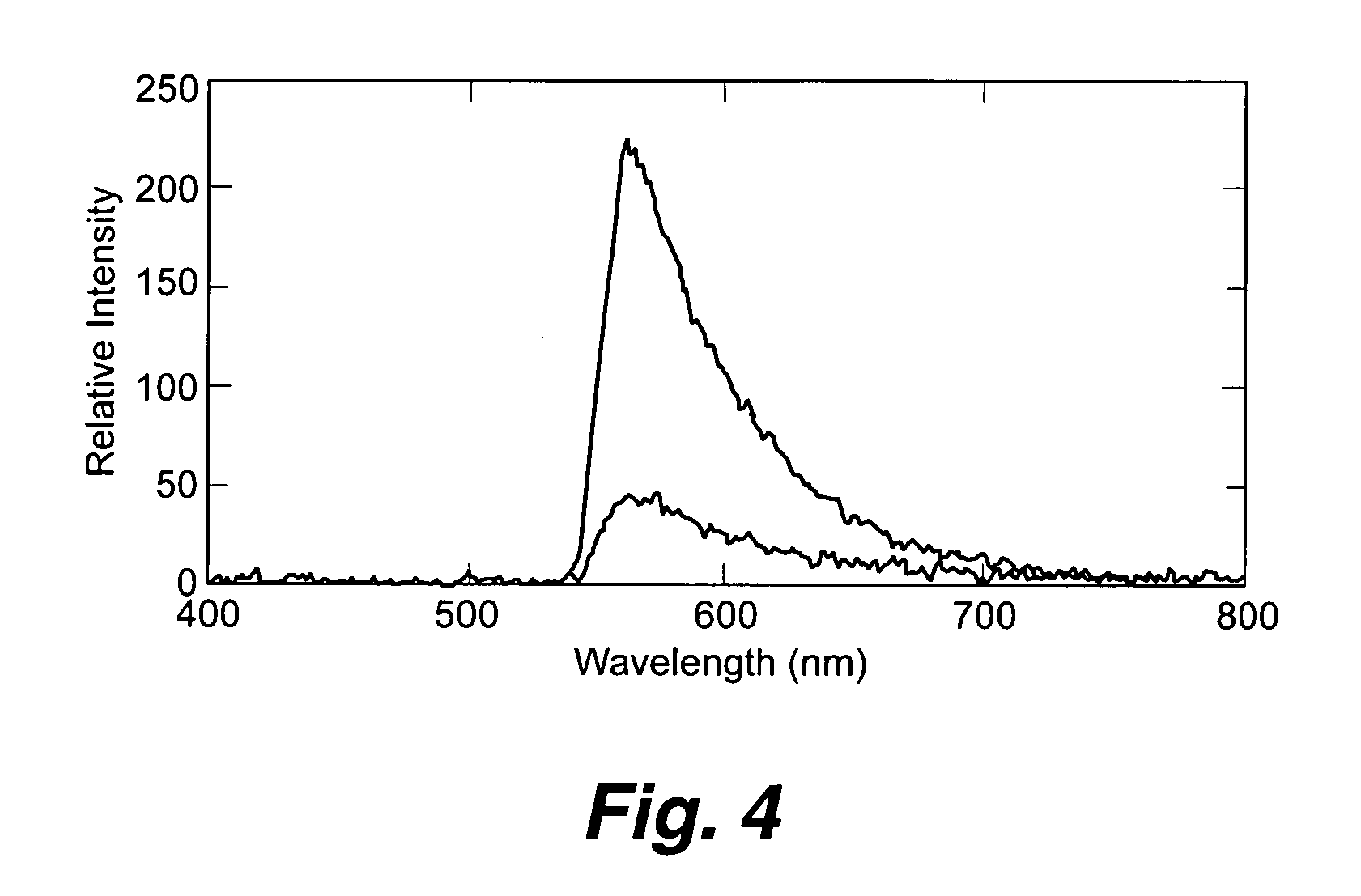
[0065] Biotinylated capture antibodies were coupled through avidin / streptavidin to biotinylated phospholipid bilayers composed of 1% to 3% biotin-phosphoethanolamine in a matrix of DOPC (1,2-Dioleoyl-sn-Glycero-3-Phosphocholine) and attached to the surface of planar optical waveguides. The performance of the immunoassay was evaluated by monitoring the response of the waveguide apparatus to varying concentrations of PA (pM to EM). No significant differences between the sensitivity of the instrumentation when PA was in buffer or in complex medium consisting of sugars, proteins, and cellular debris were observed. Although this assay has not yet been optimized for sensitivity, results from 1 pM PA (0.083 ng / mL, 0.0166 ng) in complex medium on sol-gel waveguides (FIG. 4) are shown. The top curve of this figure shows the spectral response of the excited waveguide following exposure to the complex medium spiked with 1 pM (0.083 ng / mL) PA. The lower curve is the response observed for the sa...

example 2

[0068] The present invention has also been targeted towards detection of B. anthracis cells. FIG. 6(a) shows the detection of cells using SiONx waveguides. These results show that not only can the present invention detect pathogenic antigens, but also can detect cellular components. Whether cell detritus or whole cells are being detected is not wholly clear, as the majority of a whole cell captured on a waveguide surface would be beyond the evanescent field. Moreover, it is expected that capture of whole cells at the waveguide surface will be limited owing to slow diffusion of intact cells relative to smaller cell components from disrupted cells. Again, as stated earlier, whole cell or cellular debris detection in serum might be an alternative to cell culturing and fluorescent microscopy assays commonly used as definitive diagnostic tests. To add to this B. anthracis biosensor, an assay to detect capsule, encoded by one of the virulent plasmids of B. anthracis, is being incorporated...


PUM

PropertyMeasurementUnit
Nanoscale particle sizeaaaaaaaaaa
Biological propertiesaaaaaaaaaa
Timeaaaaaaaaaa

Abstract

An assay element is described including recognition ligands bound to a film on a single mode planar optical waveguide, the film from the group of a membrane, a polymerized bilayer membrane, and a self-assembled monolayer containing polyethylene glycol or polypropylene glycol groups therein and an assay process for detecting the presence of a biological target is described including injecting a biological target-containing sample into a sensor cell including the assay element, with the recognition ligands adapted for binding to selected biological targets, maintaining the sample within the sensor cell for time sufficient for binding to occur between selected biological targets within the sample and the recognition ligands, injecting a solution including a reporter ligand into the sensor cell; and, interrogating the sample within the sensor cell with excitation light from the waveguide, the excitation light provided by an evanescent field of the single mode penetrating into the biological target-containing sample to a distance of less than about 200 nanometers from the waveguide thereby exciting the fluorescent-label in any bound reporter ligand within a distance of less than about 200 nanometers from the waveguide and resulting in a detectable signal.

Description

[0001] This application claims the benefit of U.S. Provisional Application Ser. No. 60 / 583,911 filed on Jun. 29, 2004.STATEMENT REGARDING FEDERAL RIGHTS [0002] This invention was made with government support under Contract No. W-7405-ENG-36 awarded by the U.S. Department of Energy. The government has certain rights in the invention.FIELD OF THE INVENTION [0003] The present invention relates to sandwich assay processes and to a thin film supported sandwich assay element on a single mode planar optical waveguide. Such an assay can provide a means of detecting and quantifying proteins and the like for medical diagnostics or for environmental surveillance. BACKGROUND OF THE INVENTION [0004] The detection of trace amounts of biologically significant compounds, such as steroids, drugs, selected antigens, or tumor markers, is often accomplished inexpensively by the employment of an immunoassay. Enzyme immunoassay (EIA) methods are common antigen detection techniques. One of the most common...

Claims


Application Information

Patent Timeline
IPC IPC(8): C12Q1/70C12Q1/68G01N33/554C12M1/34G01N33/569
CPCB01J2219/00497Y10S436/805B01J2219/00576B01J2219/00605B01J2219/00612B01J2219/0063B01J2219/00637B01J2219/00725B01J2219/00743B82Y15/00B82Y30/00C12Q1/6825G01N21/6428G01N21/648G01N33/54373G01N2610/00B01J2219/00527
Inventor MARTINEZ, JENNIFER S.SWANSON, BASIL I.GRACE, KAREN M.GRACE, WYANE K.SHREVE, ANDREW P.
Owner TRIAD NAT SECURITY LLC
Why Eureka
Try Eureka