2d-3d rigid registration method to compensate for organ motion during an interventional procedure

a registration method and interventional procedure technology, applied in the field of ultrasound imaging techniques, can solve the problems of requiring attention, difficult for the physician to accurately guide needles to suspicious locations, and negative biopsies, and achieve the effect of accelerating the registration

Inactive Publication Date: 2015-07-23
UNIV OF WESTERN ONTARIO +1
View PDF5 Cites 25 Cited by
  • Summary
  • Abstract
  • Description
  • Claims
  • Application Information

AI Technical Summary

Benefits of technology

[0039]In another example, the method, described above, ...

Problems solved by technology

However, with the small size of the biopsy cores taken, the presence of small, multi-focal cancers might result in negative biopsies.
Poor visibility of cancer in TRUS images and the limited anatomical context available in the 2D TRUS plane make it challenging for the physician to accurately guide needles to suspicious locations within the prostate.
Although early reports of 3D TRUS guided systems are promising, some limitations have been identified that require attention [6].
This may lead to a misalignment between the targets identified in the initially-acquired 3D image and their corresponding locations within the patient's prostate as depicted by the real-time 2D TRUS images acquired throughout the biopsy procedure.
In addition, this approach requires a ...

Method used

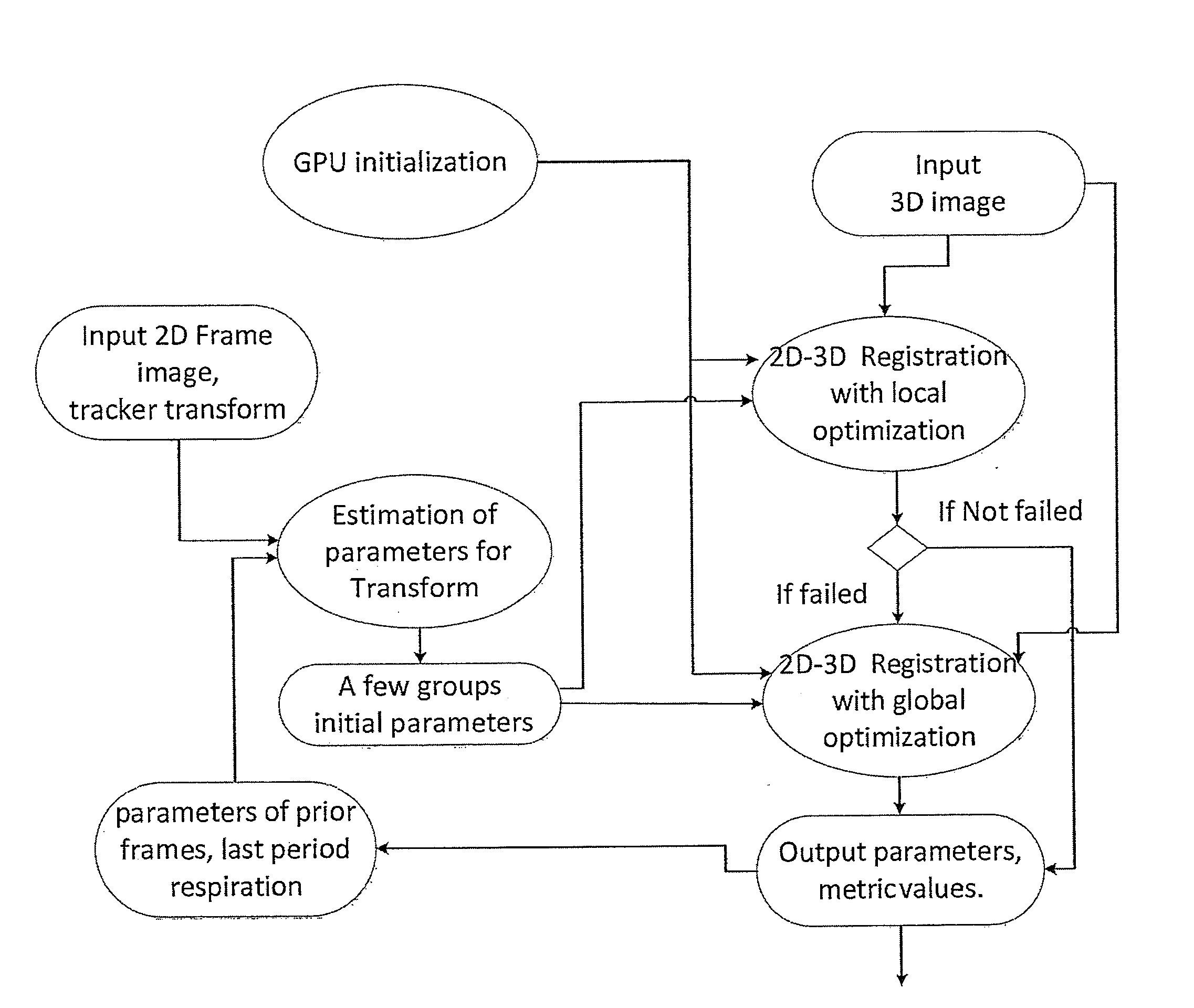
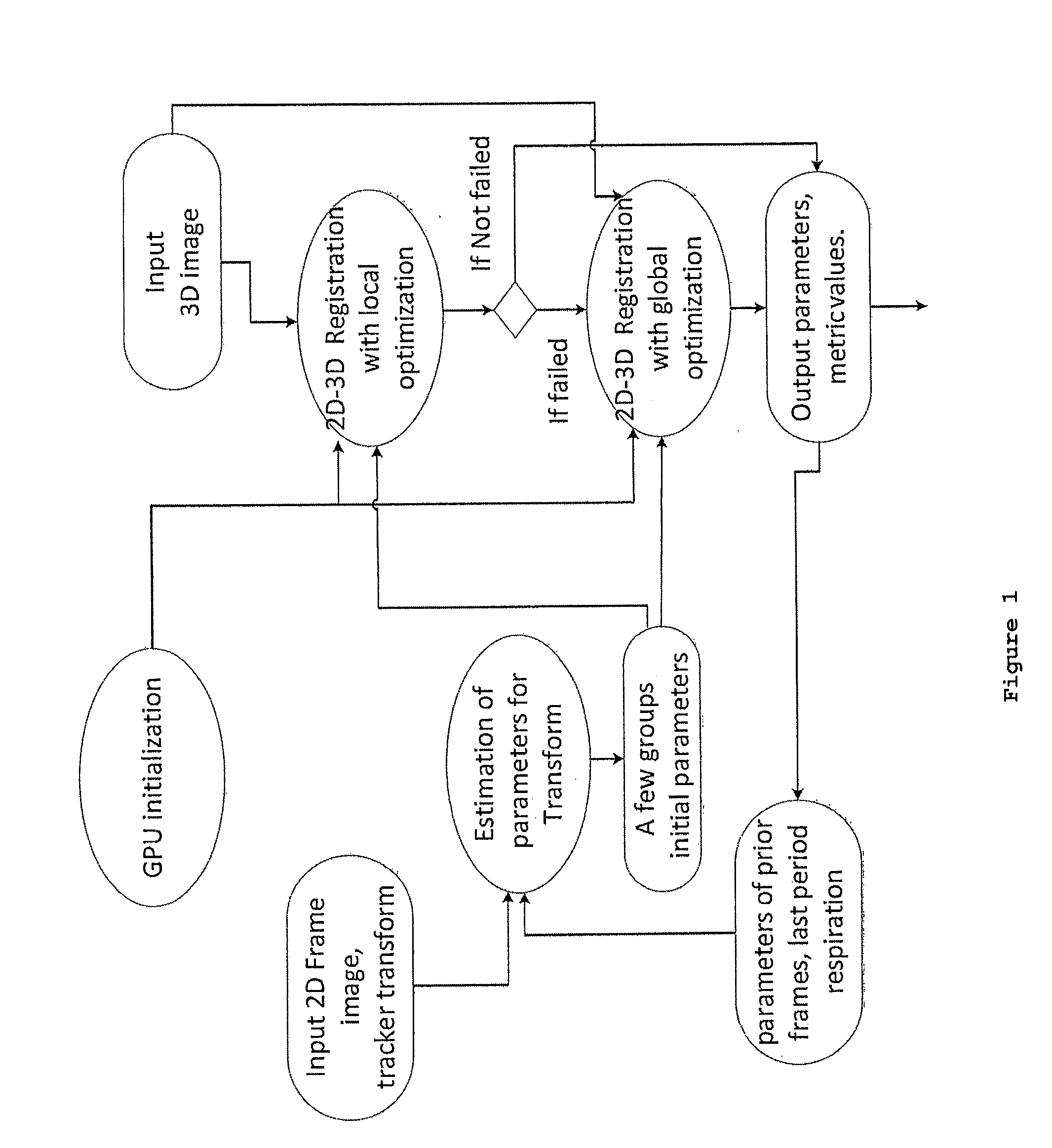
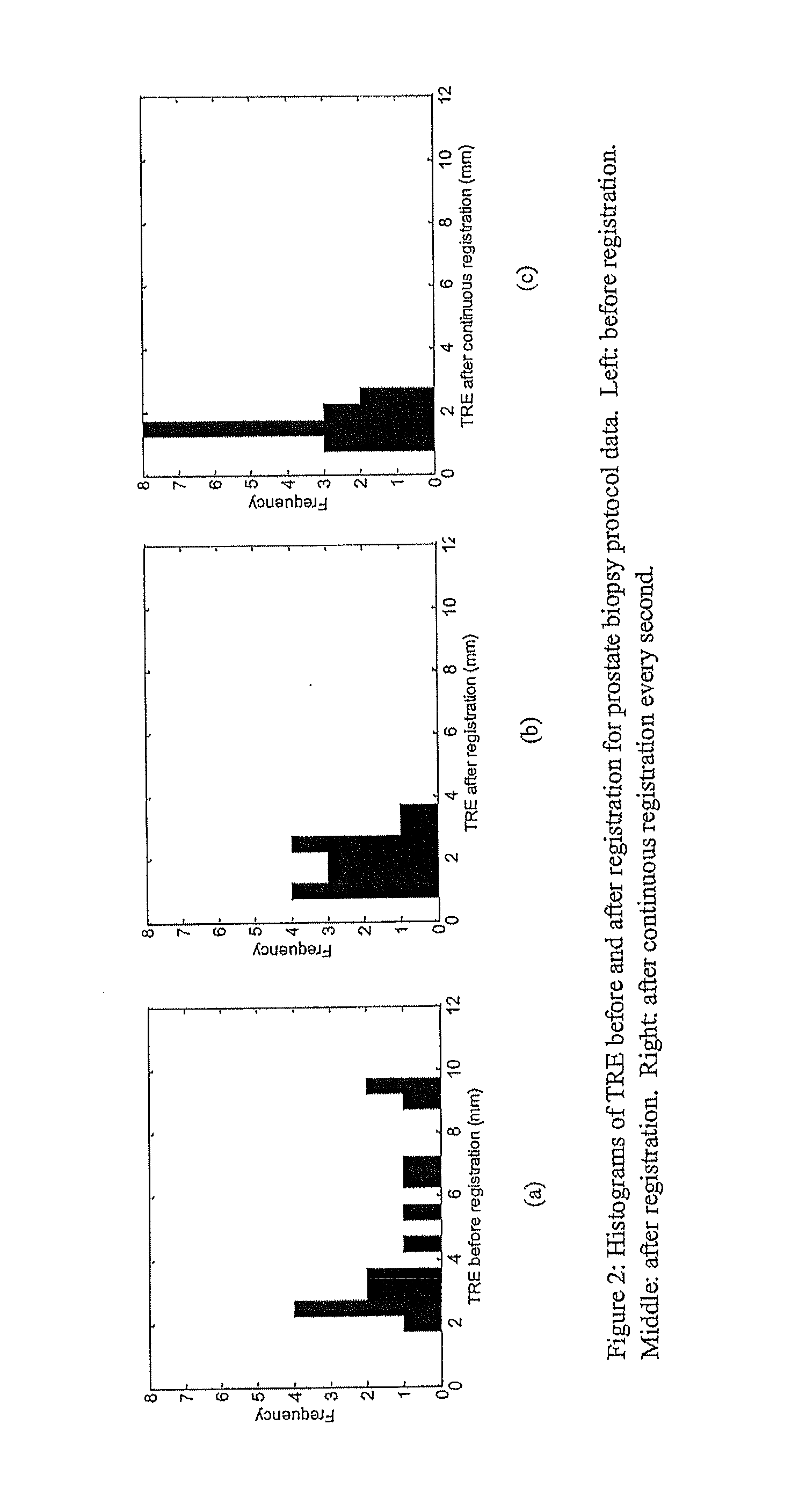

Image

  • 2d-3d rigid registration method to compensate for organ motion during an interventional procedure
  • 2d-3d rigid registration method to compensate for organ motion during an interventional procedure
  • 2d-3d rigid registration method to compensate for organ motion during an interventional procedure

Examples


Embodiment Construction

[0085]Ultrasound is a widely used imaging modality that is traditionally 2D. 2D ultrasound images remove 3D volume and three-dimensional information that allows for determining shapes, distances, and orientations. Ultrasound is used in medical, military, sensor, and mining applications

[0086]In interventional oncology, ultrasound is the preferred intra-operative image modality for procedures, including biopsies and thermal / focal ablation therapies in liver & kidney, laparoscopic liver surgery, prostate biopsy and therapy, percutaneous liver ablation, all other abdominal organs and ophthalmic intervention known to those skilled in the art. Some brain interventions also use ultrasound, although MR and CT are more common.

[0087]Ultrasound allows for “live information” about anatomical changes to be obtained with the requirement for further radiation dose to patient or physician. For image-guided interventions, it can be difficult for a surgeon to navigate surgical instruments if the ta...


PUM


Abstract

Disclosed herein is a system and method for generating a motion-corrected 2D image of a target. The method comprises acquiring a 3D static image of the target before an interventional procedure. During the procedure, a number of 2D real time images of the target are acquired and displayed. A slice of the 3D static image is acquired and registered with one 2D real time image and then the location of the 3D static image is corrected to be in synchrony with body motion. The motion corrected 2D image of the target is then displayed.

Description

TECHNICAL FIELD[0001]The present relates to ultrasound imaging techniques, and more particularly to an image-based registration algorithm for 2D to 3D rigid / affine ultrasound image registration technique.BACKGROUND[0002]Prostate cancer is the second most frequently diagnosed cancer among men in North America [1], with prostate biopsy as the clinical standard for diagnosis. During biopsy, the physician systematically obtains approximately a dozen tissue samples from different regions of the prostate to assess disease status via histopathology analysis of the extracted tissue. Prostate biopsy is usually performed under two-dimensional (2D) trans-rectal ultrasound (TRUS) guidance by inserting a needle through the patient's rectal wall. However, with the small size of the biopsy cores taken, the presence of small, multi-focal cancers might result in negative biopsies. In fact, the false negative rate of the 2D TRUS-guided biopsy procedure is reported to be as high as 30% [3]. Poor visib...

Claims


Application Information

Patent Timeline
IPC IPC(8): A61B8/08G01S7/52A61B10/04A61B8/12A61B8/00G06T7/00
CPCA61B8/5253A61B8/463A61B8/5207A61B8/466G01S7/52053A61B8/0841A61B10/04A61B8/12A61B8/483G06T7/0036G06T2207/10132A61B8/085A61B8/5261A61B8/5276G06T7/33
Inventor ZHAO, SHUANG-RENFENSTER, AARONDE SILVA, THARINDUWARD, AARON D.
Owner UNIV OF WESTERN ONTARIO
Why Eureka
Try Eureka