Cell culture of corneal endothelial cells
a cell culture and endothelial cell technology, applied in the field of cell culture, can solve the problems of limiting the number of corneal transplantations, graft failure is a problem, and the risk of graft rejection
- Summary
- Abstract
- Description
- Claims
- Application Information
AI Technical Summary
Benefits of technology
Problems solved by technology
Method used
Image
Examples
example 1
Isolation of Primary HCECs
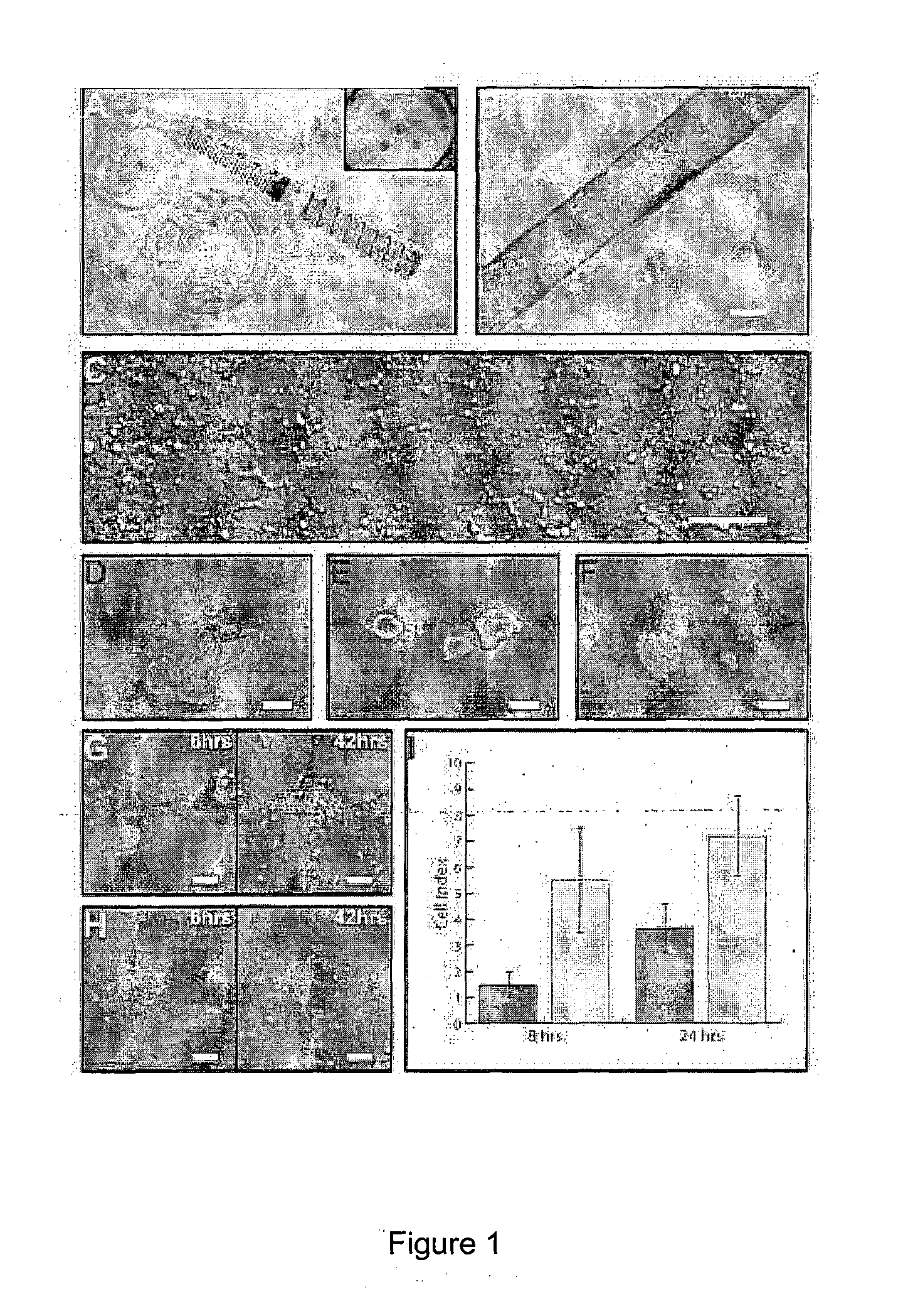
[0059]A working protocol for the isolation and establishment of primary HCECs was adapted for this study (FIG. 1). Isolation of primary HCECs involved a two-step, peel-and-digest method. Firstly, the thin corneal DM-endothelial layer was carefully peeled off under the dissecting microscope with the aid of a vacuum suction cup (or vacuum donor punch) to hold the cornea in place [FIG. 1(A)]. Much caution was taken in the peeling process to prevent the potential contamination of the posterior corneal stroma. Isolated DM-endothelial layer sheet [FIG. 1(B)] was subsequently subjected to an enzymatic digestion using collagenase (2 mg / mL) for at least 2 hours, and up to 4 hours to release the corneal endothelium from the DM [FIG. 1(D) and (E)]. Length of collagenase treatment is variable and is believed to be donor dependent. Clusters of collagenase-treated HCECs can be dissociated further with a brief treatment of TrypLE Express [FIG. 1(F)]. Following a quick rin...
example 2
Comparison of Culture Media
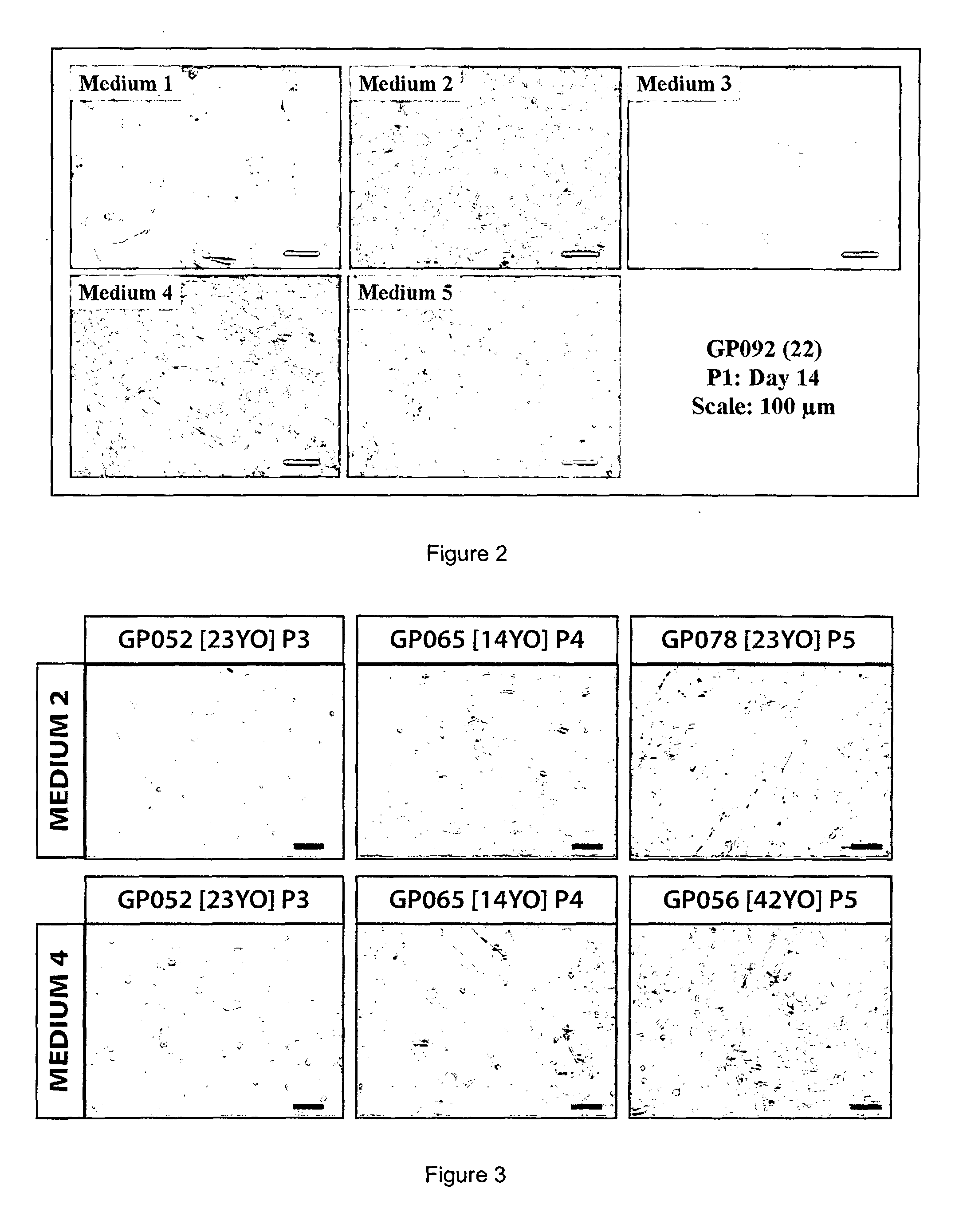
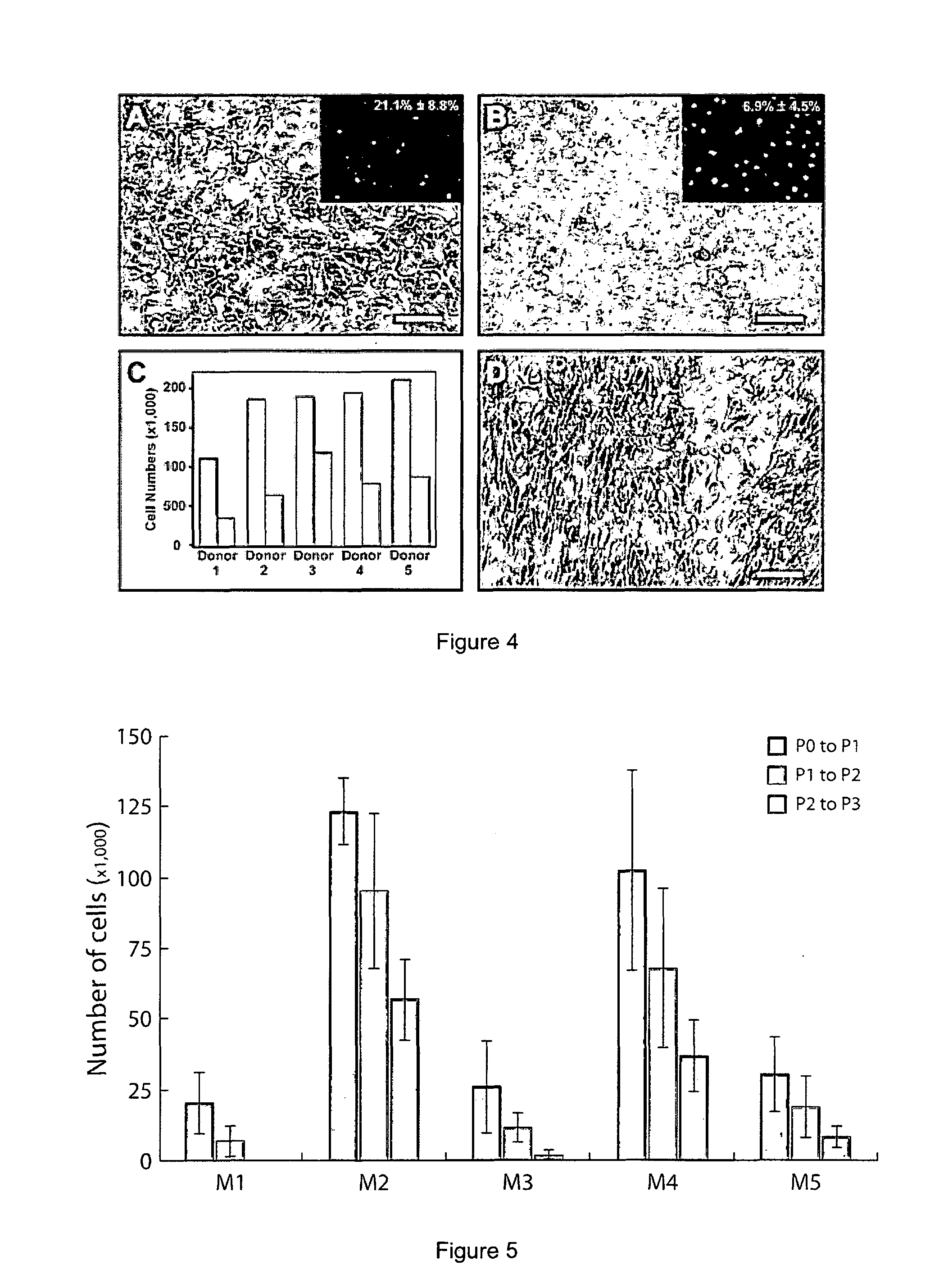
[0060]There are various serum-supplemented culture media reported for the in vitro serum-supplemented culture media were compared (Table 2), each developed from a different basal medium coded here as M1-DMEM (10% serum); M2-OptiMEM-I (8% serum); M3-DMEM / F12 (5% serum), & M4-Ham's F12 / M199 (5% serum). It was shown that cultured HCECs can be established in all four culture media [Passage 1 (P1), FIG. 2] but only M2 and M4 were able to promote and support their continual proliferation beyond the second passage. However, HCECs cultured in M2 and M4 became heterogeneous, usually by the third passage (sometimes as early as the second passage), taking up a more fibroblastic morphology [see later; FIG. 3]. In particular, although HCECs grown in M4 can be propagated beyond the second passage, the formation of fibroblastic-like cells became evident [FIG. 4(D)].
[0061]The inventors found that a maintenance medium could be used for the limited propagation of HCECs deri...
example 3
Dual Media Culture
[0063]The use of a dual media culture was then investigated. Interestingly, exposure of HCECs cultured in the proliferative medium M2 or M4 to M5 improved the morphology of HCECs. Interestingly, when Endothelial-SFM was used with either 2.5% [Endo2.5%] or 5% [Endo5%] fetal bovine serum (FBS) as the respective maintenance medium, long-term (>30 days) maintenance of HCECs is possible with a regular medium change every 48 to 72 hours.
[0064]Exposure of HCECs expanded in the proliferative medium M4 to the maintenance medium M5 in 80%-90% confluent HCECs cultured were found to be beneficial to the cultivated cells and improvement to the morphology of the HCECs were observed. For example, the use of both M4 and M5 in the culture of HCECs as a dual media approach relies on the timely incorporation of M5 as depicted in the schematic diagram in FIG. 4. When placed in M5 after culturing in proliferative medium, long-term (>30 days) maintenance of HCECs is possible with a regu...
PUM
| Property | Measurement | Unit |
|---|---|---|
| morphology | aaaaa | aaaaa |
| corneal transparency | aaaaa | aaaaa |
| cell size | aaaaa | aaaaa |
Abstract
Description
Claims
Application Information
 - R&D
- Intellectual Property
- Life Sciences
- Materials
- Tech Scout
- Unparalleled Data Quality
- Higher Quality Content
- 60% Fewer Hallucinations
Browse by: Latest US Patents, China's latest patents, Technical Efficacy Thesaurus, Application Domain, Technology Topic, Popular Technical Reports.
© 2025 PatSnap. All rights reserved.Legal|Privacy policy|Modern Slavery Act Transparency Statement|Sitemap|About US| Contact US: help@patsnap.com


