
Ct/mri integrated system for the diagnosis of acute strokes and methods thereof

a stroke and integrated system technology, applied in the field of dual-mode ct/mri integrated system, can solve the problems of permanent brain damage, painless test, and brain cells to di

Inactive Publication Date: 2015-07-30
ASPECT IMAGING
View PDF2 Cites 32 Cited by
  • Summary
  • Abstract
  • Description
  • Claims
  • Application Information

AI Technical Summary

Benefits of technology

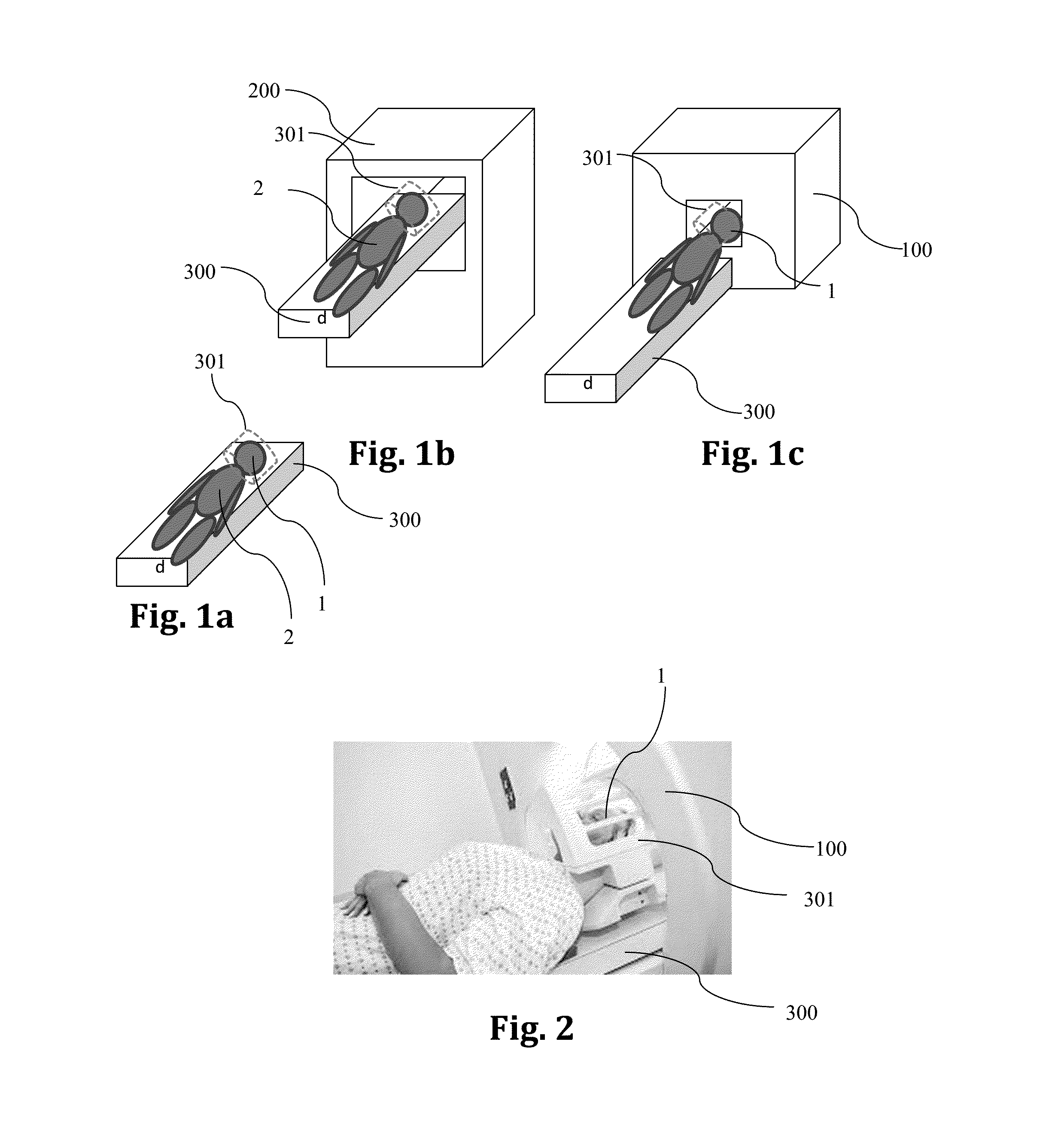
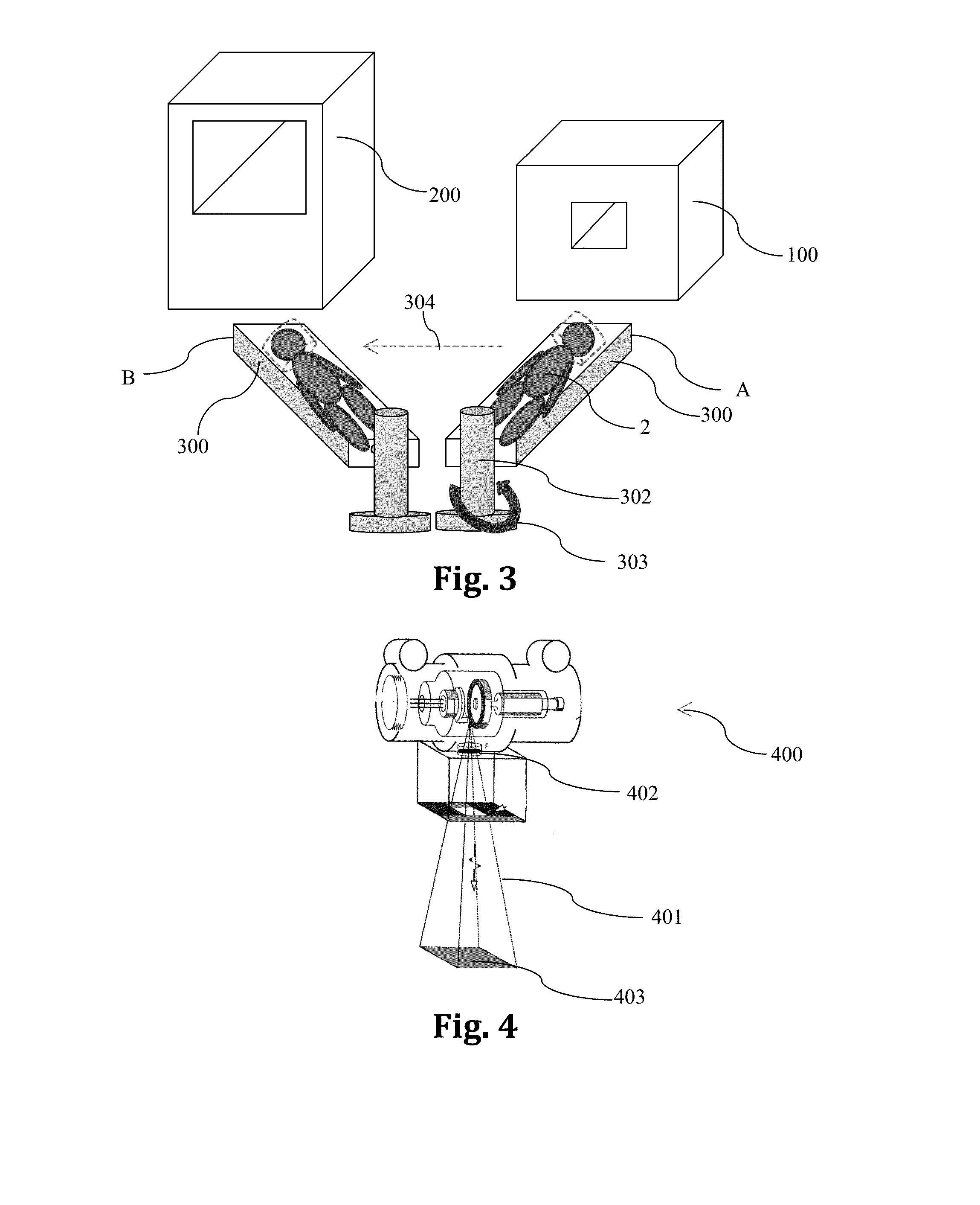
The present invention provides a two-step method of diagnosing medical conditions in a patient using a combination of a CT device and an MRI device. The method involves immobilizing the patient and either scanning the patient in the CT device and then the MRI device or placing the patient in the MRI device and then the CT device for scanning. The orientation of the patient in the CT scanning and MRI imaging is identical or retrievable. The method can be used to diagnose stroke by immobilizing the patient's head and either scanning the patient in the CT device and then the MRI device or placing the patient in the MRI device and then in the CT device for scanning. The MRI device can have at least one opening with a proximal aperture and a distal aperture, and the method can provide spatially correlated MRI / CT diagnosis by superimposing or correlating the CT scans and MRI images. The invention also provides a single-step method of diagnosing an object by using an open-bore MRI device with at least one opening and a non-MRI diagnosing effector within the opening.

Problems solved by technology

When a stroke goes untreated for too long, there can be permanent brain damage.
In both cases, part of the brain is deprived of blood and oxygen, causing the brain's cells to die.
The test is painless and usually takes about ½ hour.
If the patient has a pacemaker, he cannot have this test.
However, this is not a definitive test for a stroke.
Many methods that are used to measure these parameters, while giving accurate results, are not practical to use for continuous monitoring, or for initial diagnosis outside a hospital setting, because they are invasive, or because they require expensive and / or non-portable equipment.

Method used


Image

  • Ct/mri integrated system for the diagnosis of acute strokes and methods thereof
  • Ct/mri integrated system for the diagnosis of acute strokes and methods thereof
  • Ct/mri integrated system for the diagnosis of acute strokes and methods thereof

Examples


Embodiment Construction

[0037]X-radiation (composed of X-rays) is a form of electromagnetic radiation. Most X-rays have a wavelength in the range of 0.01 to 10 nanometers, corresponding to frequencies in the range 30 petahertz to 30 exahertz (3×10¹⁶ Hz to 3×10¹⁹ Hz) and energies in the range 100 eV to 100 keV. Fluoroscopy is an imaging technique commonly used by physicians or radiation therapists to obtain real-time moving images of the internal structures of a patient through the use of a fluoroscope. In its simplest form, a fluoroscope consists of an X-ray source and fluorescent screen between which a patient is placed. However, modern fluoroscopes couple the screen to an X-ray image intensifier and CCD video camera allowing the images to be recorded and played on a monitor. This method may use a contrast material. Examples include cardiac catheterization (to examine for coronary artery blockages) and barium swallows (to examine for esophageal disorders). The use of X-rays as a treatment is known as radi...


PUM


Abstract

A dual modality CT / MRI integrated system for the diagnosis of acute strokes and treating a patient and methods thereof. The method includes: immobilizing a patient or a portion thereof to a support; accommodating the patient or portion thereof in a CT device; scanning the same; placing the patient or portion thereof within an MRI device and imaging the same such that the orientation of the patient or portion thereof in the CT scanning and the MRI imaging is identical or at least spatially (2D or 3D) retrievable.

Description

FIELD AND BACKGROUND OF THE INVENTION[0001]The present invention, in some embodiments thereof, relates to a dual modality CT / MRI integrated system for the diagnosis of acute strokes and treating a patient and to methods thereof.[0002]A cerebrovascular accident is the medical term for a stroke. A stroke is when blood flow to a part of the patient brain is stopped either by a blockage or a rupture of a blood vessel. The more quickly the patient gets treatment, the better the prognosis. When a stroke goes untreated for too long, there can be permanent brain damage.[0003]There are two main types of cerebrovascular accident, or stroke. An ischemic stroke is caused by a blockage, and a hemorrhagic stroke is caused by a breakage in a blood vessel. In both cases, part of the brain is deprived of blood and oxygen, causing the brain's cells to die.[0004]An ischemic stroke occurs when a blood clot blocks a blood vessel, preventing blood and oxygen from getting to a part of the brain. There are...

Claims


Application Information

Patent Timeline
IPC IPC(8): A61B6/00A61N5/10A61B6/04A61B8/13A61B6/03A61B5/055
CPCA61B6/4417A61B6/032A61B5/0555A61B6/0421A61B6/5247A61B6/501A61B8/13A61B6/037A61N5/1039A61B6/508A61B5/0035A61B5/0042A61B5/055A61B6/0407A61B6/06A61N5/1049A61N2005/1052A61N2005/1055A61N2005/1061G01R33/4812
Inventor RAPOPORT, URI
Owner ASPECT IMAGING
Learn More