
System and Method for Personalized Computation of Tissue Ablation Extent Based on Medical Images

a technology of medical images and tissue ablation, applied in the field of patient-specific simulation of tissue ablation using medical imaging data, can solve the problems of reducing the efficiency of rfa, less than 25% of patients with primary tumors, and a significant challenge in the treatment of primary and metastatic tumors of the abdomen, and achieve accurate pre-ablation planning

Active Publication Date: 2015-08-27
SIEMENS HEALTHCARE GMBH
View PDF0 Cites 23 Cited by
  • Summary
  • Abstract
  • Description
  • Claims
  • Application Information

AI Technical Summary

Benefits of technology

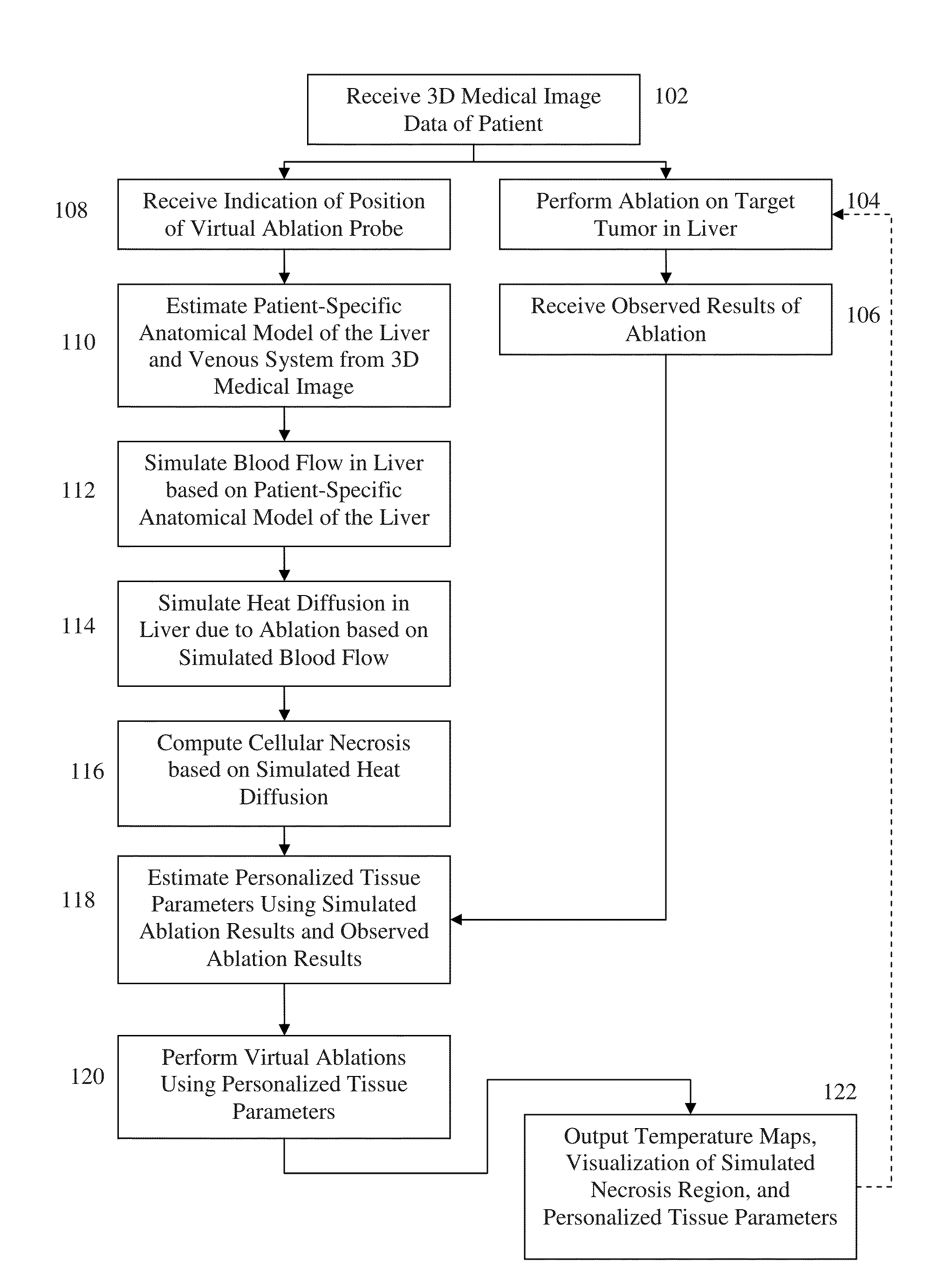
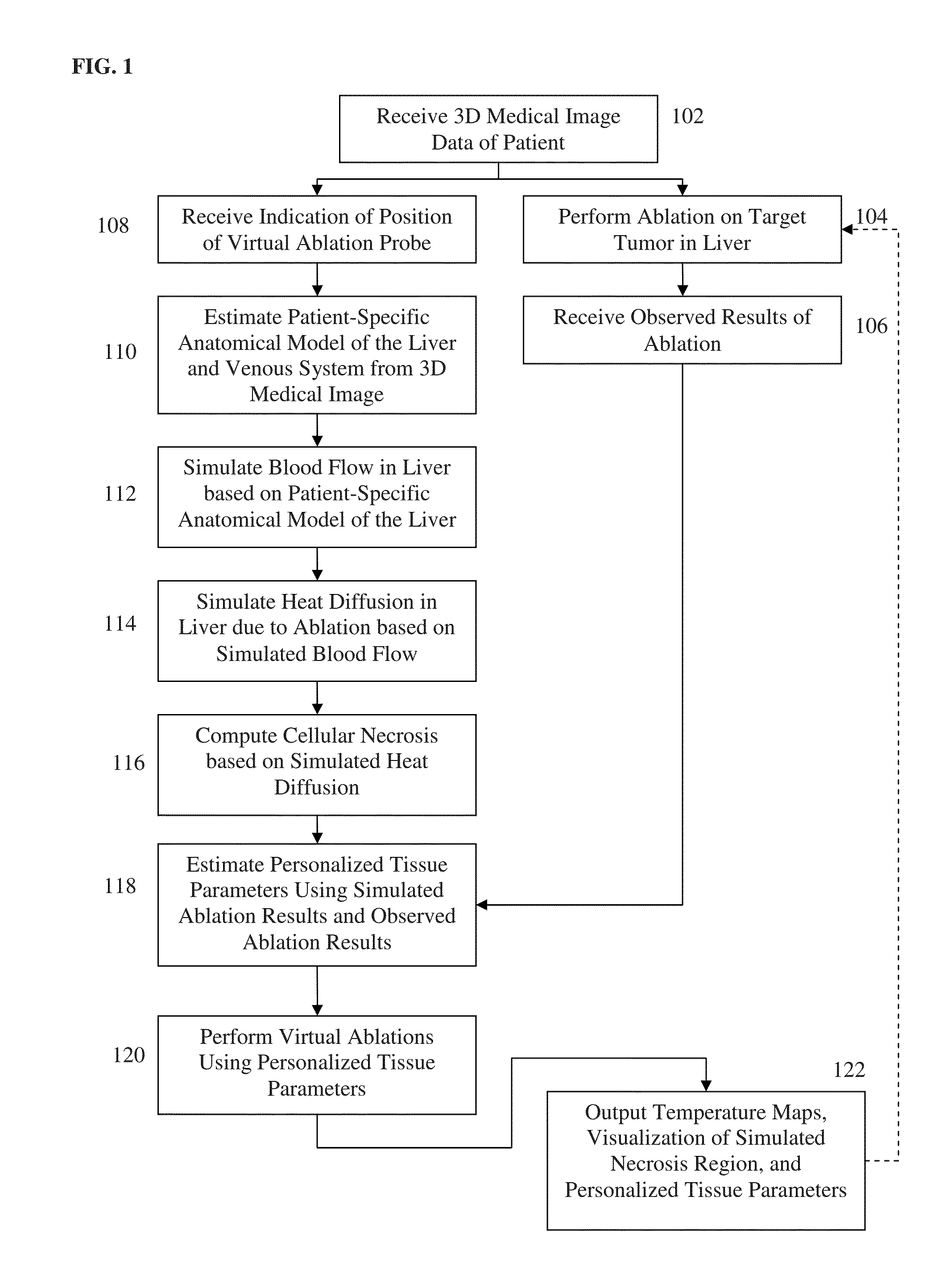
The present invention provides a method and system for personalized planning and guidance of tumor ablation using medical images. It models the effects of ablation by considering patient-specific tissue parameters and blood flow. By starting from a medical image of a patient, the method estimates the tissue parameters involved in heat transfer and performs individualized simulations of liver tumor radio-frequency ablation. The simulations can be used to plan additional ablations or refinement of the current lesion. The method also displays the estimated patient-specific tissue parameters along with patient-specific temperature diffusion maps, cellular necrosis maps, and virtual probe locations. The technical effect of the invention is that it provides a personalized approach to tumor ablation that takes into account patient-specific factors and can improve treatment outcomes.

Problems solved by technology

In spite of recent advances in cancer therapy, treatment of primary and metastatic tumors of the abdomen, including the liver, remains a significant challenge.
Unfortunately, less than 25% of patients with primary or secondary liver cancer are candidates for resection or transplantation, primarily due to tumor type, location, or underlying liver disease.
The success of ablation is further challenged by the hepatic blood vessels that dissipate heat, thus potentially reducing RFA efficiency.

Method used


Image

  • System and Method for Personalized Computation of Tissue Ablation Extent Based on Medical Images
  • System and Method for Personalized Computation of Tissue Ablation Extent Based on Medical Images
  • System and Method for Personalized Computation of Tissue Ablation Extent Based on Medical Images

Examples


Embodiment Construction

[0014]The present invention relates to patient-specific modeling and simulation of tumor ablation using medical imaging data for therapy planning and guidance. Embodiments of the present invention are described herein to give a visual understanding of the methods for patient-specific modeling and simulation using medical imaging data, exemplified on the case of liver tumor. However, the same approach could be employed to other tumors that can be treated through ablation therapy. The proposed invention could also apply to other ablation techniques that rely on heat delivery. A digital image is often composed of digital representations of one or more objects (or shapes). The digital representation of an object is often described herein in terms of identifying and manipulating the objects. Such manipulations are virtual manipulations accomplished in the memory or other circuitry / hardware of a computer system. Accordingly, is to be understood that embodiments of the present invention ma...


PUM


Abstract

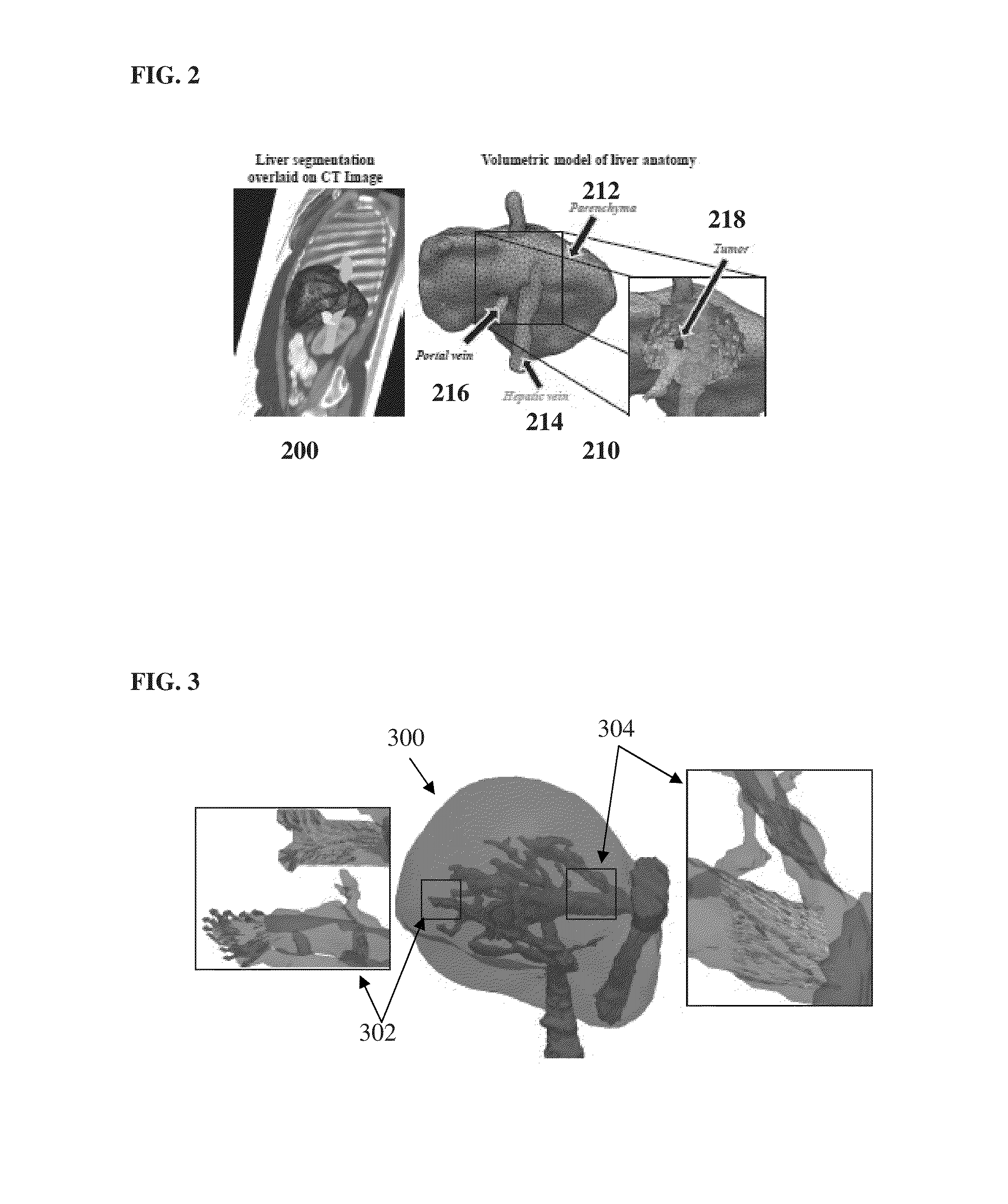
A method and system for personalized computation of tissue ablation extent based on medical images of a patient is disclosed. A patient-specific anatomical model of the liver and liver vessels is estimated from medical image data of a patient. Blood flow in the liver and liver vessels is simulated. An ablation simulation is performed that uses a bio-heat model to simulate heat diffusion due to an ablation based on the simulated blood flow and a cellular necrosis model to simulate cellular necrosis in the liver based on the simulated heat diffusion. Personalized tissue parameters of the bio-heat model and the cellular necrosis model are estimated based on observed results of a preliminary ablation procedure. Planning of the ablation procedure is then performed using the personalized bio-heat equation and the cellular necrosis model. The model can be subsequently refined as more ablation observations are obtained.

Description

[0001]This application claims the benefit of U.S. Provisional Application No. 61 / 944,713, filed Feb. 26, 2014, the disclosure of which is herein incorporated by reference.BACKGROUND OF THE INVENTION[0002]The present invention relates to ablation therapy planning, and more particularly to patient-specific simulation of tissue ablation using medical imaging data.[0003]In spite of recent advances in cancer therapy, treatment of primary and metastatic tumors of the abdomen, including the liver, remains a significant challenge. Hepatocellular carcinoma (HCC) for example is one of the most common malignancies encountered throughout the world (more than 1 million cases per year), with increasing frequency in western countries due to the changing prevalence of hepatitis C. For both primary liver cancer and hepatic metastases, liver resection (partial hepatectomy) is the current preferred option in patients having confined disease. In selected cases of early HCC, total hepatectomy with liver...

Claims


Application Information

Patent Timeline
Patent Type & Authority Applications(United States)
IPC IPC(8): G06F19/00A61B18/14A61B19/00G06F17/10G16Z99/00
CPCG06F19/3437G06F17/10A61B18/14A61B2019/501A61B2018/00988A61B2018/00577A61B19/50A61B18/12A61B2018/00791A61B2034/101A61B2034/104A61B34/10A61B2034/105A61B6/032A61B6/50A61B6/5217G16H50/50G16Z99/00
Inventor AUDIGIER, CHLOEMANSI, TOMMASORAPAKA, SAIKIRANKAMEN, ALIMIHALEF, VIORELDELINGETTE, HERVEAYACHE, NICHOLASCOMANICIU, DORIN
Owner SIEMENS HEALTHCARE GMBH
Learn More