

Visualization Method for a Human Skeleton from a Medical Scan
a visualization method and skeleton technology, applied in the field of medical imaging, can solve the problems of inability for users (e.g., medical technicians or medical doctors) to interact with the medical scan from the aforementioned systems, difficulty in navigating the medical image volume itself, and error-prone bone assessment from such medical scans, so as to improve visualization and diagnostic capabilities, the effect of increasing efficiency
- Summary
- Abstract
- Description
- Claims
- Application Information
AI Technical Summary
Benefits of technology
Problems solved by technology
Method used
Image

Examples
Embodiment Construction
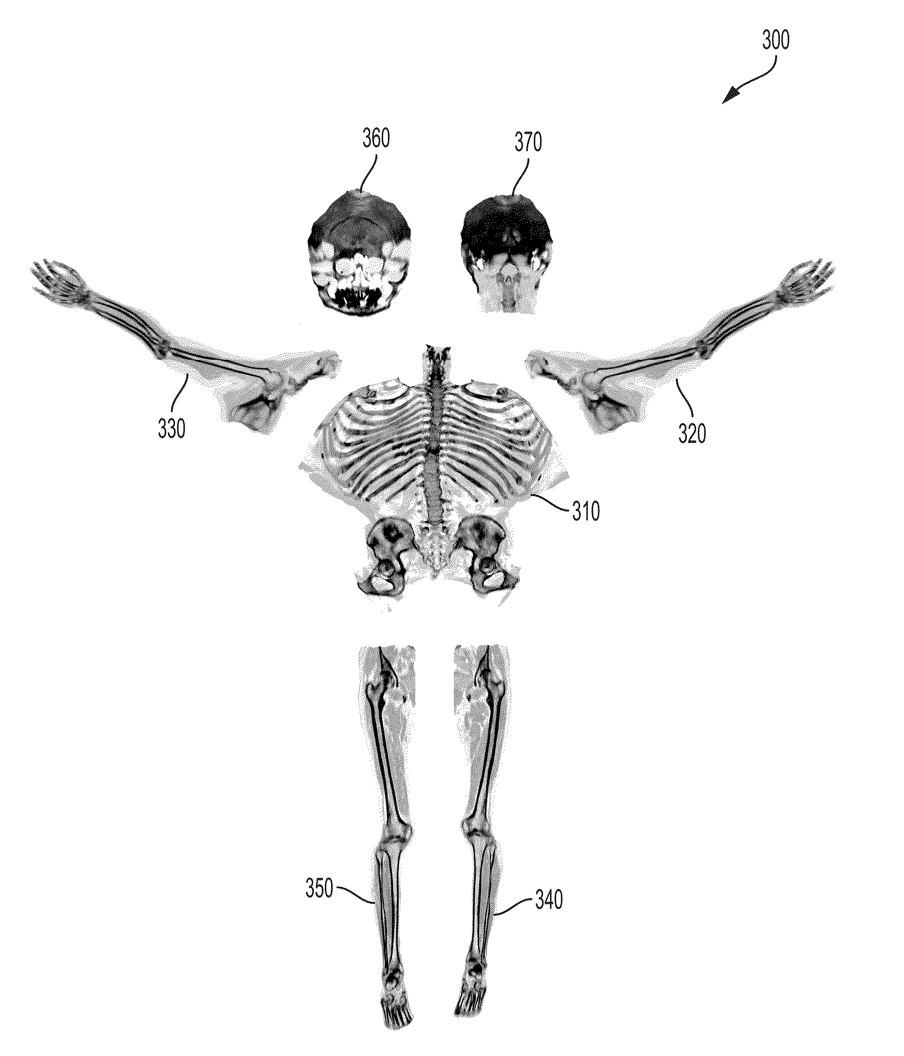
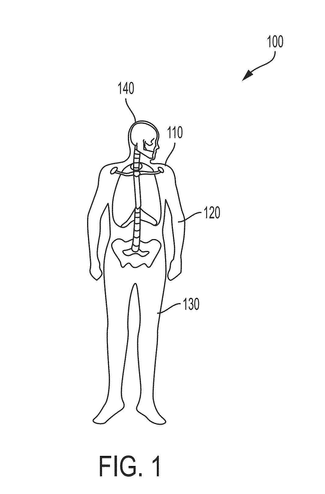
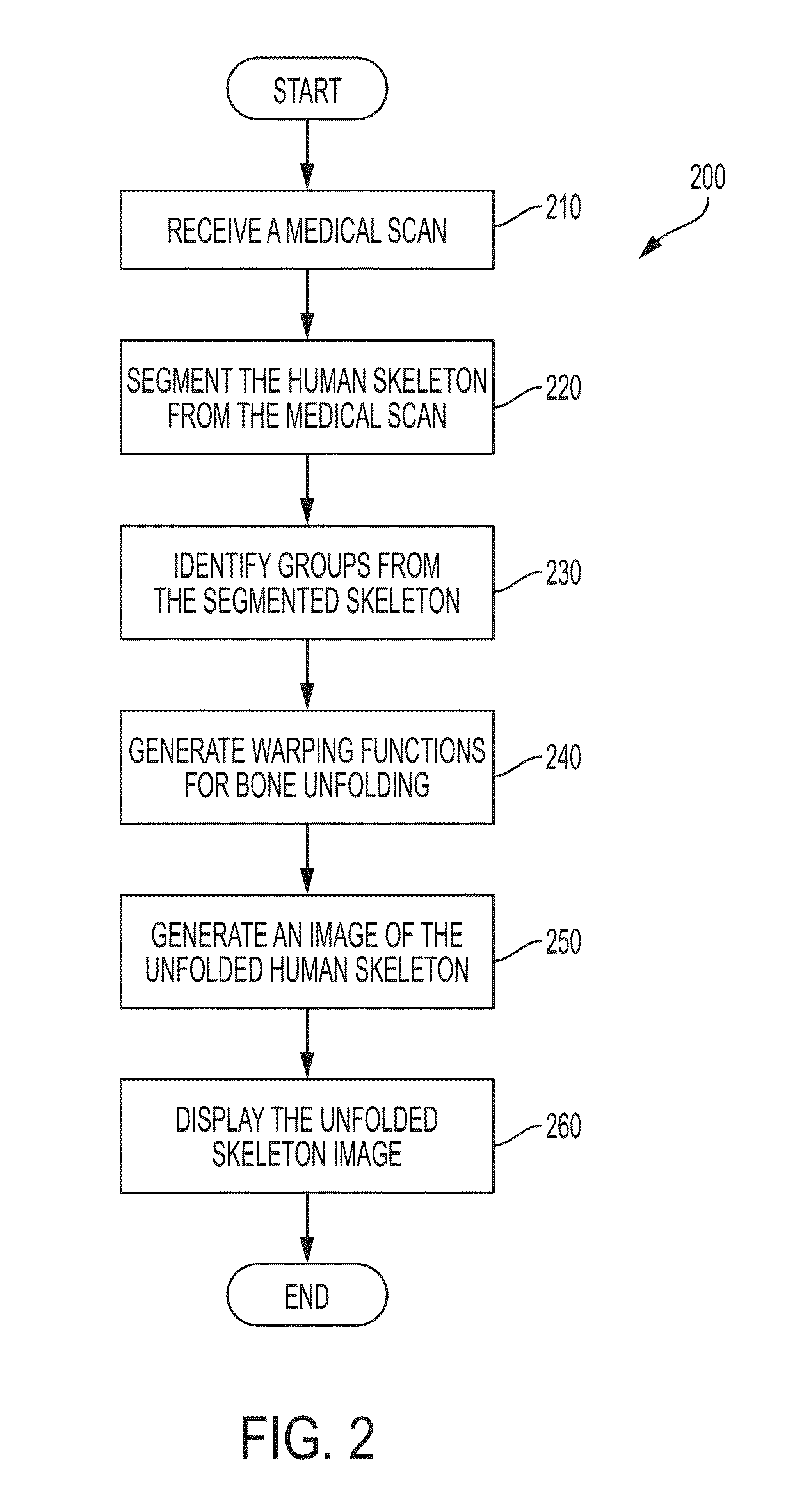
[0008]In accordance with various embodiments, a visualization method is provided that allows for the unfolding of a human skeleton from a medical image scan and providing increased efficiency for interacting with the image scan and whole body bone reading from such scans. That is, in accordance with various embodiments, a full head-to-toe unfolded skeleton view (e.g., a 2D unfolded view) is realized for improved visualization and diagnostic capabilities.
[0009]In accordance with an embodiment, given a 3D medical scan, automatic image segmentation is utilized to segment an image volume of the human skeleton (having a plurality of bones) from the medical scan into a 3D mesh or binary mask corresponding to the entire skeleton that is specific to the medical scan (i.e., the skeleton specific to the patient being diagnosed). The image volume could be of the entire skeleton itself or individual image volumes of each bone of the plurality of bones that define the skeleton. Alternatively, th...
PUM
 Abstract
Description
Claims
Application Information