Handheld Device for Identification of Microbiological Constituents in the Middle Ear

a microbiological and ear infection technology, applied in the field of optical identification of the microbiological origin of presenting ear infections, can solve the problems of ineffective treatment, overprescription of antibiotics, and inability to identify the microbiological origin of ear infections through video imaging

Inactive Publication Date: 2016-01-14
THE BOARD OF TRUSTEES OF THE UNIV OF ILLINOIS
View PDF3 Cites 33 Cited by
  • Summary
  • Abstract
  • Description
  • Claims
  • Application Information

AI Technical Summary

Problems solved by technology

While more than 75% of children have at least one episode of OM by age 3, and many have recurrent or chronic OM, a major impediment to effective treatment is the distinction of bacterial from viral infections.
Failure to distinguish bacterial from viral infection results in overprescription of, and overexposure to, antibiotics, and, more generally, to ineffective treatment.
However, current screening and diagnostic technology based on OCT and video imaging are unable to identify the microbiological origin of ear infections.
In the prior art, however, application of Raman spectroscopy to infected material in the middle ear has required withdrawal of a biological sample for analysis in the laboratory.
The Patil system, however, uses galvo scanners for transverse beam scanning, something that is particularly impractical for in vivo scattering diagnostics within the ear.
Liu, however, uses the same femtosecond fiber laser source both for Fourier-domain optical coherence microscopy (OCM) and as the excitation source for multiphoton microscopy, thereby imposing various operational limitations that are undesirable in the context of middle-ear diagnostics.
However, such technology has never been suggested in an otoscopic context, because of the anatomical peculiarities of the middle ear, insofar as scattering signals are depth-dependent, and no mechanism is provided, in the prior art, to account for the variation of Raman signal with placement.
The prior art provides no independent way to assess where the Raman probe is actually sampling.
Thus, for well over a decade, a solution has eluded practitioners as to how the specificity of Raman diagnostic techniques could be brought to bear on inner-ear problems.
The foregoing art, however, is incapable of effective point-of-care diagnostic distinction among microbiological origins of ear infections, and, thus, an instrument with such capability would be highly desirable, both in the developed and developing world.

Method used


Image

  • Handheld Device for Identification of Microbiological Constituents in the Middle Ear
  • Handheld Device for Identification of Microbiological Constituents in the Middle Ear
  • Handheld Device for Identification of Microbiological Constituents in the Middle Ear

Examples


Embodiment Construction

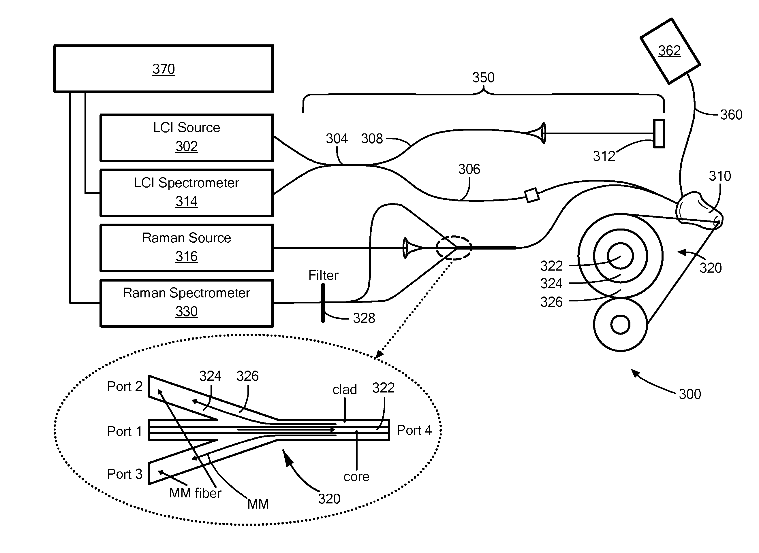
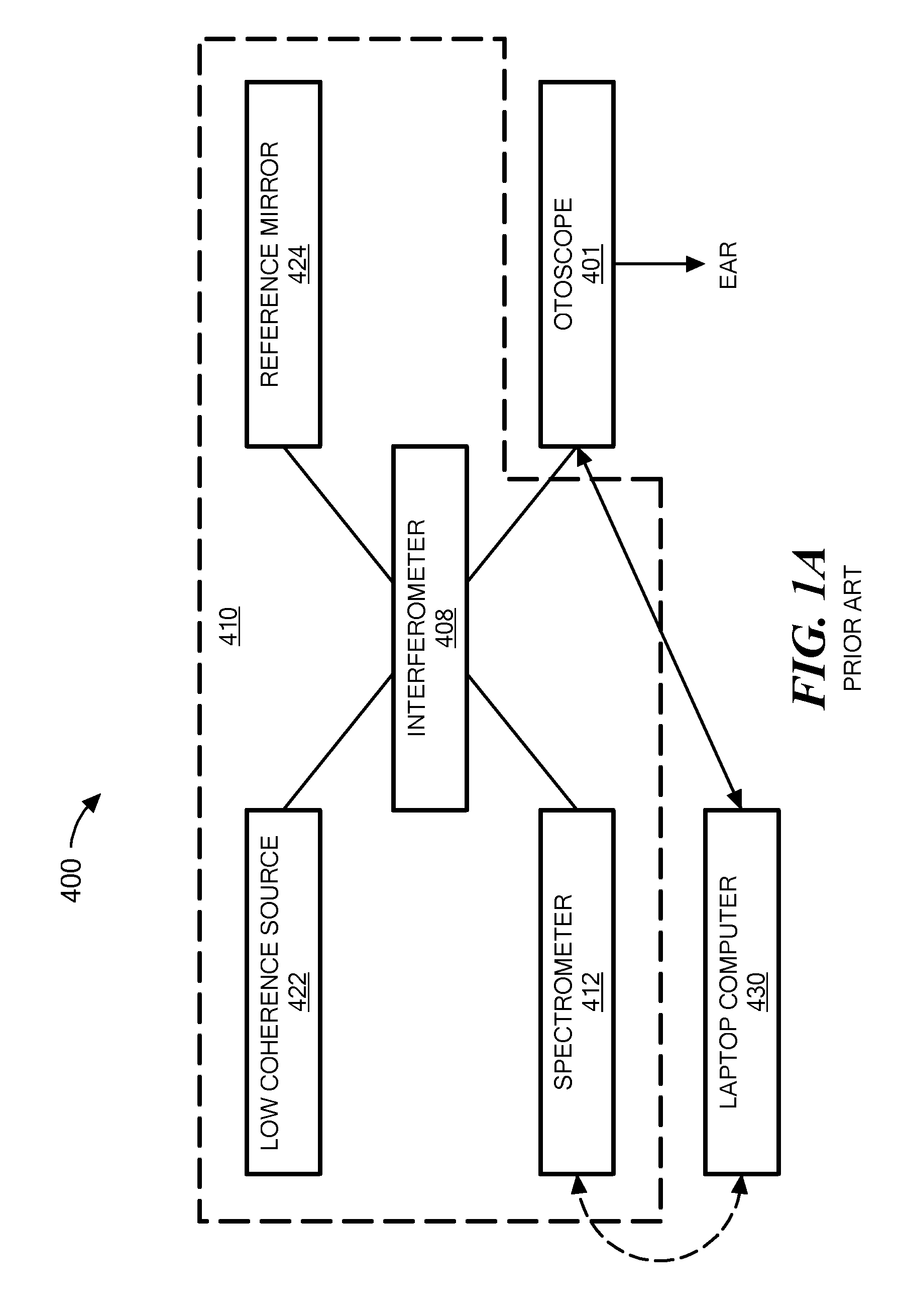
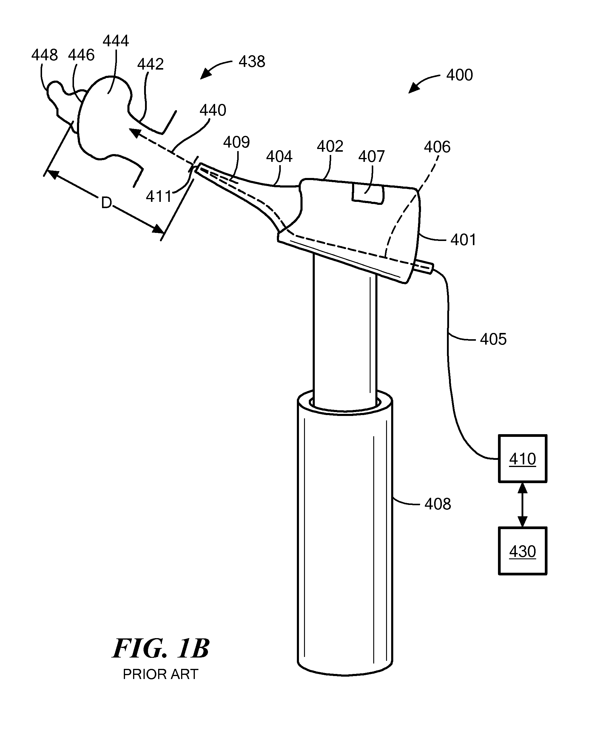
[0025]In accordance with embodiments of the invention, apparatuses and methods are provided for diagnosing and identifying microbiological material within the ear of a person. An otoscopic diagnostic system for jointly performing those functions has[0026]a. a source of substantially monochromatic excitation light;[0027]b. an otoscopic tip for abutment with the ear canal for directing the substantially monochromatic excitation light to a tympanic membrane of an ear of a person and for collecting Raman scattered light from the tympanic membrane and material behind the tympanic membrane and from material adjacent to the tympanic membrane;[0028]c. a spectrometer for receiving the Raman-scattered light, for resolving spectral features of the Raman-scattered light, and for generating a Raman signal; and[0029]d. a processor for receiving the Raman signal, and for generating therefrom a Raman spectrum of the tympanic membrane and the material behind the tympanic membrane and the material ad...


PUM


Abstract

Methods and apparatus for identifying microbiological constituents in the middle ear. A spectrometer receives Raman-scattered light from the region of the tympanic membrane and resolves spectral features of the Raman-scattered light. A processor receives the interferometry signal and the Raman signal, and generates a Raman spectrum of the tympanic membrane and material adjacent thereto. In some embodiments of the invention, low-coherence light and substantially monochromatic excitation light are directed onto a tympanic membrane of the ear of a person via an otoscopic tip that abuts the ear canal. An interferometer combines scattered low-coherence light from the ear tissue with a reference signal to generate an interferometric signal.

Description

[0001]The present application claims priority from U.S. Provisional Patent Application Ser. No. 62 / 023,052, filed Jul. 10, 2014, and incorporated herein by reference.[0002]This invention was made with government support under Grant R01 EB013723, awarded by the National Institutes of Health. The Government has certain rights in the invention.TECHNICAL FIELD[0003]The present invention relates to apparatus and methods for non-invasive otoscopy, and, more particularly, to apparatus and methods for optical identification of the microbiological origins of a presenting ear infection.BACKGROUND ART[0004]Otitis media (OM) is a highly prevalent disease, in fact, it is the most common childhood illness treated by pediatricians. While more than 75% of children have at least one episode of OM by age 3, and many have recurrent or chronic OM, a major impediment to effective treatment is the distinction of bacterial from viral infections. Failure to distinguish bacterial from viral infection result...

Claims


Application Information

Patent Timeline
Patent Type & Authority Applications(United States)
IPC IPC(8): A61B1/07A61B1/055A61B1/227
CPCA61B1/07A61B1/055A61B1/227A61B5/0075A61B5/0086A61B5/6817
Inventor BOPPART, STEPHEN A.SHELTON, RYAN L.
Owner THE BOARD OF TRUSTEES OF THE UNIV OF ILLINOIS
Why Eureka
Try Eureka