A computer implemented method for assessing vascular networks from medical images and uses thereof

Inactive Publication Date: 2016-02-11
EXPERT YMAGING SL +4
View PDF9 Cites 12 Cited by
  • Summary
  • Abstract
  • Description
  • Claims
  • Application Information

AI Technical Summary

Benefits of technology

The present invention provides a computer-based method for analyzing video sequences of ultrasound images to assess the complexity of the local vascular tree in a patient-specific manner. This method allows for the analysis of vascular networks in a more accurate and efficient way compared to existing methods. The analysis involves detecting events from the video sequences, selecting a region of interest, and computing a graph to represent the local vascular network. The graph can then be used to assess specific risk factors for disease. The method can be used in clinics to monitor the progress of disease in patients.

Problems solved by technology

Current state of the art proposals don't allow the analysis of video sequence of ultrasound images (2D / 3D) to assess the complexity of the local vascular tree into a graph model.

Method used


Image

  • A computer implemented method for assessing vascular networks from medical images and uses thereof
  • A computer implemented method for assessing vascular networks from medical images and uses thereof
  • A computer implemented method for assessing vascular networks from medical images and uses thereof

Examples


Embodiment Construction

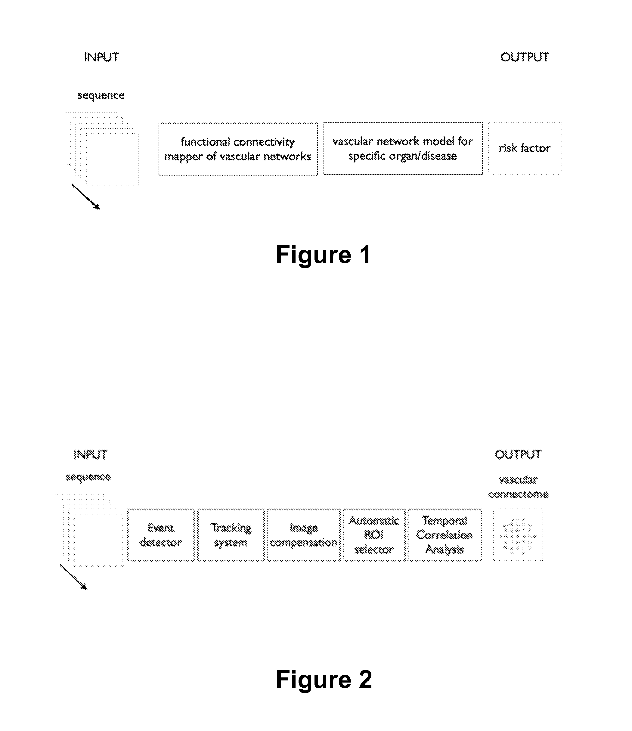
[0049]FIG. 1 shows the number of processing blocks: functional connectivity mapper of vascular networks and vascular network model for specific organ / disease, that enable to compute patient specific risk factor, according to the first aspect of the present invention.

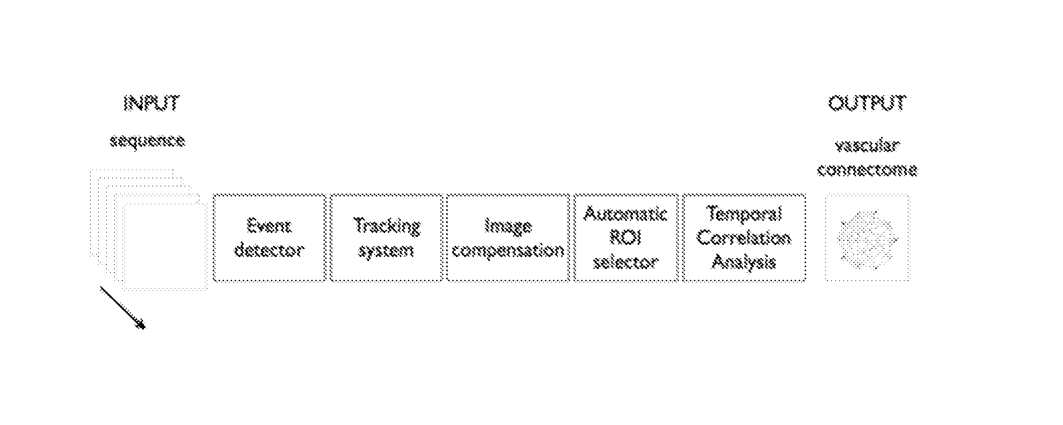
[0050]FIG. 2 shows in an embodiment, the blocks included in the functional connectivity mapper of vascular network block of FIG. 1. These blocks are: an Event detector, a Tracking system, Image compensation, Automatic ROI detection and a Temporal Correlation Analysis.

[0051]Some characterizations of the main blocks used by the proposed invention will be described in the following paragraphs in order to better explain their functions, thus allowing the analysis of the set of video sequences for further computing patient specific risk factors in the clinics.

[0052]The Event detector module is a group of signal processing techniques that detects from time series when a specific event has occurred. These approaches require tim...


PUM


Abstract

The method comprising acquiring and analysing by computer means image information of video sequences of two or more dimensions obtained from contrast-enhanced signals, for example ultrasound, coherence tomography, fluorescence images, or Magnetic Resonance Imaging, of a body part or tissue, for example of an organ, of a living subject; detecting events from said information of video sequences; selecting a Region of Interest of said body part or tissue; computing a first graph representative of a local vascular network of said image information of video sequences in which the edges of the graph are estimated by the temporal relationship among spatially remote signals of said image information of video sequences within a set of video sequences; and using said graph for assessment of vascular networks.

Description

FIELD OF THE ART[0001]The present invention generally relates to the field of imaging processing and information extraction applied to life sciences, and more particularly to a computer implemented method for assessing vascular networks from medical images by means of the analysis of medical images being enhanced by a contrast agent.[0002]The invention further relates to the use of such method for monitoring patients, for example cirrhotic, or for monitoring of therapeutic effects for specific medical conditions.PRIOR STATE OF THE ART[0003]Several diseases produce changes to the local vascular system or perfusion of a body part and techniques that are able to measure such changes are an intensive field of research as they have the potential to become useful tools for many clinical applications. Different imaging techniques (including contrast-enhanced computerized tomography, CECT, contrast-enhanced magnetic resonance imaging, CE-MRI, and contrast enhanced ultrasound, CEUS) have bee...

Claims


Application Information

Patent Timeline
IPC IPC(8): G06T7/00A61B8/08G06T11/20
CPCG06T7/0016G06T11/206A61B8/0891A61B8/481G06T2207/10096G06T2207/30056G06T2207/10132G06T2207/30101G06T2207/10016G06T2207/20104G06T2207/30024G06T2207/10121
Inventor AMAT ROLDAN, IVANBOSCH GENOVER, JAIMEBERZIGOTTI, ANNALISAGILABERT SOLE, MARIA ROSA
Owner EXPERT YMAGING SL
Why Eureka
Try Eureka