
Method for reducing biofilm formation

a biofilm and apparatus technology, applied in the field of apparatus and methods for reducing biofilm, can solve the problems of biofilm formation, impede healing, and chronic wound infection, and achieve the effect of accelerating healing

Active Publication Date: 2016-05-12
MISONIX INC
View PDF3 Cites 14 Cited by
  • Summary
  • Abstract
  • Description
  • Claims
  • Application Information

AI Technical Summary

Benefits of technology

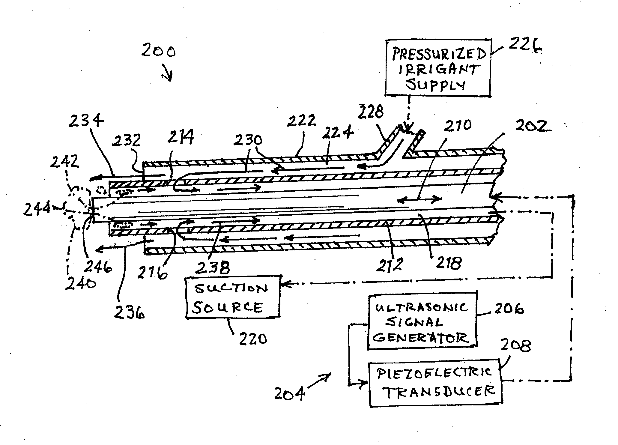
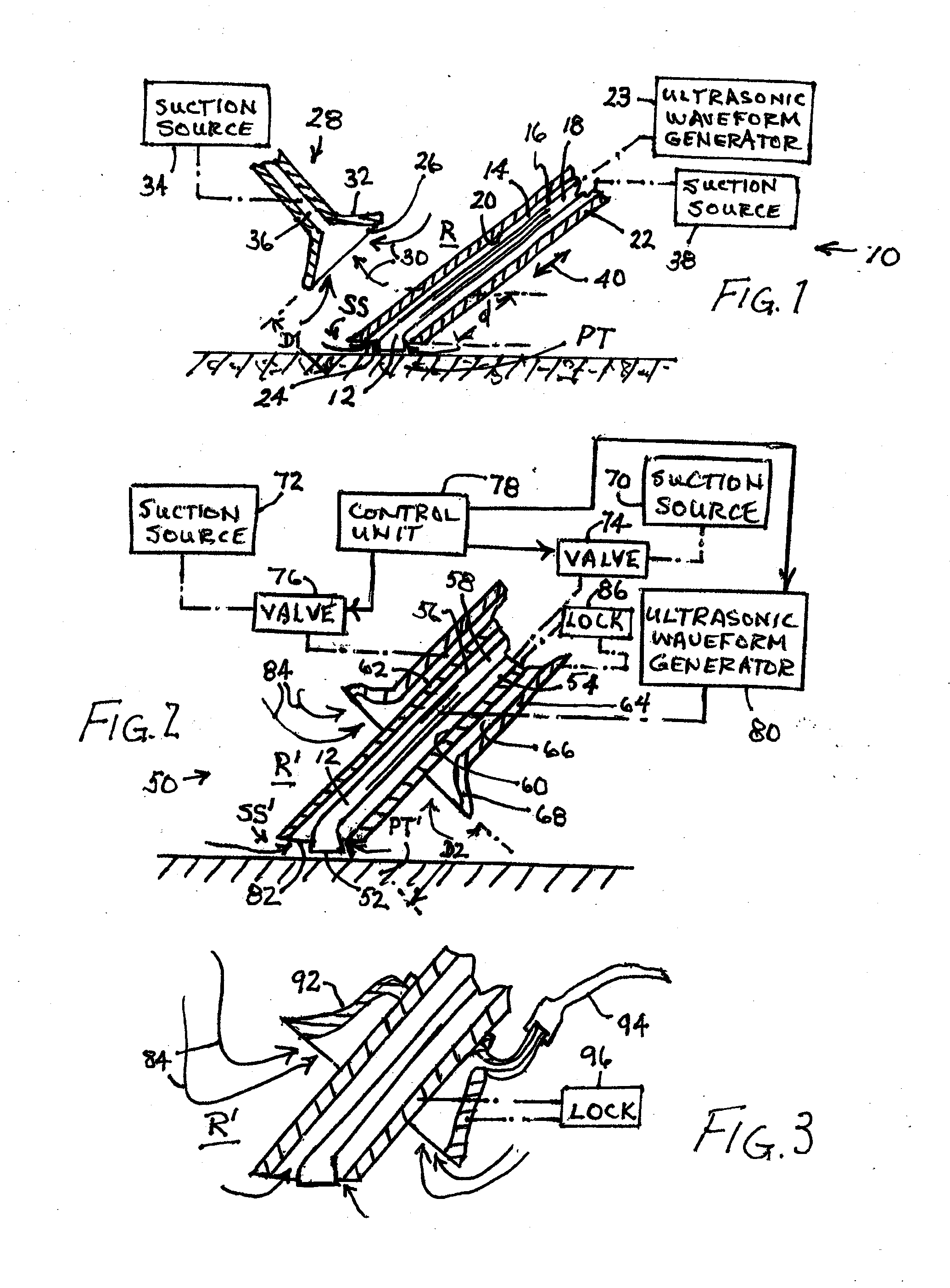
The present invention provides a method for removing biofilm and reducing the likelihood of reformation. This is accomplished through a two-step process involving the use of a suction device to capture any bacteria displaced during debridement. The suction device can be strategically placed adjacent to the treatment area or incorporated into an ultrasound applicator to capture bacteria and prevent it from causing infections. The suction device can also contain irrigation spray and be secured to the patient to create a barrier for bacteria or spray to be unable to escape.

Problems solved by technology

Chronic wound infection represents a significant healthcare problem worldwide.
Yet chronic wounds compromise a number of different and complex conditions that each interferes with the healing process.
One element of the chronic wound infection condition that impedes healing is the formation of biofilm.
Yet even in this preferred method, biofilm debris can be left behind to propagate.
Suction alone has not proven to be effective in removing biofilm, and can potentially interfere with the operation of other methods like ultrasonic debridement if applied simultaneously.
In particular this approach is limited due to the potential interference of the suction tip during the ultrasonic debridement operation.
Thus the ultrasonic probe needs to operate in a cavitation mode at the surface of a wound, causing destruction of the biofilm.
Methods of mechanical removal of biofilm in wounds alone have proven to be inadequate.

Method used


Image

  • Method for reducing biofilm formation
  • Method for reducing biofilm formation
  • Method for reducing biofilm formation

Examples


Embodiment Construction

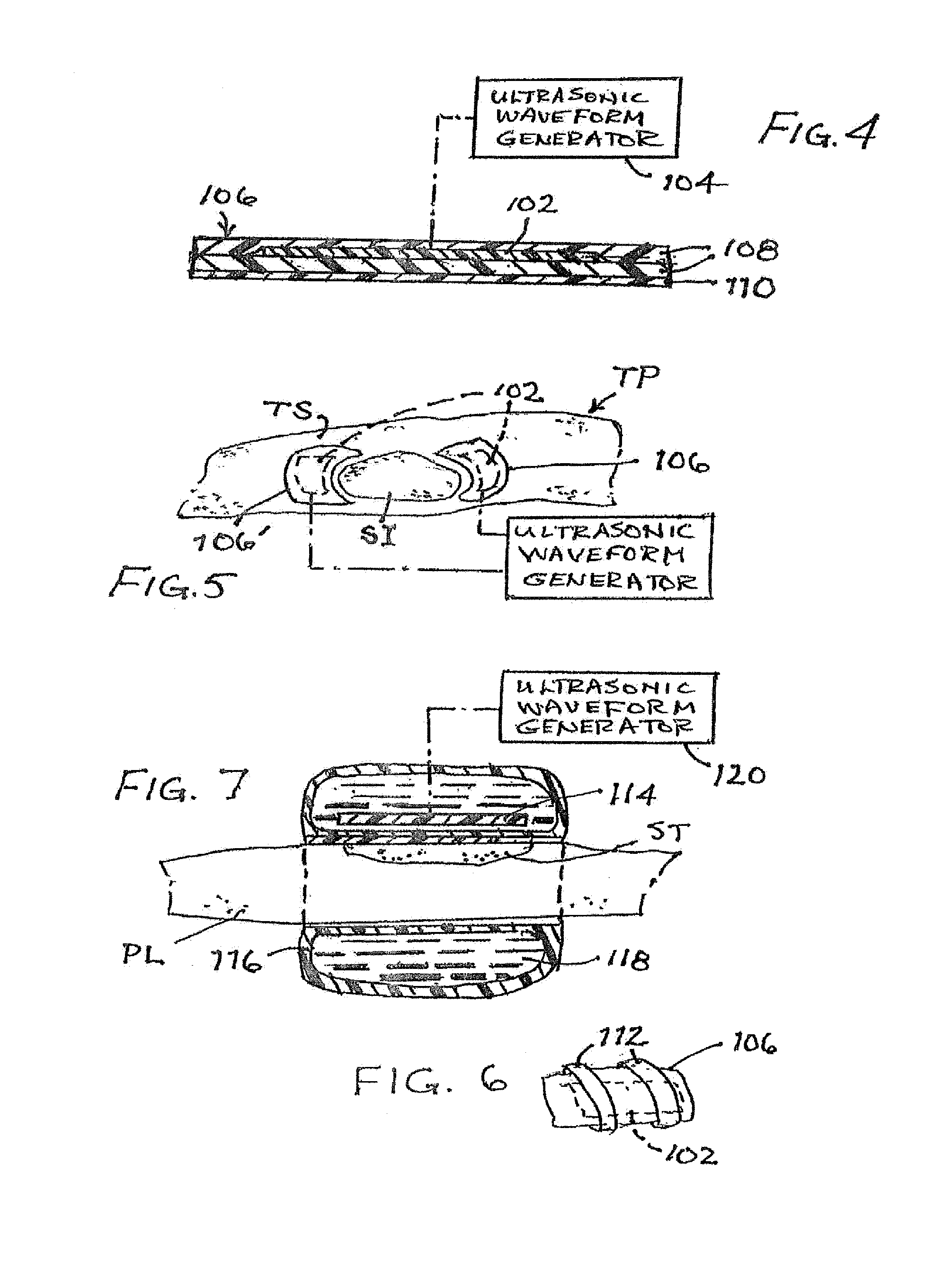
[0061]The present disclosure contemplates a two phase method for reducing the formation of biofilm. The first phase is performed where a wound site is being treated for removal of necrotic tissue, eschar or biofilm and includes an evacuation of ambient air from a region about the surgical or treatment site, to extract airborne or aerosolized bacteria ejected from the site by the treatment. The extracted bacteria are prevented from settling back onto the cleansed tissue surface, thus at least reducing colonial bacteriological growth and concomitantly exuded biofilm material. The second phase or approach for reducing biofilm involves the attachment of one or more ultrasonic transducers to the patient over or near a surgical treatment site after the surgery is terminated. Each applied ultrasonic transducer is used to vibrate the patient's tissues at the treatment site to disrupt biofilm formation. The two phases of treatment may be used separately depending on the application. Thus, ul...


PUM


Abstract

A two phase method for reducing the formation of biofilm includes an evacuation of ambient air from a region about the surgical or treatment site, to extract airborne or aerosolized bacteria ejected from the site by the treatment. The extracted bacteria are prevented from settling back onto the cleansed tissue surface, thus at least reducing colonial bacteriological growth and concomitantly exuded biofilm material. A second phase involves the attachment of one or more ultrasonic transducers to the patient over or near a surgical treatment site after the surgery is terminated. Each applied ultrasonic transducer is used to vibrate the patient's tissues at the treatment site to disrupt biofilm formation.

Description

BACKGROUND OF THE INVENTION[0001]This invention relates to apparatus and method for reducing biofilm on a wound site particularly a wound site that is being debrided to remove necrotic tissue.[0002]Chronic wound infection represents a significant healthcare problem worldwide. Often the end objective of wound healing is the objective for new therapeutic options. Yet chronic wounds compromise a number of different and complex conditions that each interferes with the healing process. For example, a chronic wound can comprise necrotic tissue in need of debridement, bacterial infection in need of antimicrobial agents and compromised vasculature that impedes the normal healing process.[0003]One element of the chronic wound infection condition that impedes healing is the formation of biofilm. Biofilm is the result of planktonic bacteria forming together and secreting exopolysaccharide (EPS) to adhere and protect the colonizing community. At the height of formation, EPS can make up between ...

Claims


Application Information

Patent Timeline
Patent Type & Authority Applications(United States)
IPC IPC(8): A61B17/22
CPCA61B17/22004A61B2217/005A61B2017/22005A61B2017/320084A61M2205/058A61B2017/320072A61B2017/00761A61M1/85A61B17/320068
Inventor MIKUS, PAULISOLA, SCOTTVOIC, DANLUDECKER, SCOTTDARIAN, ALEXANDER
Owner MISONIX INC
Learn More