Medicament comprising Anti-phospholipase d4 antibody

Inactive Publication Date: 2016-06-16
SBI BIOTECH CO LTD
View PDF0 Cites 1 Cited by
  • Summary
  • Abstract
  • Description
  • Claims
  • Application Information

AI Technical Summary

Benefits of technology

[0050]The present invention provides a therapeutic method attributable to suppression of activated B cells using an antibody specifically recognizing PLD4 and a fragment thereof, and a medicament having its therapeu

Problems solved by technology

As a result of the same analysis in Europe, however, significant correlation with PLD4 has not been found and strong

Method used


Image

  • Medicament comprising Anti-phospholipase d4 antibody
  • Medicament comprising Anti-phospholipase d4 antibody
  • Medicament comprising Anti-phospholipase d4 antibody

Examples


Example

Example 1

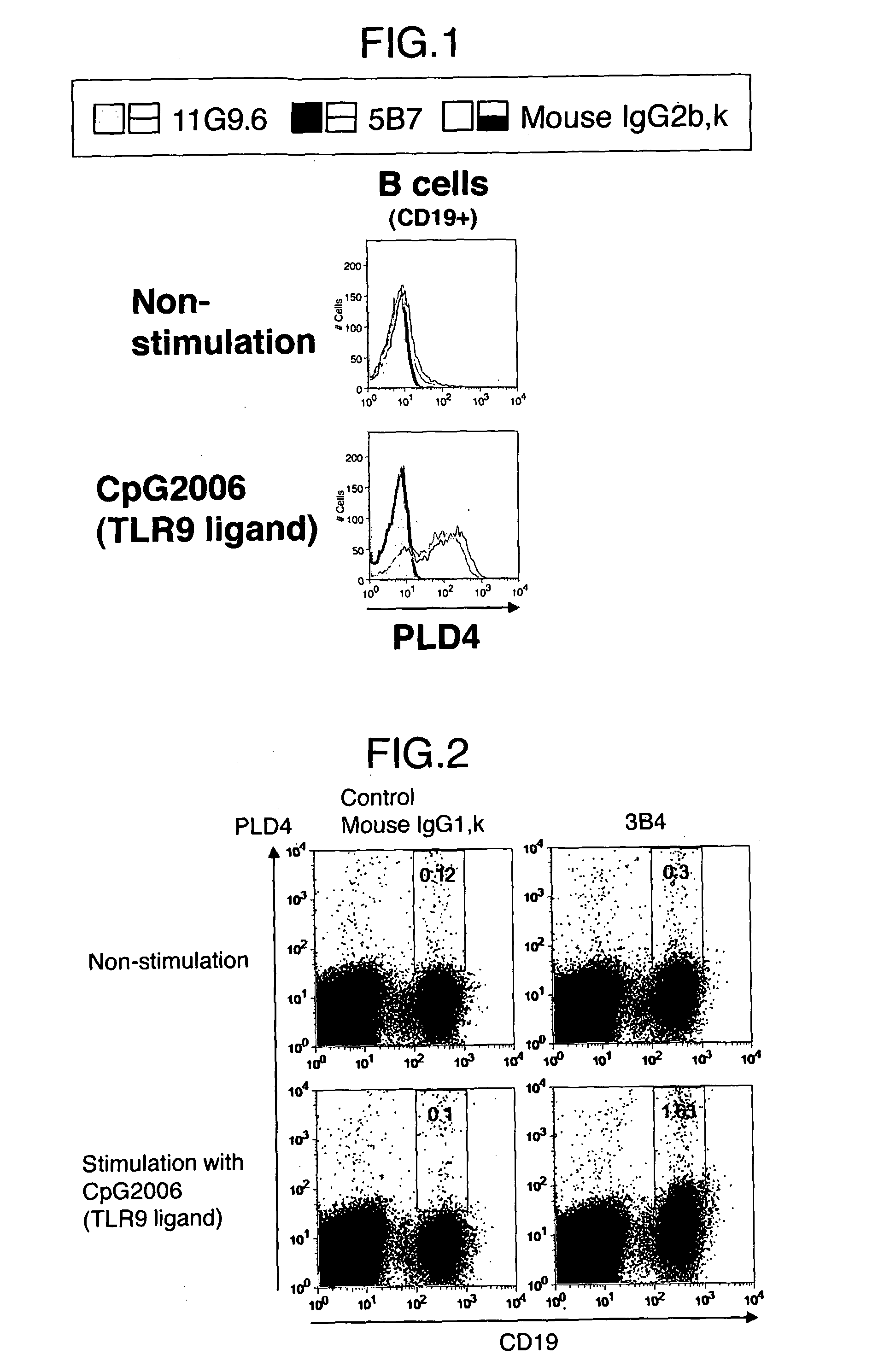
[0119]Human PBMC (1×10⁷ cells / ml) was stimulated by CpG2006, a ligand of TLR9, (a final concentration of 1 LM) and incubated in a 24 well plate in a CO2 incubator (37° C., 5% CO2) for about 20 hours. In parallel, human PBMC (1×10⁷ cells / ml) which was not stimulated was also cultured in a CO2 incubator (37° C., 5% CO2) for about 20 hours.

[0120]Human PBMC was treated with FcR Blocking Reagent (Miltenyi), which was diluted 5-fold with FACS buffer (1% FBS / PBS), at 4° C. for 20 minutes. After washing, staining was carried out with 5B7, 11G9.6 or mouse IgG2b, κ, a primary antibody, (each 10 μg / ml) at 4° C. for 15 minutes. A secondary antibody and subsequent antibodies were diluted with FACS buffer so that FcR Blocking Reagent would be diluted 25-fold. PE-labeled anti-mouse Ig (BD), a secondary antibody, was diluted 100-fold and the solution was added thereto and mixed. Besides, to fractionate B cells on FACS, an APC-labeled anti-human CD 19 antibody (Biolegend) was diluted 30-fol...

Example

Example 2

Binding Test to B Cells by Each Monoclonal Antibody

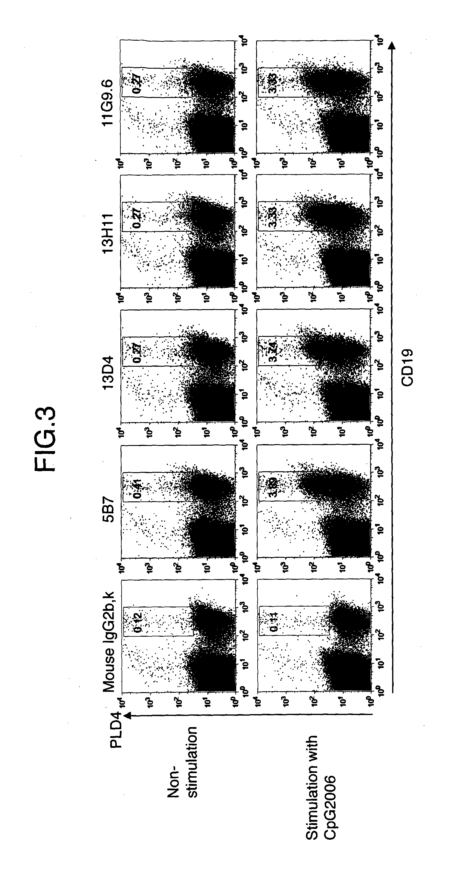
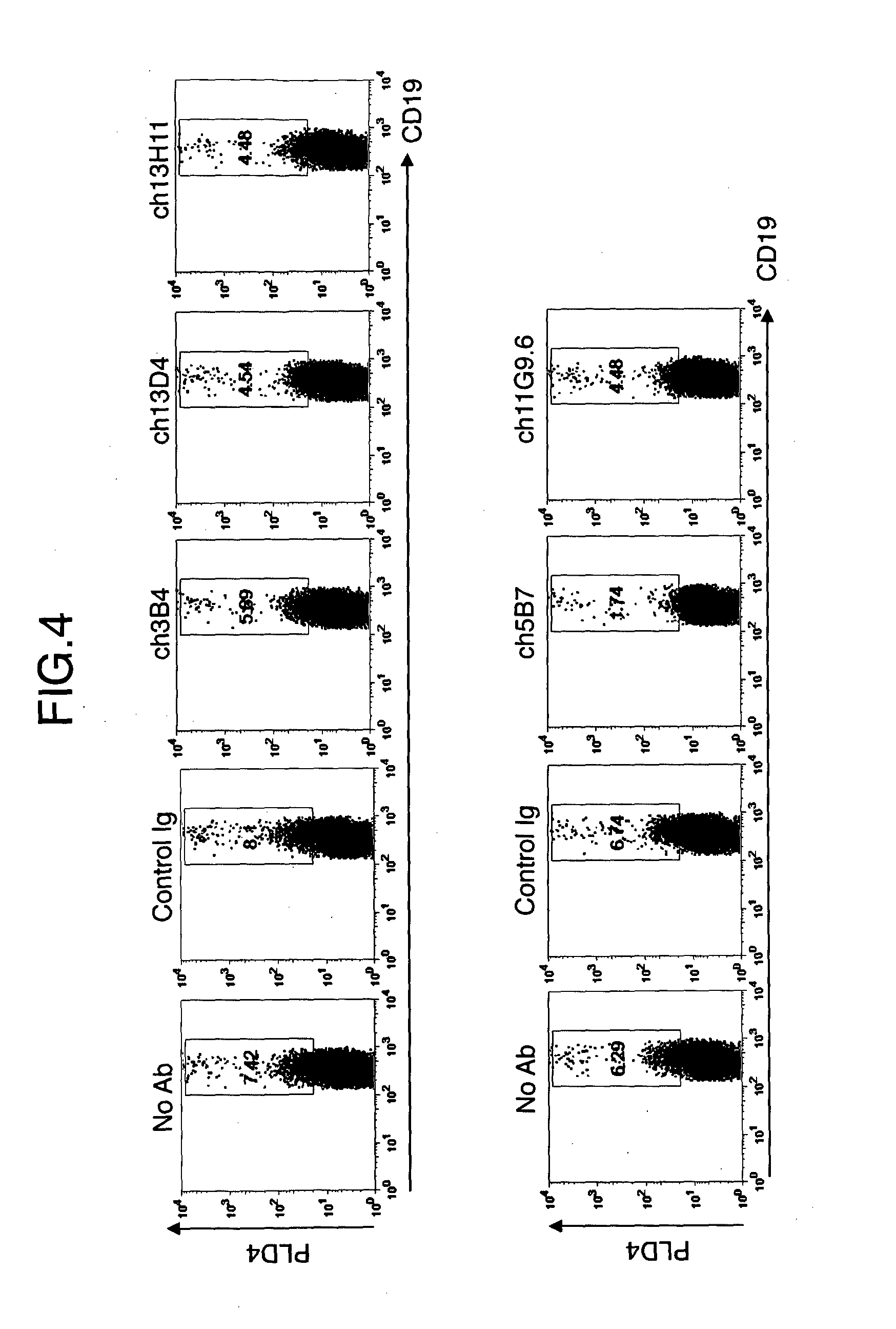
[0121]Human PBMC was stimulated with CpG2006 with a final concentration of 1 μM for about 20 hours. Cells were collected and treated with FcR Blocking Reagent at 4° C. for 20 minutes. After washing, staining was carried out with each 10 μg / ml of 3B4, 5B7, 13D4, 13H11, 11G9.6, mouse IgG1, K or mouse IgG2b, κ, a primary antibody, at 4° C. for 15 minutes. Staining was carried out with PE-labeled anti-mouse Ig, a secondary antibody, at 4° C. for 15 minutes. For gating of a B cell group, double staining was carried out with an APC-labeled anti-human CD19 antibody at 4° C. for 15 minutes. A living cell group on the dot plot of the X axis: FSC and the Y axis: SSC was analyzed by binding of anti-PLD4 antibody to CD19+ B cells (FIG. 2 and FIG. 3) Consequently, all of the tested anti-PLD4 monoclonal antibodies were bound to B cells stimulated by TLR9. That is, it was verified that by all anti-PLD4 monoclonal antibodies, expression of...

Example

Example 3

Cytotoxic Activity of Anti-PLD4 Chimeric Antibodies Against Activated B Cells

[0122]Frequency of PLD4+ activated B cells induced by stimulation with TLR9 ligand (1 μM) was used as an index. Human PBMC was cultured with CpG2006 and each anti-PLD4 chimeric antibody or control Ig for about 16 hours. As a medium, RPM11640 (SIGMA) was used (including 10% FBS (Equitech-bio), 5 ml of 200 mML-Glutamine (GIBCO), 5 ml of Pen-Strep (GIBCO), 5 ml of Sodium Pyruvate (GIBCO), and 0.5 ml of 50 mM 2-ME (SIGMA)). The cells were collected and treated with FcR Blocking Reagent at 4° C. for 20 minutes. After washing, the cells were further stained by 5B7 or 13D4, 3B4 or mouse IgG2b, K, a primary antibody, at 4° C. for 15 minutes (each 10 g / ml). A sample in which PBMC was treated with a chimeric 3B4 antibody (ch3B4), a chimeric 3D4 antibody (ch3D4), or a chimeric 13H11 antibody (ch13H11) was stained with 5B7, and a sample in which PBMC was treated with a chimeric 5B7 antibody (ch5B7) or a chimer...


PUM


Abstract

The present application provides the medicaments comprising the antibodies binding to phospholipase D4 (PLD4) as well as a method using said medicaments for detecting and suppressing activated B cells. The present application is further directed to therapy of auto-immune diseases and allergosis, resulting from the active-repressing function. In order to solves these problems, the present application provides that a monoclonal antibody binding to the extracellular domain of phospholipase D4 (PLD4) protein, or a fragment containing an antigen-binding region thereof as an active ingredient.

Description

TECHNICAL FIELD[0001]The present invention relates to a use of an antibody binding to phospholipase D4. Hereinafter, “phospholipase D” may be abbreviated as PLD and “phospholipase D4” and the like may be abbreviated as PLD4 and the like.BACKGROUND ART[0002]PLD is an enzyme which catalyzes a reaction to produce phosphatidic acid and choline by hydrolyzing phosphatidyl choline and causes various intracellular signaling. It has been believed that the produced phosphatidic acid functions as a lipid signal molecule.[0003]PLD1 and PLD2 have been known as two types of mammal PLD, which have been previously known, and contain a phosphatidyl inositide-binding Phox homology domain (PX domain) and a phosphatidyl inositide-binding pleckstrin homology domain (PH domain) in the N-terminal region thereof. Both domains are involved in membrane localization of PLD.[0004]PLD1 and PLD2 further contain two His-x-Lys-x-x-x-x-Asp sequences (HKD motifs). The HKD motifs are essential domains for PLD activi...

Claims


Application Information

Patent Timeline
IPC IPC(8): C07K16/40G01N33/573
CPCC07K16/40G01N33/573C07K2317/73G01N2333/916C07K2317/732C07K2317/76C07K2317/24A61K2039/505A61P37/00A61P37/06A61P37/08A61P43/00A61K39/395
Inventor YAMAZAKI, TOMOHIDEENDO, MAYUKIISHIDA, KOJI
Owner SBI BIOTECH CO LTD
Why Eureka
Try Eureka