Device and method for acquiring fusion image

a technology of fusion image and imaging device, which is applied in the field of medical image technique for diagnosis, analysis and treatment, can solve the problems of not having the technical means to acquire various kinds of image information simultaneously for clinical trials, and achieve the effect of simple user manipulation and easy analysis of images

Inactive Publication Date: 2016-07-07
SOGANG UNIV RES FOUND
View PDF1 Cites 7 Cited by
  • Summary
  • Abstract
  • Description
  • Claims
  • Application Information

AI Technical Summary

Benefits of technology

[0024]The embodiments of the present disclosure allows easier analysis of images by providing a probe structure, which may utilize various medical imaging techniques using an ultrasound image, a photoacoustic image and a fluorescent image simultaneously, provide pathologic information, anatomical information and functional information for a single observation area in a multilateral way by generating a fusion image based on planar image information and depth image information having different probing planes with respect to the observation target, and generate a fusion image through simple user manipulation at a medical site.

Problems solved by technology

At present, there has been reported one-shot investigation methods in which images acquired using different imaging devices for a single lesion area are comparatively investigated for experiments or studies, but there has been proposed no technical means to acquire various kinds of image information simultaneously for the utilization in clinical trials.

Method used


Image

  • Device and method for acquiring fusion image
  • Device and method for acquiring fusion image
  • Device and method for acquiring fusion image

Examples


Embodiment Construction

[0035]Hereinafter, a basic idea adopted in embodiments of the present disclosure will be described briefly, and then detailed technical features will be described in order.

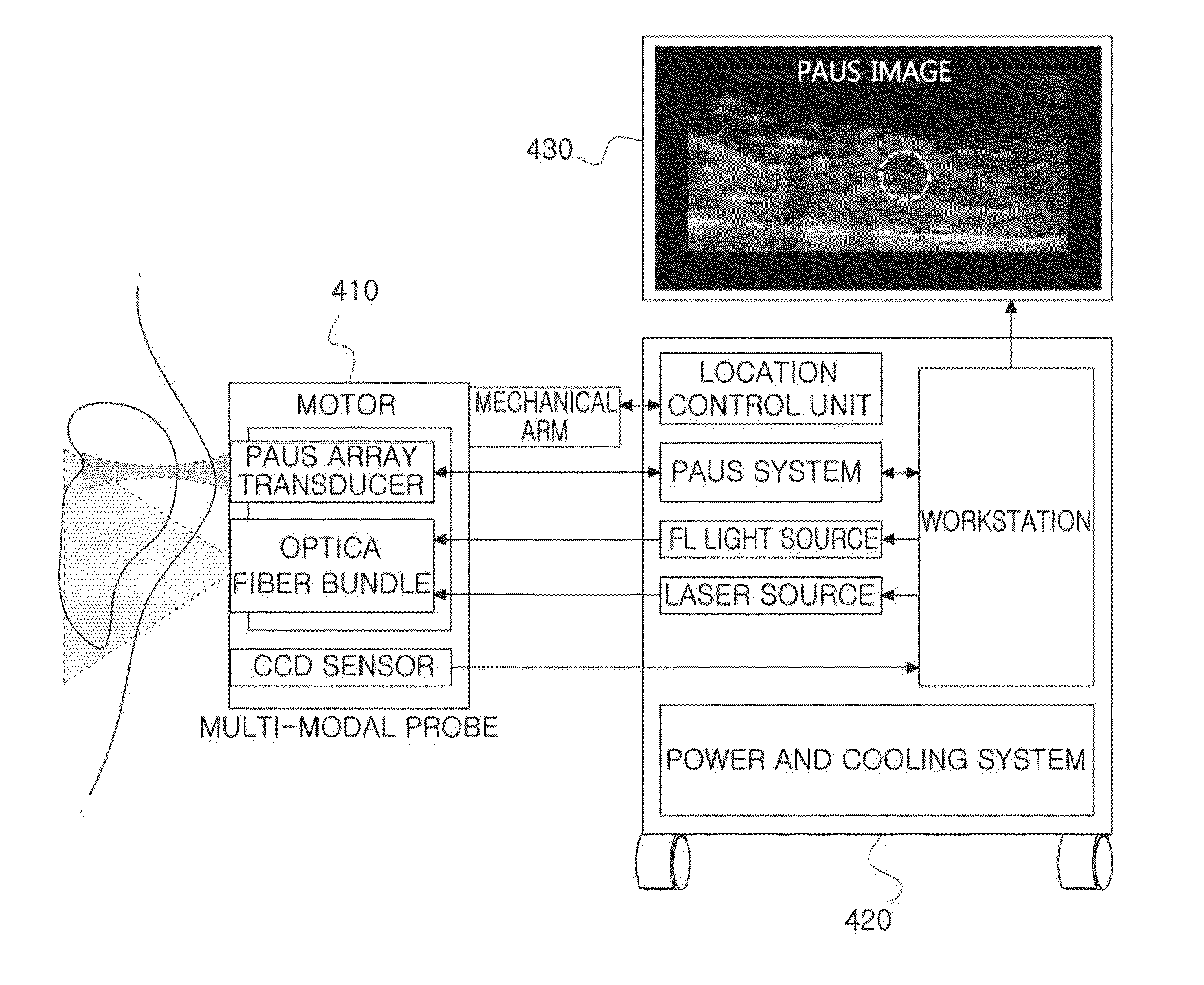
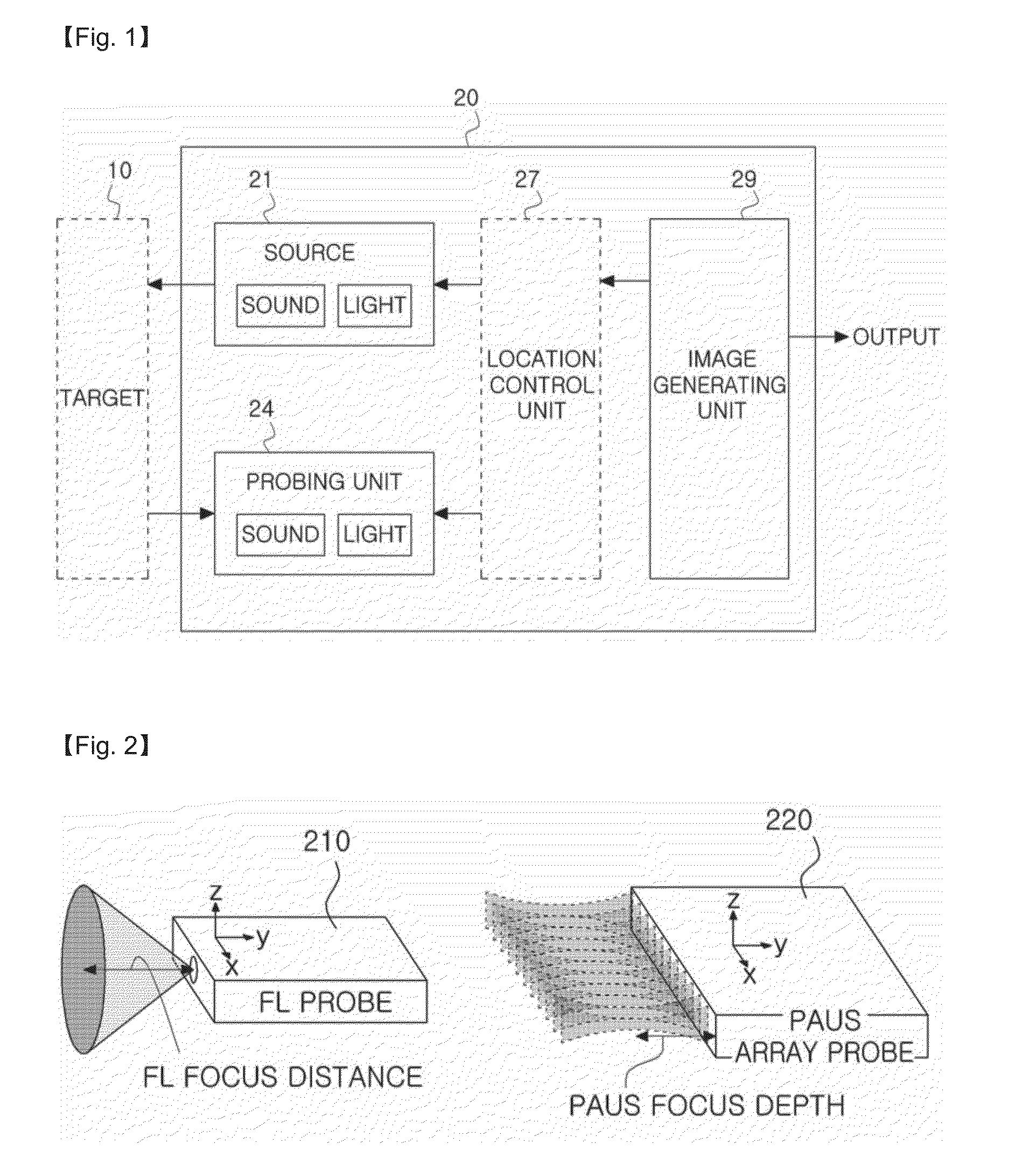
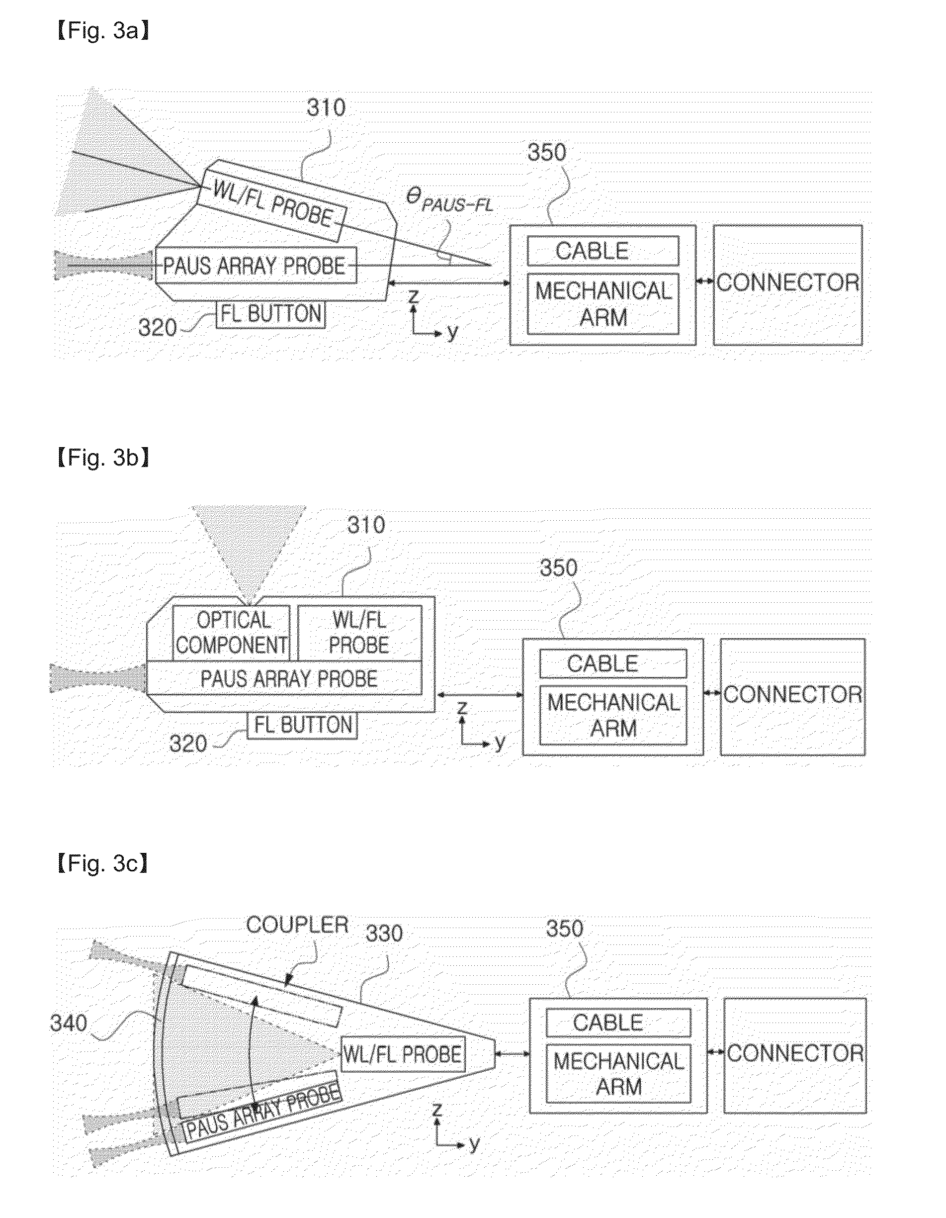
[0036]A biological tissue causes a radiative process and a nonradiative process through a photoacoustic coefficient, and a fluorescent image and a photoacoustic image are formed by means of different process bases through absorbed optical energy. The embodiments of the present disclosure allow observing the degree of light absorption and the generation of radiative / nonradiative process of a tissue, thereby proposing a system structure which may obtain an optical characteristic of the tissue as a more accurate quantitative index and provide elastic ultrasound image (elastography) and color flow imaging by processing the ultrasound signal. In addition, the embodiments of the present disclosure may provide a quantitative index with high contrast in an application using a contrast agent which is reactive with a single...


PUM


Abstract

Disclosed is a device and method for acquiring a fusion image, which applies an ultrasound signal for an ultrasound image, an optical signal for a photoacoustic image and a fluorescent image to a target, receives an ultrasound signal, a photoacoustic signal and an optical signal from the target, and generates a fusion image including image information with different probing planes with respect to the target by using at least two signals of the received ultrasound signal, the received photoacoustic signal and the received optical signal.

Description

TECHNICAL FIELD[0001]This disclosure relates to a medical image technique for diagnosis, analysis and treatment, and more particularly, to a probe structure, an imaging device and an imaging method for acquiring a fusion image capable of providing pathologic and anatomical information simultaneously based on various medical image techniques.BACKGROUND ART[0002]An ultrasound (US) imaging device is equipment for imaging structure and characteristics of an observation area by applying an ultrasound signal to an observation area in a human body with an ultrasound probe, receiving a returning ultrasound signal reflected by tissues, and extracting information included in the signal. The US imaging device may advantageously obtain an image in real time without any harm to the human body at low costs in comparison to other medical imaging systems such as X-ray, CT, MRI, PET or the like.[0003]A photoacoustic (PA) imaging device applies photon to an observation area in a human body, receives ...

Claims


Application Information

Patent Timeline
Patent Type & Authority Applications(United States)
IPC IPC(8): A61B5/00A61B8/08
CPCA61B5/0035A61B8/5207A61B5/0059A61B5/0095A61B5/0071A61B8/4416A61B8/5246H04N5/2624
Inventor CHANG, JIN HOSONG, TAI-KYONGYOO, YANG MOKANG, JEEUNWILSON, BRIANKIM, KANGHAN, SEUNG HEE
Owner SOGANG UNIV RES FOUND
Why Eureka
Try Eureka