Method for inducing dopaminergic neuron progenitor cells

a dopaminergic neuron and progenitor cell technology, applied in the direction of embryonic cells, non-embryonic pluripotent stem cells, drug compositions, etc., can solve the problems of high infection risk, method needs improvement, formation of benign tumors, etc., and achieve high survival rate

Active Publication Date: 2016-07-28
KYOTO UNIV +2
View PDF1 Cites 9 Cited by
  • Summary
  • Abstract
  • Description
  • Claims
  • Application Information

AI Technical Summary

Benefits of technology

[0050]According to the present invention, dopaminergic neuron progenitor cells which are useful for therapeutic agents for Parkinson's disease and the like and suitable for transplantation, and have a high survival rate, can be efficiently obtained.

Problems solved by technology

Fetal mesencephalic grafting is problematic from an ethical point of view because of its source of supply, and the risk of infection is high in this treatment.
However, it has been pointed out that transplantation of neural cells prepared by differentiation induction may cause formation of a benign tumor, and dyskinesia which is thought to be due to cells other than the dopaminergic neural cells of interest.
However, these methods still need improvement in the process of selection of markers.

Method used


Image

  • Method for inducing dopaminergic neuron progenitor cells
  • Method for inducing dopaminergic neuron progenitor cells
  • Method for inducing dopaminergic neuron progenitor cells

Examples


example 1

Cells and Culture

[0147]Human ES cells (KhES-1) were obtained from Institute for Frontier Medical Sciences, Kyoto University (Suemori H, et al. Biochem Biophys Res Commun. 345:926-32, 2006). 404C2 and 836B3, which are human iPS cells produced by introducing Oct3 / 4, Sox2, Klf4, L-MYC, LIN28, and p53shRNA to human fibroblasts using an episomal vector, were received from Prof. Yamanaka at Kyoto University (Okita et al., Nat Methods. 8: 409-412, 2011).

[0148]The ES cells and the iPS cells were cultured by the method according to the method described in Miyazaki T et al. (Nat Commun. 3: 1236, 2012). Briefly, these cells were cultured in 6-well plates coated with Laminin 511E8.

[0149]The thus obtained ES cells or iPS cells were dissociated using TrypLE CTS (Life Technologies), and transferred to a 6-well plate coated with Laminin 511E8 (iMatrix-511, Nippi) in an amount of 4×10⁴ cells per well. The cells were then cultured by the above-described culture method for four days, and, after confir...

example 2

Cell Culture

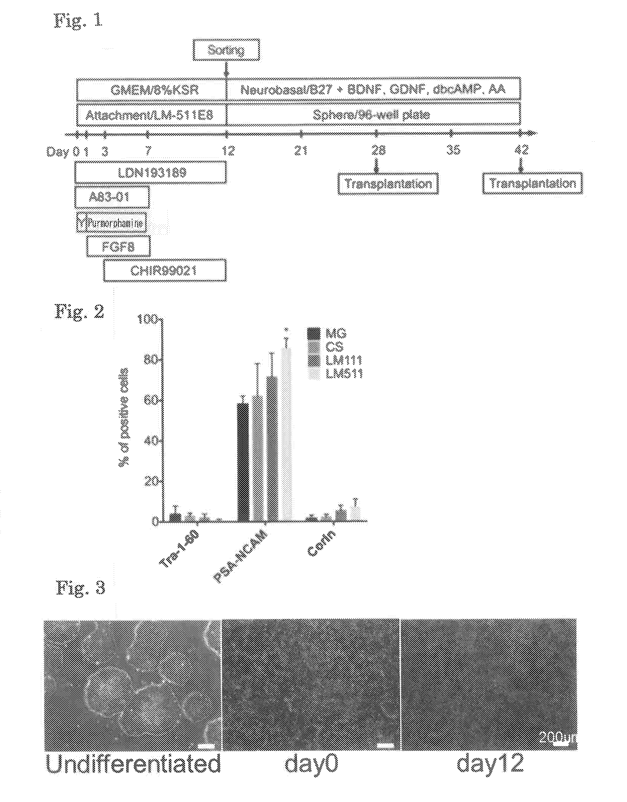
[0168]ES cells (Kh-ES1) were dissociated using TrypLE CTS (Life Technologies), and the whole cells were transferred to a 6-well plate coated with Laminin 511E8. The cells were then cultured in Basal Medium A (GMEM supplemented with 8% KSR, 1 mM sodium pyruvate, 0.1 mM MEM non-essential amino acid, and 0.1 mM 2-mercaptoethanol) supplemented with 10 μM Y-27632, 0.1 μM LDN193189, and 0.5 μM A83-01. One day after the beginning of the culture (Day 1), the medium was replaced with Basal Medium A supplemented with 0.1 μM LDN193189, 0.5 μM A83-01, 2 μM purmorphamine, and 100 ng / mL FGF8. Three days after the beginning of the culture (Day 3), the medium was replaced with Basal Medium A supplemented with 0.1 μM LDN193189, 0.5 μM A83-01, 2 μM purmorphamine, 100 ng / mL FGF8, and 3 μM CHIR99021. Seven days after the beginning of the culture (Day 7), the medium was replaced with Basal Medium A supplemented with 0.1 μM LDN193189 and 3 μM CHIR99021. On Day 12 (12 days after the beginning ...

example 3

Cell Culture

[0173]ES cells (Kh-ES1) were dissociated using TrypLE CTS (Life Technologies), and 4×10⁵ cells were then transferred to a 6-well plate coated with Laminin 511E8. The cells were then cultured in StemFit medium (Ajinomoto) supplemented with 10 μM Y-27632. Four days later, the medium was replaced with the above-described Basal Medium A supplemented with 0.1 μM LDN193189 and 0.5 μM A83-01 (Day 0). One day after the beginning of the culture (Day 1), the medium was replaced with Basal Medium A supplemented with 0.1 μM LDN193189, 0.5 μM A83-01, 2 μM purmorphamine, and 100 ng / mL FGF8. Three days after the beginning of the culture (Day 3), the medium was replaced with Basal Medium A supplemented with 0.1 μM LDN193189, 0.5 μM A83-01, 2 μM purmorphamine, 100 ng / mL FGF8, and 3 μM CHIR99021. Seven days after the beginning of the culture (Day 7), the medium was replaced with Basal Medium A supplemented with 0.1 μM LDN193189 and 3 μM CHIR99021. On Day 14 (14 days after the beginning of...


PUM

PropertyMeasurementUnit
concentrationaaaaaaaaaa

Abstract

The present invention provides a method for producing dopaminergic neuron progenitor cells from pluripotent stem cells, which method comprises the steps of: (i) performing adherent culture of pluripotent stem cells on an extracellular matrix in a medium containing a reagent(s) selected from the group consisting of BMP inhibitor, TGFβ inhibitor, SHH signal-stimulating agent, FGF8, and GSK3β inhibitor; (ii) collecting Corin- and / or Lrtm1-positive cells from the cells obtained in Step (i) using a substance which binds to Corin and / or a substance which binds to Lrtm1; and (iii) performing suspension culture of the cells obtained in Step (ii) in a medium containing a neurotrophic factor.

Description

TECHNICAL FIELD[0001]The present invention relates to a method for producing dopaminergic neuron progenitor cells.BACKGROUND ART[0002]Parkinson's disease is a neurodegenerative disease caused by loss of dopaminergic neural cells in the mesencephalic substantia nigra, and about 4 million people in the world are currently suffering from this disease. For treatment of Parkinson's disease, pharmacotherapy with L-DOPA or a dopamine agonist; coagulation or deep brain stimulation by stereotaxy; fetal mesencephalic grafting; or the like has been carried out.[0003]Fetal mesencephalic grafting is problematic from an ethical point of view because of its source of supply, and the risk of infection is high in this treatment. Thus, a therapeutic method using neural cells prepared by differentiation induction from pluripotent stem cells such as embryonic stem cells (ES cells) or induced pluripotent stem cells (iPS cells) has been proposed (Non-patent Document 1). However, it has been pointed out t...

Claims


Application Information

Patent Timeline
Patent Type & Authority Applications(United States)
IPC IPC(8): C12N5/0797A61K35/30
CPCC12N5/0623A61K35/30C12N2501/13C12N2501/119C12N2533/52C12N2506/03C12N5/0619C12N2501/15C12N2501/155C12N2501/41C12N2501/415C12N2501/727C12N2506/02C12N2506/45A61P25/16
Inventor TAKAHASHI, JUNDOI, DAISUKESAMATA, BUMPEISEKIGUCHI, KIYOTOSHIONO, YUICHI
Owner KYOTO UNIV
Why Eureka
Try Eureka