Methods, compositions and systems for the analysis of nucleic acid molecules

a nucleic acid and composition technology, applied in the field of molecular diagnostics, can solve the problems of reducing the sensitivity and substantial sample-to-sample variability in both absolute and relative mirna levels, affecting the detection accuracy of rt-pcr inhibitors, and reducing so as to reduce or eliminate the loss of small rna, reduce the number of steps for preparation, and minimize the variability of rna recovery

Inactive Publication Date: 2016-07-28
SOMAGENICS INC
View PDF4 Cites 6 Cited by
  • Summary
  • Abstract
  • Description
  • Claims
  • Application Information

AI Technical Summary

Benefits of technology

[0044]In some embodiments, the TSP comprises a blocking group at the 3′-end. The blocking group may prevent the enzymatic extension of the 3′ end, e.g., 3′-p, or 3′-amino, or 2′, 3′-dideoxy nucleoside (ddN), or 3′-inverted 3′-3′ deoxy nucleoside (idN). The blocking group may enable the TSP to to avoid interference with RT-PCR reactions and / or prevent false-positive amplification reactions in the RNA molecule. The blocking group may prevent the TSP from serving as a primer. The blocking group may prevent extension of the TSP.
[0047]In some embodiments, the TSPs further comprise a linker. The linker may be a single-stranded overhang at the end of TSP. The linker may comprise a sequence that is non-complementary to RNA molecule or primers. The linker may comprise one or more nucleotides, non-nucleotides, or a combination thereof. The linker may be used to distance the RNA-specific portion of the TSP from the surface of the solid support. The linker may enable hybridization or attachment of the TSP to the solid support. The linker can improve the efficiency of hybridization between TSP and RNA molecule. The linker may comprise an anchor group or hapten. The linker may enable capture of the TSP or TSP-hybridized RNA molecule.
[0064]In some instances, the provided methods decrease or eliminate the loss of small RNAs that usually occurs under standard total RNA isolation conditions, reduce the number of steps for preparation of miRNAs for RT-qPCR analysis to streamline the procedure, minimize the variability of RNA recovery from different samples, and / or reduce the RNA recovery time and facilitate its automation. In some instances, the provided methods increase the accuracy in determining absolute RNA copy numbers, and / or enable expression profiling of small RNAs of interest (target RNAs) while removing inhibitors of amplification reactions and depleting irrelevant small RNAs, including degradation products of larger RNAs or DNA. The latter features increase the efficiency of amplification of specific RNA sequences and reduce background amplification of non-specific sequences, thereby increasing the sensitivity (signal-to-noise ratio) and multiplexing capability of RT-PCR.

Problems solved by technology
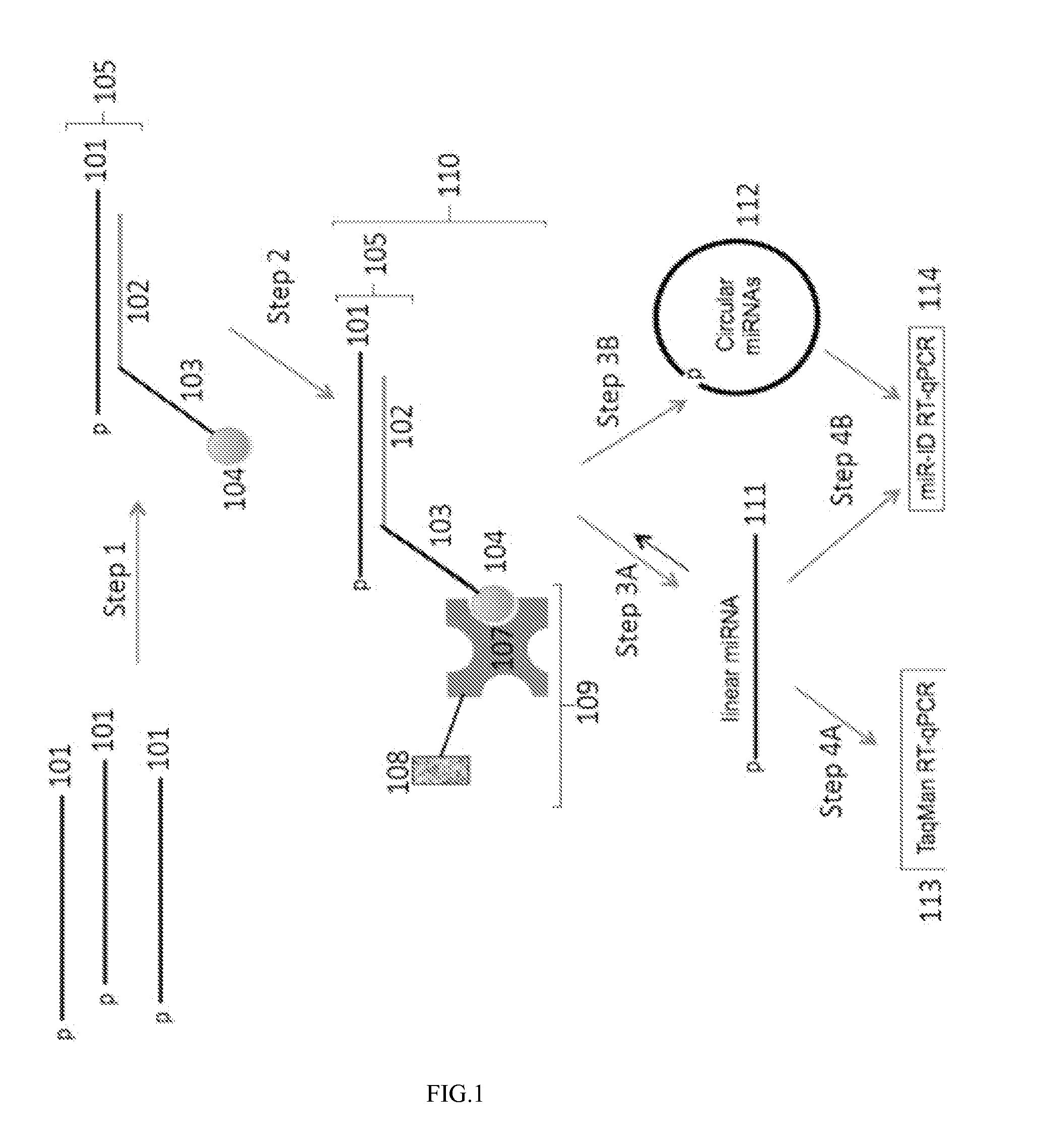
2010), but they are hampered by challenges associated with the requirement for isolating total RNA prior to analysis.
2012) but can also hinder their detection.
These procedures result in reduced sensitivity and substantial sample-to-sample variability in both absolute and relative miRNA levels because of incomplete and inconsistent miRNA recovery, a consequence of the small size of miRNAs and the low concentrations at which they are typically found in plasma and serum (Etheridge et al.
Normalizing results to a synthetic miRNAs spike-in control does not solve this variability problem (McDonald et al.
Also, with standard RNA isolation procedures, it is difficult to eliminate RT-PCR inhibitors that co-purify with total RNA and, as a result, sensitivity cannot be increased by scaling up the quantity of RNA used in each sample assay (Kim et al.
In addition, the isolated total RNA or enriched small RNA fractions contain abundant fragments of unrelated RNAs (such as ribosomal RNA and tRNA); these can serve as primers for reverse transcription (RT) and PCR and can also compromise detection of low abundant miRNAs by specific primers added exogenously.
However, these “solid-phase” RT-PCR methods cannot be applied directly to small RNA molecules that have no poly(A)-tail and are nearly the same size as an ordinary RT or PCR primer, since two PCR primers are required for exponential amplification.

Method used


Image

  • Methods, compositions and systems for the analysis of nucleic acid molecules
  • Methods, compositions and systems for the analysis of nucleic acid molecules
  • Methods, compositions and systems for the analysis of nucleic acid molecules

Examples


example 1

Direct Quantification of miRNAs in Plasma without Capture Using TaqMan or miR-ID Detection

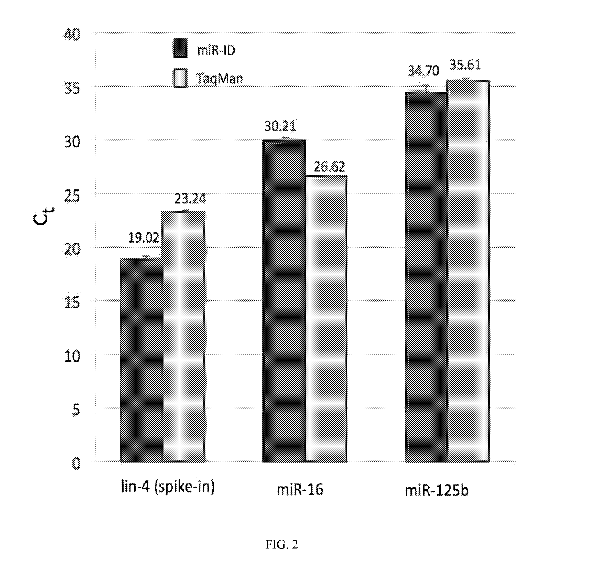
[0210]Frozen plasma specimens obtained from Biological Specialty Co. were thawed and filtered through a 1.2-mm filter to remove cells and cellular debris as recommended by Bryant et al. (2012). 24 μl of plasma was mixed with 1 μl of an RNAse inhibitor and incubated at 60° C. for 10 min. Next, an equal volume (25 μl) of a release / dissociation buffer and 1 fmol of synthetic lin-4 miRNA (as a spike-in control) was added and incubated at 25° C. for 1 hour. The samples were subsequently heated at 95° C. for 5 min and then centrifuged at 16,000 g for 4 min at 25° C. The supernatant was collected and 4 μl aliquots (each corresponding to 2 μl of the original plasma) were analyzed by miR-ID (Kumar et al. 2011) and TaqMan (Chen et al. 2005) RT-qPCR assays specific for hsa-miR-16-5p (miR-16), hsa-miR-125b-5p (miR-125b), cel-lin-4 (lin-4) and cel-miR-39 (cel-39) shown in Table 1. The results are shown in F...

example 2

Quantification of miRNAs in Different Volumes of Plasma by miR-Direct using miR-ID Detection

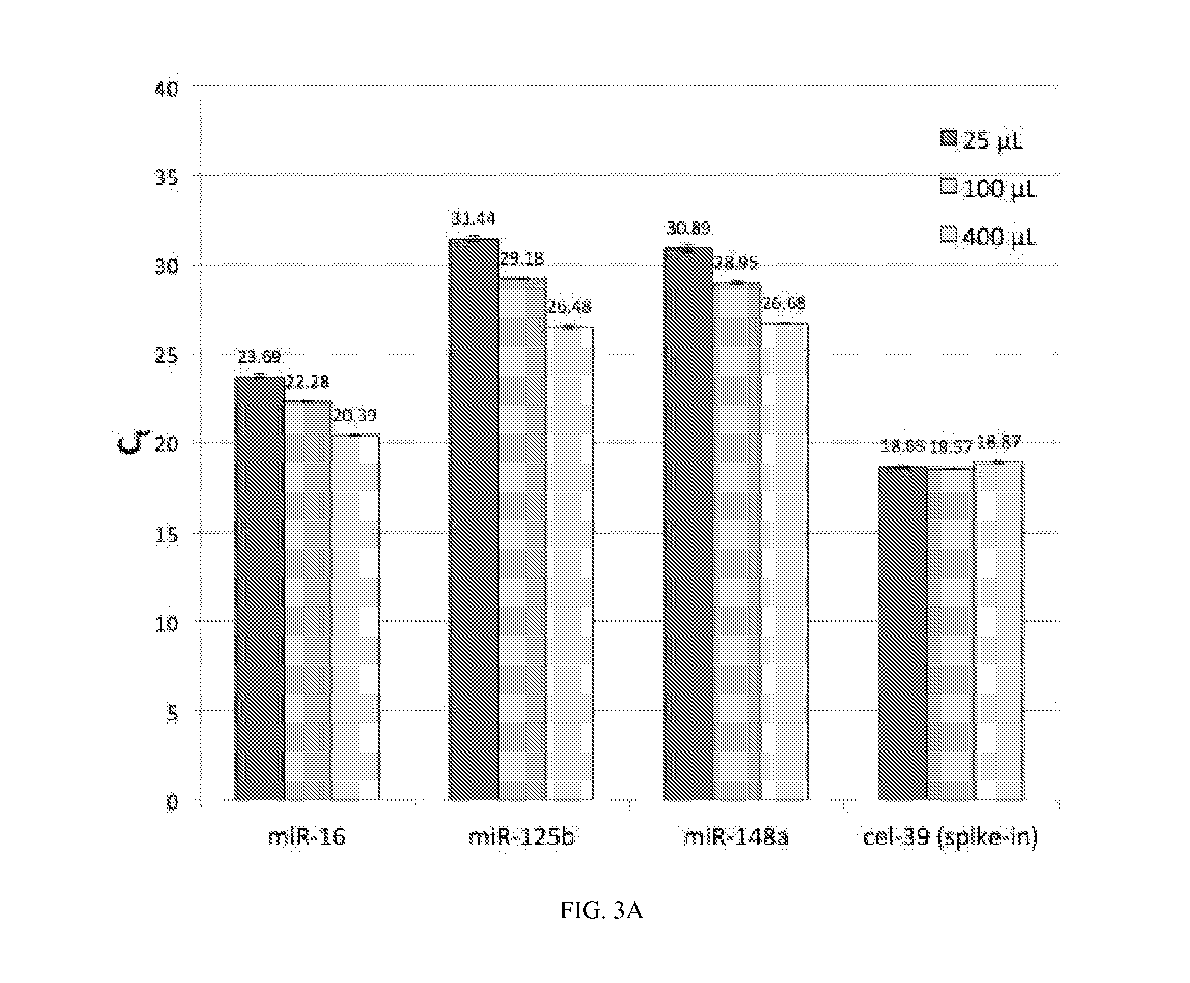
[0214]Various volumes (25, 100 or 400 μl) of plasma sample #55, which was collected in an EDTA-containing tube from one individual, or 200 μL of plasma sample #M7259 collected either in heparin- or EDTA-containing tubes from a second individual (provided Biological Specialty Co.) were treated as follows to release the circulating miRNAs into solution. An equal volume of a lysis buffer was added to each plasma sample. Then a carrier RNA and 1 fmol cel-39 spike-in miRNA were added sequentially. The entire mixture was incubated at 25° C. for 1 hour. Following this incubation, 16 pmol of each of a set of target-specific oligonucleotide probes (TSPs) biotinylated at their 3′ ends (3-BioTEG, IDT), which were specific for miRNAs present in human blood (miR-16, miR-125b, miR-148a, let-7d, let-7g, miR-15b, miR106a, miRl42, miR-191 and miR-301a) or spiked-in miRNA cel-miR-39 (cel-39) (Table 3) were add...

example 3

Quantification of miRNAs in Various Plasma Samples by miR-Direct using miR-ID Detection

[0218]400 μl of each of plasma sample from 11 healthy donors were analyzed using the miR-Direct capture with miR-ID detection specific for the circulating miRNAs hsa-miR-16, hsa-miR-125b and hsa-miR-148a as well as a spike-in control, cel-miR-39 as described in Example 2.

[0219]In this example, we observed that the levels of the circulating miRNAs varied by 1-3 Ct units among the various plasma samples while the spike-in control remained constant (FIG. 4). To verify the Ct variations measured by miR-Direct, we compared the results obtained by miR-Direct and conventional miR-ID, which uses column-purified total RNA (see Example 4 and FIG. 5).


PUM

PropertyMeasurementUnit
temperatureaaaaaaaaaa

Abstract

Methods, systems and compositions are provided for analyzing one or more nucleic acid molecules. The methods, systems and compositions may comprise one or more target specific-oligonucleotide probes (TSPs). The TSPs may hybridize to nucleic acid molecules that are less than or equal to 200 nucleotides in length. The nucleic acid molecules may be small RNA molecules (e.g., miRNA, ncRNA, siRNA, shRNA). The methods, systems and compositions fmd use in a number of applications, for example, isolation of nucleic acid molecules, analysis of low abundance nucleic acid molecules, and / or enrichment of nucleic acid molecules.

Description

CROSS REFERENCE[0001]This application claims the benefit of U.S. provisional application Ser. No. 61 / 771,543, filed Mar. 1, 2013; which is incorporated by reference in its entirety.FIELD OF THE INVENTION[0002]The present invention is in the field of molecular diagnostics. More specifically, it concerns methods, systems and compositions useful for identification, detection, quantification, expression profiling and stabilizing of small RNAs, both naturally occurring and man-made. The present invention finds use in a variety of genomic research and diagnostic applications, in fields including medicine, agriculture, food, and biodefense. The RNA(s) of interest may represent biomarker(s) correlating to specific types of cancer or other diseases such as genetic and metabolic disorders and infectious disease.BACKGROUND OF THE INVENTION[0003]The discovery of microRNAs (miRNAs) and other short RNAs such as small interfering RNAs (siRNA), and short non-coding RNAs (snRNA) has led to a rapid e...

Claims


Application Information

Patent Timeline
Patent Type & Authority Applications(United States)
IPC IPC(8): C12Q1/68
CPCC12Q1/6837C12Q2521/501C12Q2525/155C12Q2525/204C12Q2525/207C12Q2525/307C12Q2531/125C12Q2563/131
Inventor KAZAKOV, SERGEI A.DALLAS, ANNEILVES, HEINIJAYASENA, SUMEDHAJOHNSTON, BRIAN H.
Owner SOMAGENICS INC
Why Eureka
Try Eureka