BioRoot(R) anatomic endosseous dental implant

a dental implant and anatomic technology, applied in dental implants, dental surgery, medical science, etc., can solve the problems of root socket bone fracture or breakage, restricted use of such devices, and inability to use buccal/lingual sides, etc., to minimize shear forces during function, relieve a great deal of stress, and be convenient to us

Inactive Publication Date: 2016-09-22
PEARSON THOMAS STEWART
View PDF0 Cites 8 Cited by
  • Summary
  • Abstract
  • Description
  • Claims
  • Application Information

AI Technical Summary

Benefits of technology

[0016]The instant implant is made to clearly satisfy the growing dental implant field with a truly anatomic tooth root implant that can be utilized quickly and simply, by hand insertion with a mallet and driver as needed, and with its unique macro retentive devices, offers initial stability and retention by the simultaneous BIC of all four tooth faces (mesial / distal, buccal / lingual), of the tooth root socket. By utilizing the instant invention, one can avoid all the invasive surgical procedures previously noted which will relieve a great deal of stress from both practitioner and patient, allowing more time to be spent focusing on the implant process instead of the pain, suffering, and adverse effects from any possible missteps during the implant procedure. The BioRoot® anatomic endosseous dental implant with its custom built abutment and retentive devices can be inserted within fourteen days of manufacture to avoid remodeling of the tooth root socket and bone resorption.
[0017]One additional advantage to the instant implant is a more even distribution of compressive forces and to minimize shear forces during function. By carefully manufacturing each implant to fit the existing tooth root structure of a recently extracted tooth, compressive forces are transferred along all four tooth root socket walls via the unique retentive devices, which will also reduce bone resorption, instead of focusing forces on the alveolar bone at the implant apex as occurs with rotationally inserted screw-type implants. U.S. Pat. No. 8,287,279 (Pirker), 2012, discloses retention devices of various shapes and sizes but the location of such devices are “strictly limited” to the interdental (mesial / distal), areas, leaving buccal / lingual sides non-utilized. At the opposite spectrum is U.S. Pat. No. 2,210,424 (Morrison), 1940, that discloses circular ring retentive devices that encircle the implant body without regard to any tooth face which (could these implants have been manufactured), would have resulted in root socket bone fracture or breakage, and excess bone resorption, ultimately ending in implant failure.

Problems solved by technology

U.S. Pat. No. 8,287,279 (Pirker), 2012, discloses retention devices of various shapes and sizes but the location of such devices are “strictly limited” to the interdental (mesial / distal), areas, leaving buccal / lingual sides non-utilized.
At the opposite spectrum is U.S. Pat. No. 2,210,424 (Morrison), 1940, that discloses circular ring retentive devices that encircle the implant body without regard to any tooth face which (could these implants have been manufactured), would have resulted in root socket bone fracture or breakage, and excess bone resorption, ultimately ending in implant failure.
tooth face which (could these implants have been manufactured), would have resulted in root socket bone fracture or breakage, and excess bone resorption, ultimately ending in implant failure.

Method used


Image

  • BioRoot(R) anatomic endosseous dental implant
  • BioRoot(R) anatomic endosseous dental implant
  • BioRoot(R) anatomic endosseous dental implant

Examples


Embodiment Construction

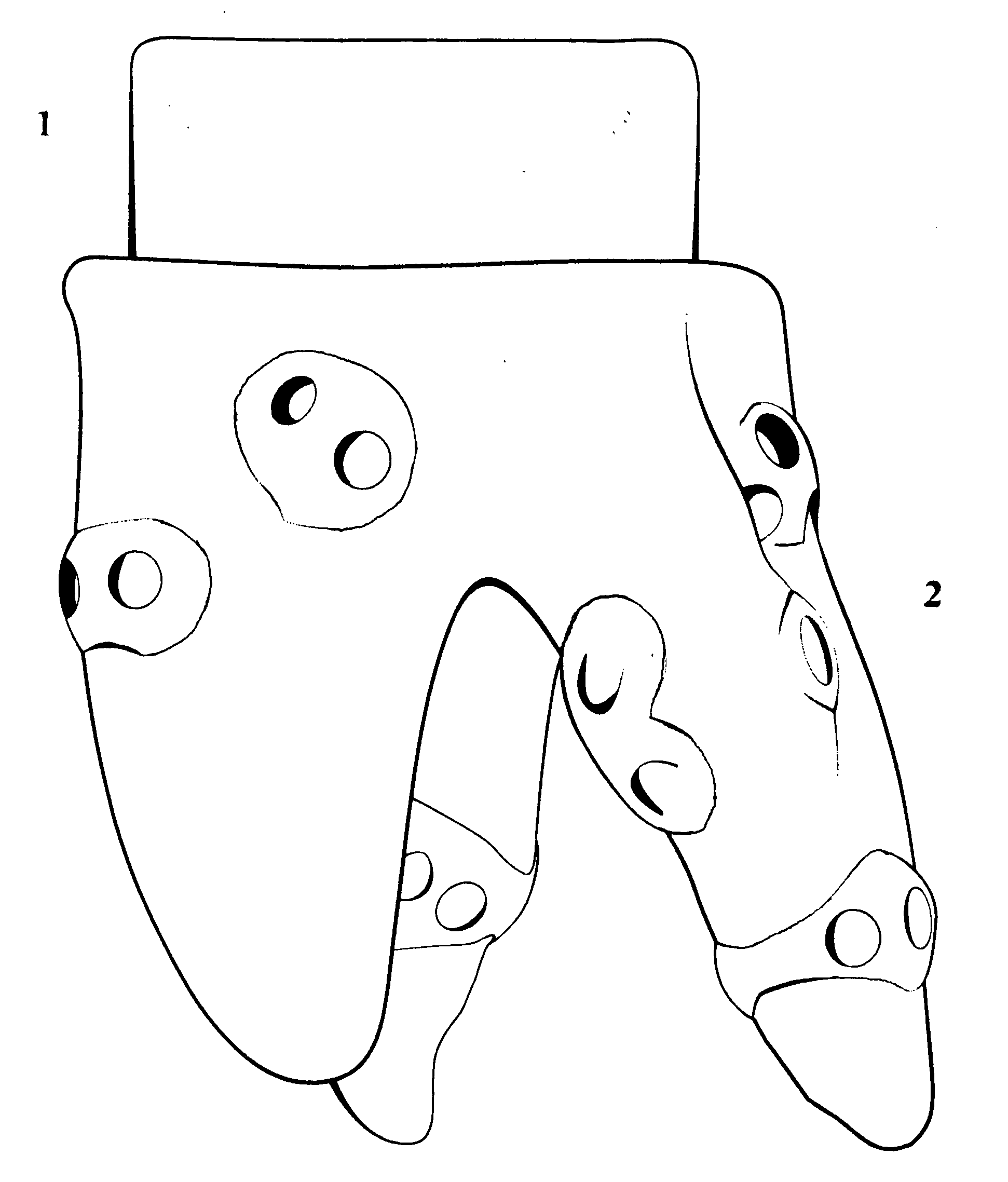
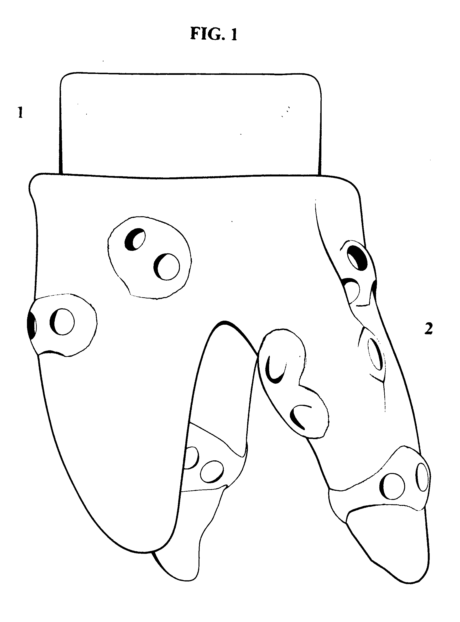
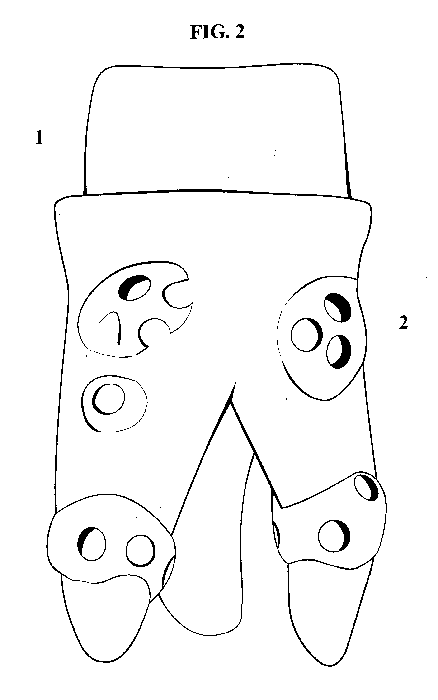
[0022]BioRoot® anatomic endosseous dental implant begins as a solid block of 3% MOL yttria-stabilized, zirconia oxide (Y3ZrO2), a biocompatible ceramic material while in the green state (non-sintered). Utilizing acquired imaging ranging from a visual exam, x ray, cone beam scan, or MRI, exacting tooth and tooth root socket measurements are made and the data is fed into a computer along with the actually-extracted tooth as a model, and by way of a 3D scanner, the operator will make manipulations to the virtual tooth by removing minor surface defects and adding retentive devices (See drawings FIG. 1, FIG. 2, FIG. 3A, and FIG. 3B). These retentions will be placed on all four tooth faces: mesial, distal, and buccal, lingual, ranging in height from 2 nm to 2 mm. When the data has been fully manipulated, it is fed into milling machines that will then mill the implant blank to produce the BioRoot® anatomic endosseous dental implant with a custom built abutment and a number of retentions wi...


PUM


Abstract

The BioRoot® anatomic endosseous dental implant begins as a block of yttria-stabilized, zirconia oxide (MOL3% Y3ZrO2) that is milled and processed into a single piece dental implant with a custom built abutment to which a dental prosthesis can be attached after a three to four month osseointegration period, with unique retention devices that can be round, ovoid, or oblong-shaped, of any size desired with a varied number of holes (See Drawings FIGS. 1, 1 and 2, FIGS. 2, 1 and 2, and FIGS. 3, 1 and 2) which through the osseointegration process will become anchors between the implant surface and the alveolar walls of the extracted tooth root socket that minimize bone resorption, increase bone-to-implant contact, increase initial implant stability and enhance overall osseointegration.

Description

[0001]The present invention is referred to as BioRoot® anatomic endosseous dental implant.CROSS-REFERENCE TO RELATED APPLICATIONS[0002]Not Applicable.STATEMENT REGARDING FEDERALLY SPONSORED RESEARCH OR DEVELOPMENT[0003]Not Applicable.REFERENCE TO SEQUENCE LISTING[0004]None.BACKGROUND OF THE INVENTION[0005]The invention described herein is in the field of dental implants, the history of which has brought innumerable designs and theories, composition and installation, but more precisely, this application describes how to manufacture and how to place an implant that follows natural human physiology and avoid unnecessary invasive dental procedures that result in direct and indirect failures, unnecessary suffering, lengthy healing periods, and financial expenditures.[0006]Typically the dental implant process in today's market involves extensive invasive procedures because of mass marketing implant designs that force practitioners to remodel a patient's jaw due to the implants' shape. It ...

Claims


Application Information

Patent Timeline
Patent Type & Authority Applications(United States)
IPC IPC(8): A61C8/00A61C8/02
CPCA61C8/0036A61C8/0043A61C2008/0046A61C8/0006A61C8/0075
Inventor PEARSON, THOMAS STEWART
Owner PEARSON THOMAS STEWART
Why Eureka
Try Eureka