
Spatial multiplexing for multisignal cellular imaging

a multi-signal, imaging technology, applied in the field of high-content, single-cell resolution, spatial multiplex cell imaging system, can solve the problems of compromising the successful use of imaging methods in living cells, not being able to monitor more than one or two signals in a living cell,

Inactive Publication Date: 2016-10-20
MASSACHUSETTS INST OF TECH
View PDF0 Cites 0 Cited by
  • Summary
  • Abstract
  • Description
  • Claims
  • Application Information

AI Technical Summary

Benefits of technology

The patent describes methods and compounds for spatial multiplex imaging in cells. These methods involve expressing in a cell one or more effector compounds, each comprising a sensor molecule with an indicator molecule and an effector molecule that specifically binds to a predetermined anchor site in the cell. The sensor molecule and effector molecule bind to different anchor sites in the cell, allowing for the detection of the indicator molecule and the effector molecule. The methods can be used to detect the activity or signal of the cell. The invention also provides effector compounds that include a sensor molecule and an effector molecule that binds to a specific chromosomal DNA site in the cell. Overall, the invention provides tools for studying cellular activity and signals in a more comprehensive and accurate way.

Problems solved by technology

Currently it is not possible to monitor more than one or two signals in a living cell at the same time.
The difficulty, is at least in part because the spectra of fluorescent biological reporters are limited to a few colors, e.g. emitting in green or red, which compromises the successful use of imaging methods in living cells.

Method used


Image

  • Spatial multiplexing for multisignal cellular imaging
  • Spatial multiplexing for multisignal cellular imaging
  • Spatial multiplexing for multisignal cellular imaging

Examples


example 1

Methods:

Bioinformatics Design in DNA Binding Sites:

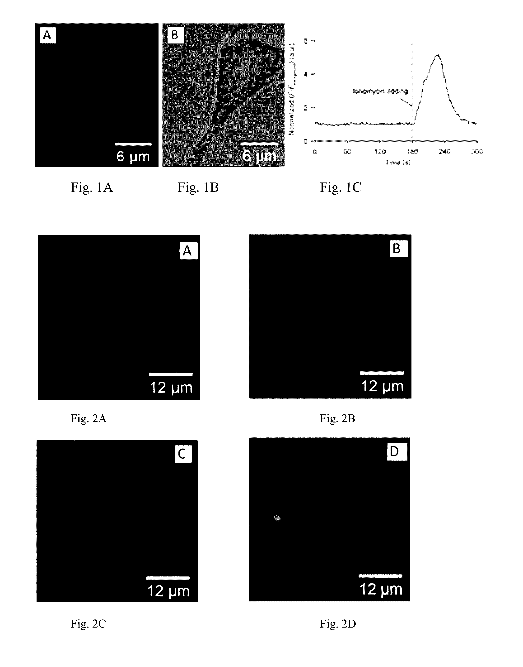
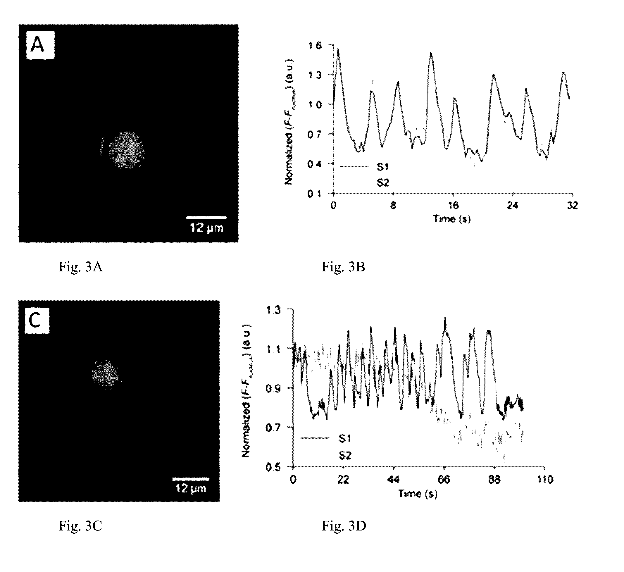
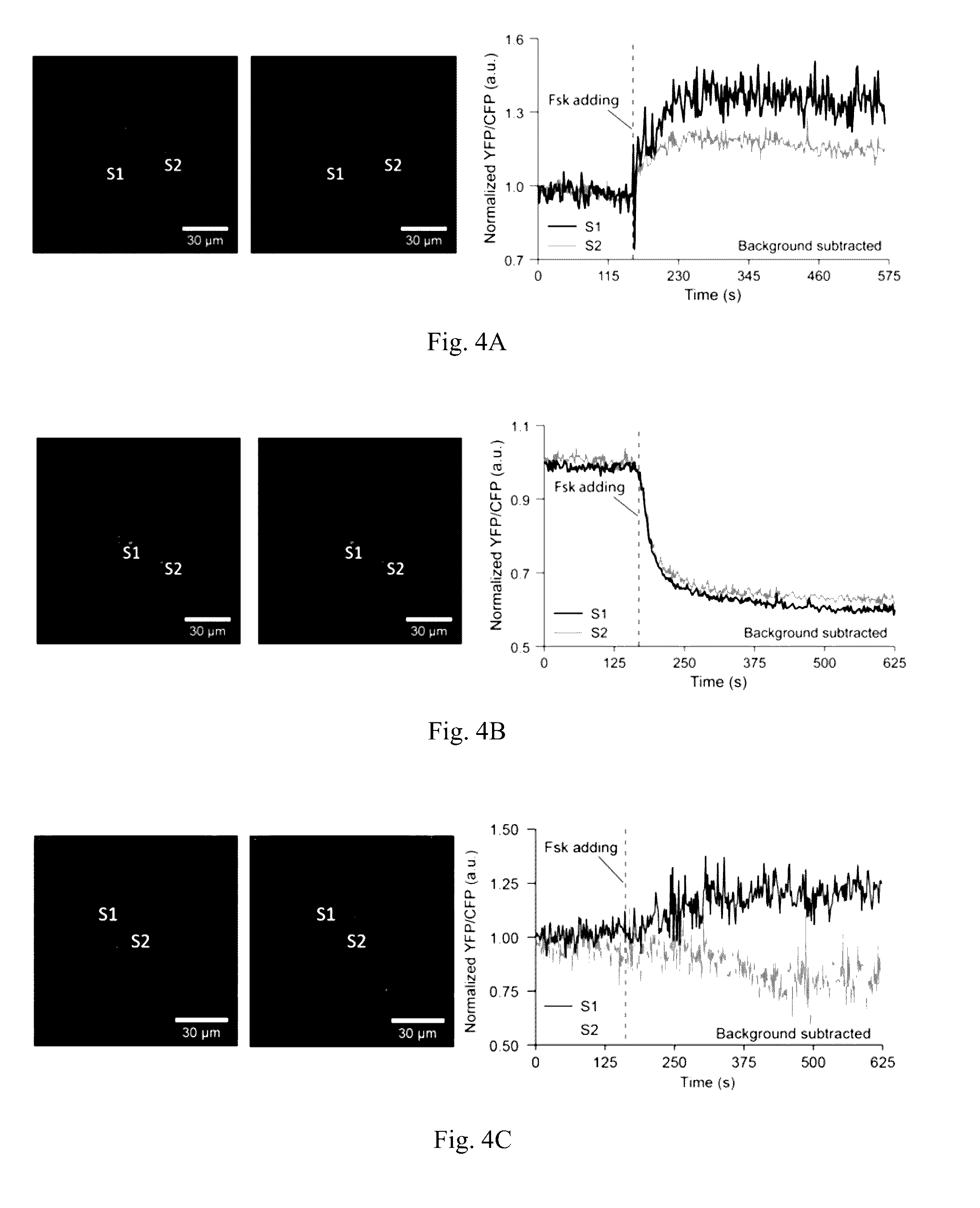
[0064]TALE binding sites were searched in hg20 (human) and mm 10 (mouse) genome database, respectively. Each binding site contained more than 20 repetitive binding sequences, with each sequence was 20 bp in length and started with a thymine. The selected binding sites were assured to be chromosome specific, and at least 5000 by away from the active genes. The selected binding sites allow at least 20 copies of TALE-fused sensors clustering with sufficient brightness for imaging. The selected sites, because they do not overlap with active genes, minimize the chance of expressed TALE proteins regulating the gene expression.

Construct Cloning:

[0065]The genes encoded the GCaMP6f, GEICS1.3, AKAR4, and ICUE3 proteins fused to nuclear localization signal (NLS) and corresponding antibody tag were subcloned into the modified pCI vector backbone (Promega) using NheI and EcoRI sites. The pCI vector was modified by removing BsaI site from bla gen...


PUM

PropertyMeasurementUnit
Pharmaceutically acceptableaaaaaaaaaa
Fluorescenceaaaaaaaaaa

Abstract

The invention, in some aspects, relates to methods, systems, and components of a high-content, single-cell resolution, spatial multiplex cell imaging system.

Description

RELATED APPLICATIONS[0001]This application claims benefit under 35 U.S.C. §119(e) of U.S. Provisional application Ser. No. 62 / 147,225 filed Apr. 14, 2015, the disclosure of which is incorporated by reference herein in its entirety.GOVERNMENT INTEREST[0002]This invention was made with government support under the NSF CBET 1344219 grant awarded by the National Science Foundation. The Government has certain rights in the invention.FIELD OF THE INVENTION[0003]The invention, in some aspects, relates to methods, systems, and components of a high-content, single-cell resolution, spatial multiplex cell imaging system.BACKGROUND OF THE INVENTION[0004]Currently it is not possible to monitor more than one or two signals in a living cell at the same time. The difficulty, is at least in part because the spectra of fluorescent biological reporters are limited to a few colors, e.g. emitting in green or red, which compromises the successful use of imaging methods in living cells.SUMMARY OF THE INVE...

Claims


Application Information

Patent Timeline
IPC IPC(8): C12N15/10
CPCC12N15/1086C12N15/102
Inventor XU, GUANGYUBOYDEN, EDWARDPIATKEVICH, KIRYL D.ADAMALA, KATARZYNA
Owner MASSACHUSETTS INST OF TECH
Learn More