
High-throughput single cell sorting using microbubble well arrays

a microbubble well and array technology, applied in the field of microfabricated devices and methods for high throughput single cell screening, can solve the problems of affecting the self-conditioning of cells in wells when seeded under limiting dilution, and affecting the quality of microbubble wells

Inactive Publication Date: 2017-03-16
NIDUS BIOSCI LLC
View PDF3 Cites 4 Cited by
  • Summary
  • Abstract
  • Description
  • Claims
  • Application Information

AI Technical Summary

Benefits of technology

The present invention is a microfabricated device that can be used for cell culture, sorting, and analysis in both in situ and ex situ (removing from the body) settings. The device has curvilinear microbubble well cavities embedded in a non-glass substrate, creating a microenvironmental niche where cells can be seeded and cultured for a period of time. The device allows for control over the number of cells seeded per cavity and can readily condition with autocrine or paracrine secreted factors to support cell survival and clonal or colony proliferation.

Problems solved by technology

For example, use of the 96-well plate format imposes the constraint of a high media volume to surface area ratio [16], which hinders cell self-conditioning of wells when seeded under limiting dilution conditions [17].
While this method is effective, the process is laborious, time consuming and costly.
Moreover, relatively few (˜103) of the hybridoma cells produced can be tested and therefore potentially high affinity mAbs may be missed.
While the aforementioned techniques make vast improvements over the conventional ELISA cell screening process, they still suffer from various drawbacks.
In shallow well architectures the cells may be easily dislodged by turbulent fluid flow, creating uncertainty in being able to recover the specific cell of interest.
Cells in MB wells are not easily displaced by fluid flow and in fact it takes several hours for nonadherent cells to exit the wells of inverted chips [29].

Method used


Image

  • High-throughput single cell sorting using microbubble well arrays
  • High-throughput single cell sorting using microbubble well arrays
  • High-throughput single cell sorting using microbubble well arrays

Examples


experimental examples

[0104]The invention is further described in detail by reference to the following experimental examples. These examples are provided for purposes of illustration only, and are not intended to be limiting unless otherwise specified. Thus, the invention should in no way be construed as being limited to the following examples, but rather, should be construed to encompass any and all variations which become evident as a result of the teaching provided herein.

[0105]Without further description, it is believed that one of ordinary skill in the art can, using the preceding description and the following illustrative examples, make and utilize the present invention and practice the claimed methods. The following working examples therefore, specifically point out the preferred embodiments of the present invention, and are not to be construed as limiting in any way the remainder of the disclosure.

Cells and Cell Lines

[0106]Three cell lines were selected for demonstrating the diffusion assay proof...

example 1

Detection of IgG by Immunoprecipitation

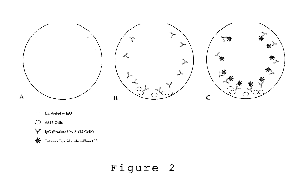
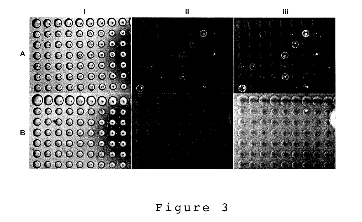
[0108]In one embodiment, the present invention describes a single cell screening method using microbubble well arrays where cells are by what they secrete using immunoprecipitation detection. For immunoprecipitation to take place the immunoglobin present must be able to bind a multivalent reporter (antigen or antibody) to form large polymerized agglutinates. Once a sufficient size threshold is reached the agglutinates settle forming a precipitate [33]. For example, SA13 hybridoma cells were used to demonstrate the accumulation of IgG secretions in the MB well using immuno-precipitation detection. Following the seeding of SA13 cells into the MB array, a rabbit α-human IgG-FITC reporter (Rockland antibodies and assays #209-4202, USA) was added to the culture media. The chips were incubated with the reporter over 3-4 days and imaged daily using an inverted fluorescent microscope (Olympus IX70 with Qlmaging Retiga EXi camera). Images were taken in ...

example 2

Tetanus Toxoid Antigen Specific Immunoglobin Detection by Immunoprecipitation Using a Nanoparticle Multivalent Reporter Construct

[0110]Nanoparticle Synthesis

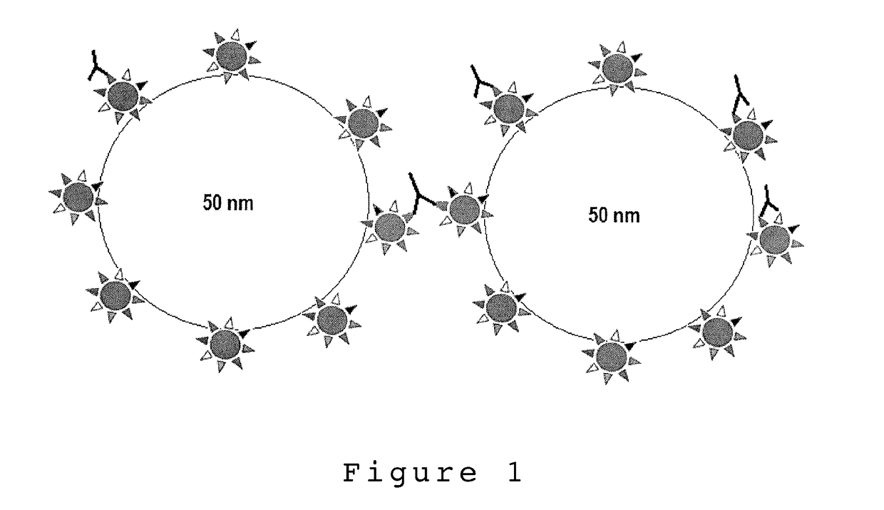
[0111]Clostridium tetani Tetanus Toxoid (Pierce Antibodies HYB 278-17-02, Thermo scientific, USA) was conjugated to carboxyl terminated polystyrene nanoparticles with an average size of 50 nm (Polylink Polysciences, Inc., USA) through covalent coupling via EDC. The conjugation was done by the manufactures specifications with some procedural alterations. Due to cost associated with tetanus toxoid, 3 ug of tetanus toxoid was added to the NP suspension, this was approximately 100× less antigen than the procedure called for to obtain complete PS-NP antigen saturation. Additionally, the toxoid-NP incubation was done over the course of 2 hours with light mixing. This was in contrast to the manufactures recommendation of a 1 hour incubation period. This was done to increase likely hood of NP-toxoid conjugation due to the relatively low...


PUM


Abstract

The present invention provides a microfabricated device and methods for high throughput single cell screening of a heterogeneous population. The present invention is partly based upon but not limited to sorting by monitoring cell secreted factors that accumulate in time (hours, days, weeks) as cells are cultured in the microbubble well niche the architecture of which facilitate the accumulation. In certain embodiments, the device and method comprises a means to identify effective drugs for personalize therapeutics such as but not limited to discovery of monoclonal antibody therapeutics.

Description

CROSS REFERENCE TO RELATED APPLICATION[0001]This application claims priority to Provisional U.S. Application 62 / 191,666, filed Jul. 13, 2015, which is hereby incorporated by reference in its entirety.STATEMENT REGARDING FEDERALLY SPONSORED RESEARCH OR DEVELOPMENT[0002]This invention was made with government support under The University of Rochester Clinical and Translational Science Institute that is supported in part by grants UL1 TR000042, KL2 TR000095, and TL1 TR000096 from the National Center for Advancing Translational Sciences (NCATS), a component of the National Institutes of Health (NIH). The government has certain rights in the invention.FIELD OF THE INVENTION[0003]The present invention provides a microfabricated device and methods for high throughput single cell screening of a heterogeneous population. The present invention is partly based upon but not limited to sorting by monitoring cell secreted factors that accumulate in time (hours, days, weeks) as cells are cultured ...

Claims


Application Information

Patent Timeline
IPC IPC(8): G01N33/543G01N33/68
CPCG01N33/54366G01N33/54346G01N33/6854C12M23/12C12M47/04G01N33/56966C12M3/00
Inventor DELOUISE, LISA A.KOBIE, JAMES J.
Owner NIDUS BIOSCI LLC
Learn More