Automated analysis of vasculature in coronary angiograms

an angiogram and vasculature technology, applied in the field of fully automated detection of coronary vessels, to achieve the effect of accurate calculation of the diameter of each branch and reduction of human estimation errors

Active Publication Date: 2017-05-25
RGT UNIV OF MICHIGAN
View PDF4 Cites 17 Cited by
  • Summary
  • Abstract
  • Description
  • Claims
  • Application Information

AI Technical Summary

Benefits of technology

[0008]The present techniques apply advanced digital image processing and machine learning methods after this pre-processing and frame selection, to segment vessels from the processed angiogram data. From the segmentation, the present techniques are able to more accurately calculate the diameter of each branch in the vasculatures.
[0009]In some embodiments, the techniques further include a decision support system that may aid cardiologists in diagnosing diseases using calculated quantitative parameters of the vasculatures. These diagnoses include identification of stenoses and the extent of stenoses. As a result, as we show, the present techniques reduce human estimation error by providing an automatically executing baseline determining, standardization system for measuring vessel characteristics and pathologies.

Problems solved by technology

There are different kinds of artifacts in angiogram videos, making it a challenge to segment vessels accurately.

Method used


Image

  • Automated analysis of vasculature in coronary angiograms
  • Automated analysis of vasculature in coronary angiograms
  • Automated analysis of vasculature in coronary angiograms

Examples


Embodiment Construction

[0020]The present techniques provide an automated system to analyze angioplasty and other images. Along with new image acquisition techniques that provide better resolution and quality, new image processing techniques help the physician perform accurate diagnosis.

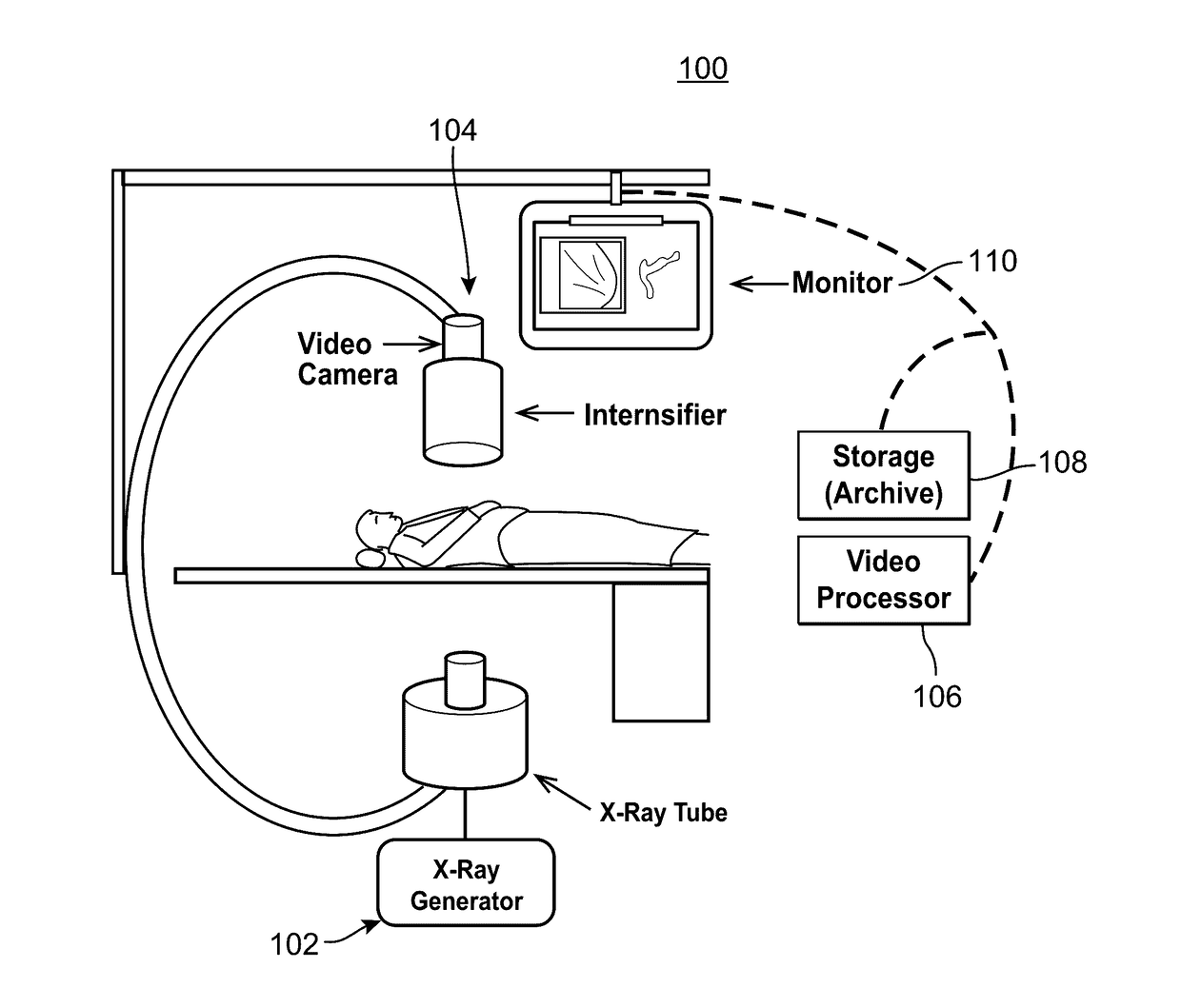
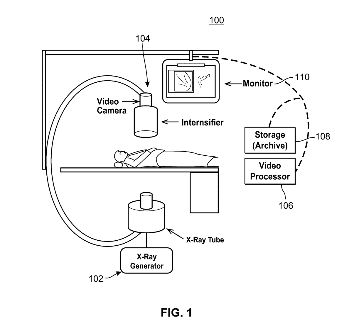
[0021]FIG. 1 illustrates a schematic diagram of an example of angiography system 100 having an X-ray source 102, an X-ray image detector 104, a digital video image processor 106, data recorder and storage 108, and display system 110 providing convention angiogram image collection and processing. While the examples herein are described in reference to an angiography system, the techniques may be applied to any number of medical imaging modalities and image types. Angiogram video data has been selected because of its standard use in stenosis detection.

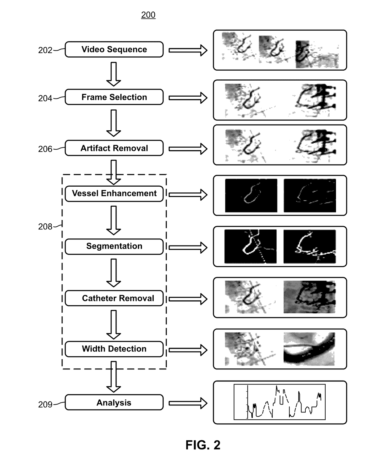
[0022]The digital video image collected by the system 100 is provided to a quality assessment system 200 (FIG. 2) configured to examine the received image or video data, whether...


PUM


Abstract

A system analyzes angiogram image data, including video data, in order to extract vasculature information. Advanced image processing and machine learning techniques are used in pre-processing, frame selection, and vasculature segmentation to remove classes of artifacts from angiogram videos, and specifically from frames selected as having a sufficient amount of image data. From segmentation, accurate vasculature diameters are calculated, and, in some examples, stenoses and / or the extent of stenosis is automatically determined and displayed.

Description

CROSS REFERENCE TO RELATED APPLICATION[0001]This application claims the benefit of U.S. Provisional Application No. 62 / 251,175, filed Nov. 20, 2015, entitled “Automated Analysis of Vasculature in Coronary Angiograms,” which is hereby incorporated by reference in its entirety.FIELD OF THE INVENTION[0002]The invention generally relates to the fully automated detection of coronary vessels and their branches in angiograms and, more particularly, to the calculation of a diameter of such vessels, detection of stenosis and determination of the percent narrowing of stenosis.BACKGROUND[0003]The background description provided herein is for the purpose of generally presenting the context of the disclosure. Work of the presently named inventor, to the extent it is described in this background section, as well as aspects of the description that may not otherwise qualify as prior art at the time of filing, are neither expressly nor impliedly admitted as prior art against the present disclosure.[...

Claims


Application Information

Patent Timeline
Patent Type & Authority Applications(United States)
IPC IPC(8): G06T7/00A61B5/02A61B5/00G06T5/00
CPCG06T7/0012G06T5/002G06T7/0083A61B5/02007G06T2207/10116A61B5/725G06T2207/20081G06T2207/10016G06T2207/30101A61B5/7203A61B6/504A61B6/5217A61B6/5258A61B2576/023G06T5/005G06T2207/30168G06T7/11G06T7/62G16H50/30
Inventor NAJARIAN, KAYVANSOROUSHMEHR, SAYEDMOHAMMADREZAWARD, KEVIN R.NALLAMOTHU, BRAHMAJEE K.
Owner RGT UNIV OF MICHIGAN
Why Eureka
Try Eureka