Microfluidic Device and Method for Analysis of Tumor Cell Microenvironments

a microfluidic device and microenvironment technology, applied in tumor/cancer cells, laboratory glassware, instruments, etc., can solve the problems of inability to accurately represent tumor heterogeneity or resistance to drug penetration, current preclinical tools to investigate cancer biology and potential tumor treatments have significant problems, and can not achieve the effect of realistic drug response and high throughput generation

Active Publication Date: 2017-07-13
NORTHEASTERN UNIV
View PDF0 Cites 17 Cited by
  • Summary
  • Abstract
  • Description
  • Claims
  • Application Information

AI Technical Summary

Benefits of technology

[0003]The invention provides a microfluidic platform for high throughput generation and analysis of clearly defined 3D cell spheroids with uniform geometry and versatility in terms of the spheroid sizes and cell composition. The cell spheroids of the invention resemble the tumor microenvironment, including pathophysiological gradients, cell composition and heterogeneity of the tumor mass mimicking the resistance to drug penetration providing more realistic drug response.

Problems solved by technology

However, current preclinical tools to investigate cancer biology and potential tumor treatments still have significant problems.
Available two-dimensional (2D) tissue culture models and monolayer cultures lack the realistic complexity of tumor microenvironments and cannot represent tumor heterogeneity or the resistance to drug penetration, which are critical parameters.
It has been suggested that the presence of immune cells allows tumor cells to be more aggressive and that, without the presence of immune cells, cancer stem cells may not efficiently progress.
Unfortunately, some so-called spheres and even spheroids described in the literature are no more than loose aggregates of cancer cells that easily detach, cannot be manipulated or transferred, and may lack both cell—cell and cell—matrix interactions, but also lack a true spherical geometry.

Method used


Image

  • Microfluidic Device and Method for Analysis of Tumor Cell Microenvironments
  • Microfluidic Device and Method for Analysis of Tumor Cell Microenvironments
  • Microfluidic Device and Method for Analysis of Tumor Cell Microenvironments

Examples


example 1

Formation of Cell Spheroids in a Microfluidic Device

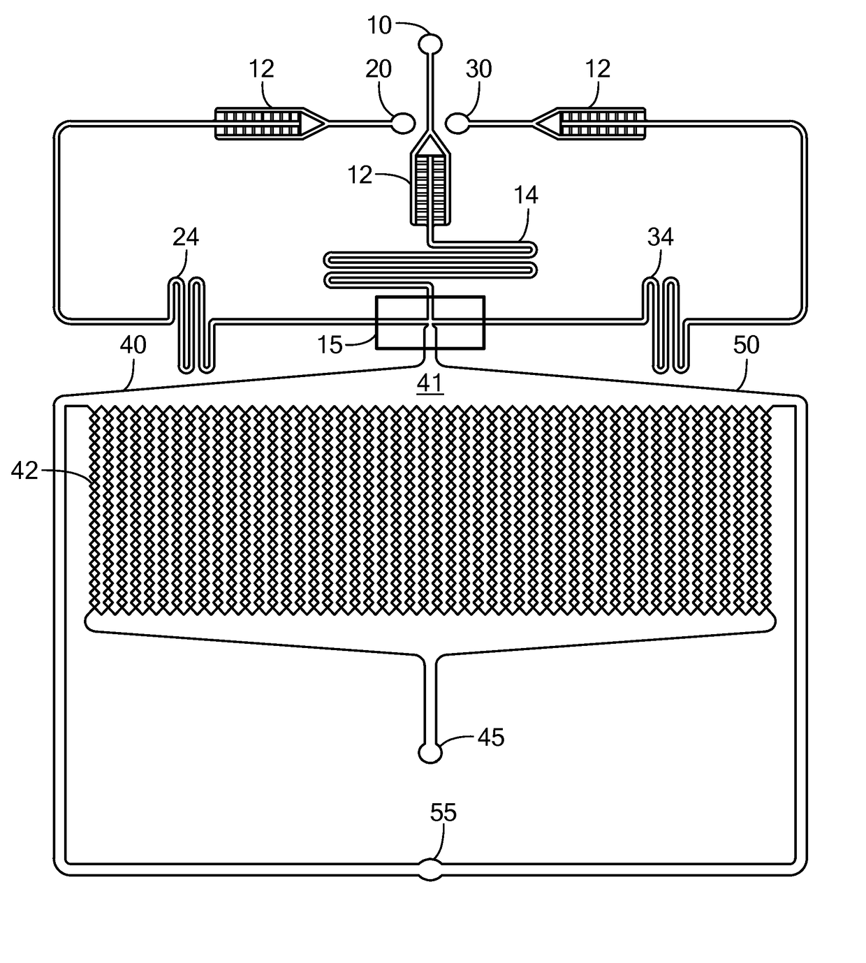
[0095]The microfluidic device depicted in FIG. 1A was used to prepare spheroids of MCF7 breast cancer cells. The three inlets of the device were simultaneously fed with mineral oil containing 3% v / v of Span 80 (a surfactant), a suspension of MCF7 cells at 7-10 million cells / mL and containing 2% w / v sodium alginate in Dulbecco's Modified Eagle Medium (DMEM) containing 10% v / v fetal bovine serum and 1% v / v antibiotic antimycotic solution, and a 4% w / v CaCl2 solution. The solutions were introduced simultaneously into the device using syringe pumps. The flow rates were 300 μL / hr for the oil, 75 μL / hr for the cell suspension, and 10 μL / hr for the calcium solution. The size of the microchambers was 200 microns, and the average size of the cell spheroids produced was 170+ / −25 microns. After the spheroids were produced, the flow of oil, cell suspension, and CaCl2 solution was stopped, the incubation chamber of the device was continuously p...

example 2

Cell Viability

[0096]The viability of cells in cell spheroids incubated in a microfluidics device of the invention was determined using the LIVE / DEAD viability / cytotoxicity assay for mammalian cells by Life Technologies (Cat No: L-3224). The kit included two dye components: calcein-AM as an indicator of live cells and ethidium homodimer-1 as an indicator of dead cells. For live cells, calcein-AM was cleaved by esterase enzymes to form the green fluorescent dye calcein in the cytoplasm of the cells. For dead cells, the compromised cell membranes allowed ethidium homodimer-1 to permeate and bind to the nucleic acids in the nuclei of the cells, which then emitted a red fluorescence.

[0097]3D spheroids of MCF7 adriamycin sensitive cells, a breast cancer cell line, were formed in a microfluidic device of the invention as described in Example 1. The cell spheroids were housed in the microfluidic device for 14 days and continuously perfused with fresh cell culture medium, and the cell viabil...

example 3

Sensitivity of Cell Spheroids to Antitumor Agents

[0098]The cell viability assay described in Example 2 was used to ascertain the sensitivity to doxorubicin of MCF7 breast cancer cells in cell spheroids present in a microfluidic device of the invention.

[0099]FIGS. 6A and 6B show adriamycin resistant MCF7 cell spheroids at 96 hr incubation without doxorubicin (FIG. 6A) and at 48 hr incubation with 12.8 μM doxorubicin (FIG. 6B). The red fluorescence in FIG. 6B indicates cell toxicity of the doxorubicin. FIGS. 6C and 6D show adriamycin sensitive MCF7 cell spheroids at 96 hr incubation without doxorubicin (FIG. 6C) and at 48 hr incubation with 12.8 μM doxorubicin (FIG. 6D). Comparing the red fluorescent nuclei staining in FIGS. 6B and 6D, it can be seen that adriamycin sensitive MCF7 cells were more susceptible to doxorubicin than the adriamycin resistant cells. FIGS. 6E and 6F show cell spheroids containing a mixture of adriamycin sensitive MCF7 cells and HS5 fibroblasts. FIG. 6E shows ...


PUM

PropertyMeasurementUnit
diameteraaaaaaaaaa
diameteraaaaaaaaaa
diameteraaaaaaaaaa

Abstract

A microfluidic device provides high throughput generation and analysis of defined three-dimensional cell spheroids with controlled geometry, size, and cell composition. The cell spheroids of the invention mimic tumor microenvironments, including pathophysiological gradients, cell composition, and heterogeneity of the tumor mass mimicking the resistance to drug penetration providing more realistic drug response. The device is used to test the effects of antitumor agents.

Description

BACKGROUND[0001]Numerous cancer model systems are now available to investigate disease mechanisms and to screen therapies. However, current preclinical tools to investigate cancer biology and potential tumor treatments still have significant problems. To investigate tumor cell biology and drug efficiency, it would be advantageous to utilize an ex vivo cell culture system that mimics the microenvironment of a tumor. Available two-dimensional (2D) tissue culture models and monolayer cultures lack the realistic complexity of tumor microenvironments and cannot represent tumor heterogeneity or the resistance to drug penetration, which are critical parameters. The tumor microenvironment plays a critical role in cell differentiation, growth and metastatic processes and greatly impacts therapeutic efficacy. Secretory growth factors from cancer-associated fibroblasts have been shown to stimulate proliferation of tumor cells and appear to play a crucial role in disease progression and establi...

Claims


Application Information

Patent Timeline
Patent Type & Authority Applications(United States)
IPC IPC(8): G01N33/50C12N5/09B01L3/00
CPCG01N33/5011B01L2300/087C12N5/0693G01N33/5044B01L3/502784B01L3/50273B01L2300/0867B01L2200/0673B01L2300/18B01L2300/168C12N2502/11C12N2533/74B01L2400/0478C12N2513/00B01L2200/0647B01L3/502761B01L2200/0668B01L2300/0816B01L2300/0864B01L2300/0883C12M23/16C12M25/01C12M25/14C12M29/10
Inventor KONRY, TANIACOHEN, NOASABHACHANDANI, POOJA
Owner NORTHEASTERN UNIV
Why Eureka
Try Eureka