Multimodality Multi-Axis 3-D Imaging With X-Ray

a multi-axis, 3d imaging technology, applied in the direction of material analysis using wave/particle radiation, material analysis by secondary emission, instruments, etc., can solve the problems of compromising the utility of handheld fluorescence devices for margin assessment at the surgical cavity, closed spaces and hidden linings, and significant challenges for the use of over-head imaging systems, etc., to improve the understanding of tumor outlin

Active Publication Date: 2017-10-26
LI COR
View PDF0 Cites 19 Cited by
  • Summary
  • Abstract
  • Description
  • Claims
  • Application Information

AI Technical Summary

Benefits of technology

[0007]A display device can be used to present images rendered using information recorded in each of the multiple imaging modalities. The presentation can simultaneously or sequentially display images from multiple modalities or from multiple angles relative to the sample. This multimodal and multi-axis imaging can, for example, offer a novel way to visualize resected tissue samples, providing surgeons with an improved understanding of tumor outlines and tissue characteristics.

Problems solved by technology

However, in many cancer surgeries deep surgical cavities with closed spaces and hidden linings pose significant challenges for the use of over-head imaging systems.
Discharging bio-fluids and small fields of view also can compromise the utility of handheld fluorescence devices for margin assessment at the surgical cavity.

Method used


Image

  • Multimodality Multi-Axis 3-D Imaging With X-Ray
  • Multimodality Multi-Axis 3-D Imaging With X-Ray
  • Multimodality Multi-Axis 3-D Imaging With X-Ray

Examples


Embodiment Construction

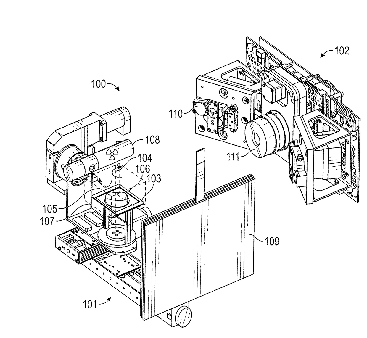
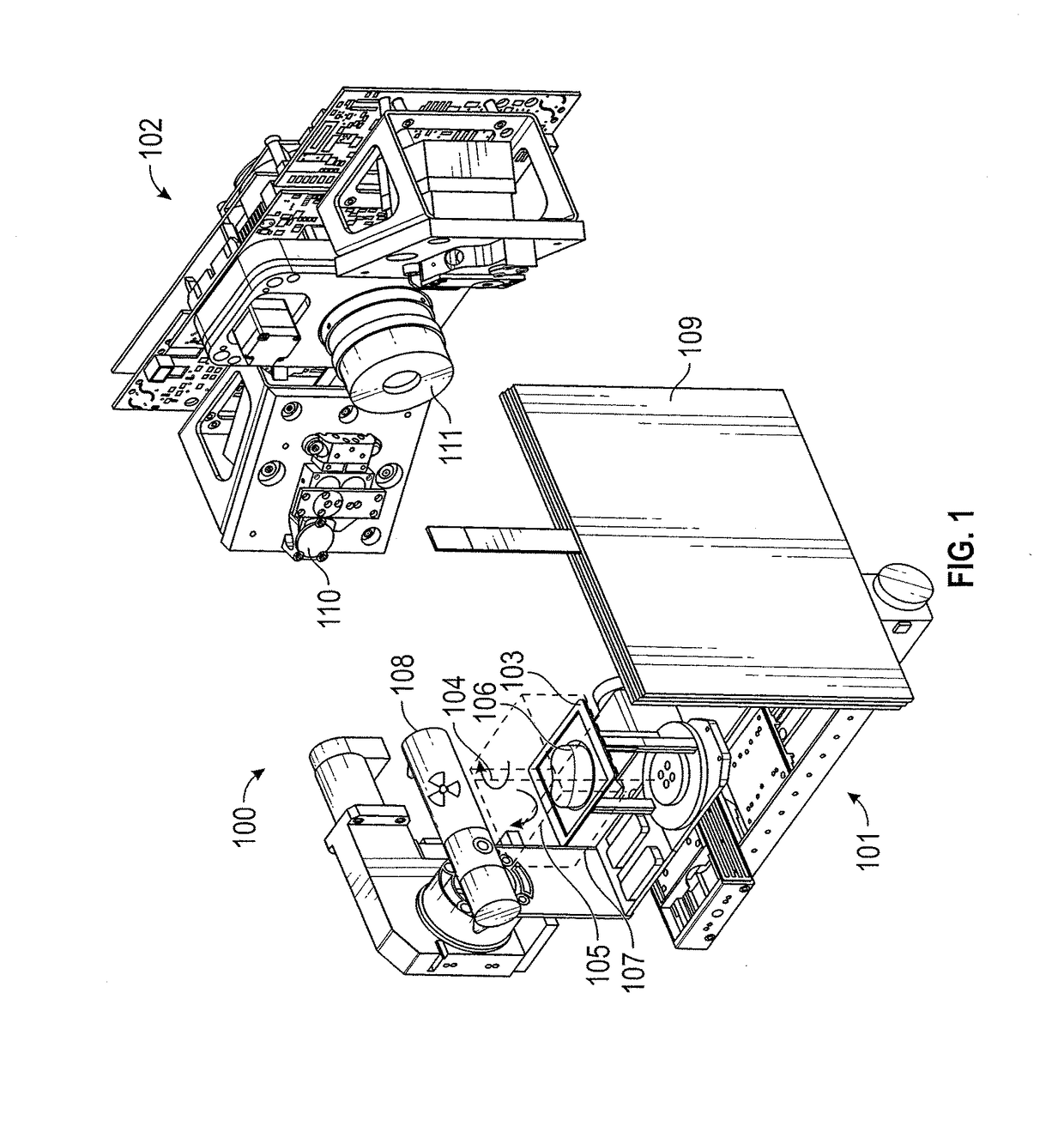
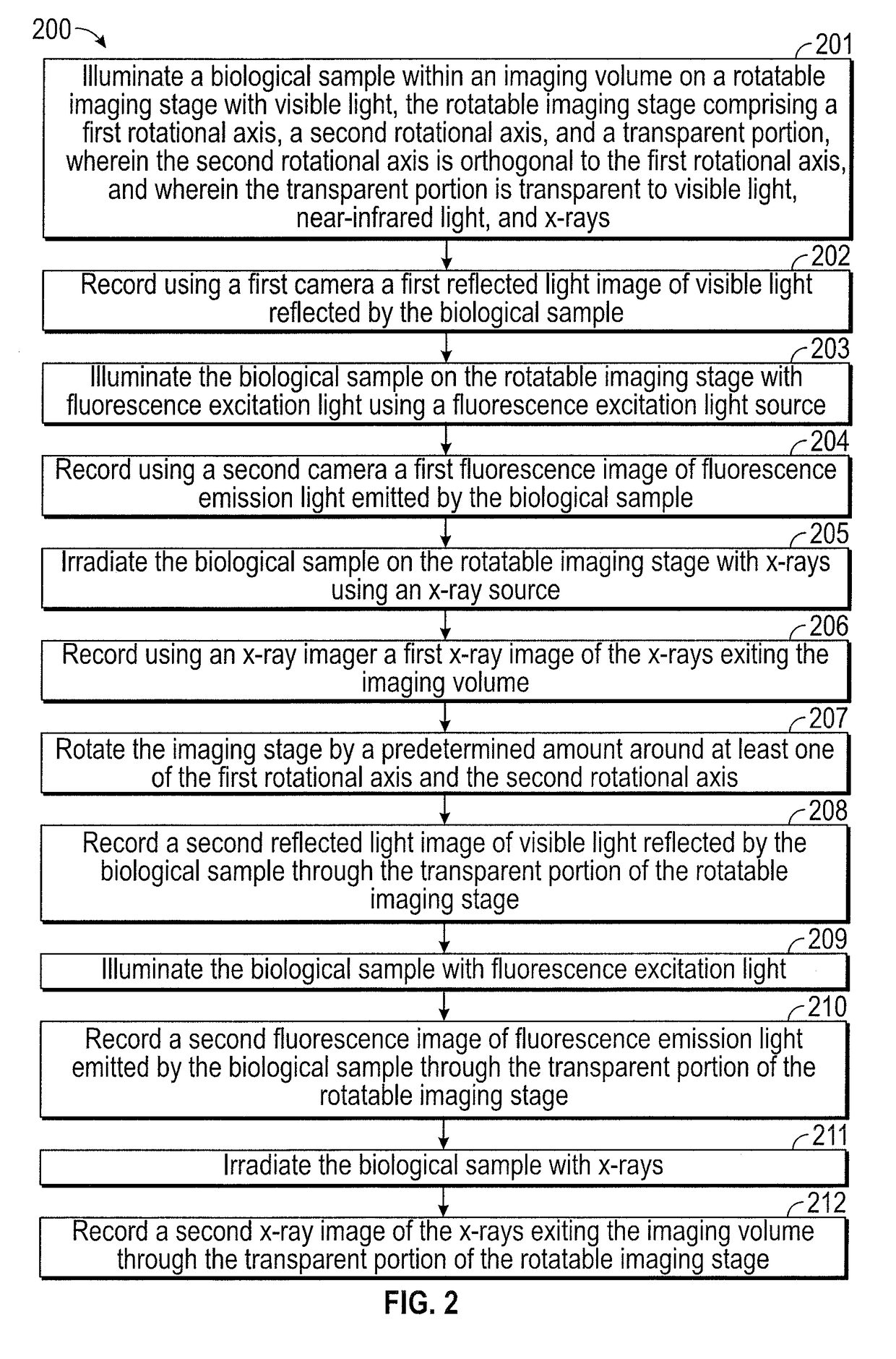
[0043]The present invention relates in part to multimodal and multi-axis three-dimensional imaging devices and methods for visualizing samples. The devices and methods can be used to record and display multimodal images of a biological sample representing views of the sample from any position rotated about the sample in three-dimensional space.

[0044]A technical advantage of the embodiments described herein is that a surgeon can have enhanced access to visualized information regarding the location and characteristics of diseased and healthy cells and tissue within a resected biopsy sample. This can allow a surgeon to more accurately assess tumor margins during surgical procedures, which can in turn increase the probability of a successful surgery and the survival rate of the patient.

[0045]By combining multi-axis rotation three-dimensional imaging with multiple imaging modalities, the inventors have made the surprising discovery of a novel way to look at a resected tissue sample. For ...


PUM

PropertyMeasurementUnit
tilting angleaaaaaaaaaa
wavelengthsaaaaaaaaaa

Abstract

Methods and devices are disclosed for the imaging of a biological sample from all rotational perspectives in three-dimensional space and with multiple imaging modalities. A biological sample is positioned on an imaging stage that is capable of full 360-degree rotation in at least one of two orthogonal axes. Positioned about the stage are imaging modules enabling the recording of a series of images in multiple modalities, including reflected visible light, fluorescence, X-ray, ultrasound, and optical coherence tomography. A computer can use the images to construct three-dimensional models of the sample and to render images of the sample conveying information from one or more imaging channels. The rendered images can be displayed for an operator who can manipulate the images to present additional information or viewing angles of the sample. The image manipulation can be with touch gestures entered using a sterilizable or disposable touch pen.

Description

CROSS-REFERENCES TO RELATED APPLICATIONS[0001]This application claims the benefit of U.S. Provisional Application No. 62 / 325,588, filed Apr. 21, 2016, which is incorporated in its entirety herein for all purposes.STATEMENT AS TO RIGHTS TO INVENTIONS MADE UNDER FEDERALLY SPONSORED RESEARCH AND DEVELOPMENT[0002]NOT APPLICABLEBACKGROUND[0003]Assessment of tumor margin during surgery is essential to the optimal outcome of many oncologic procedures. Tumor margins are the healthy tissue surrounding the tumor, more specifically, the distance between the tumor tissue and the edge of the surrounding tissue removed along with the tumor. Ideally, the margins are selected so that the risk of leaving tumor tissue within the patient is low.[0004]Fluorescence image-guided systems can be used in conjunction with a series of imaging agents to visualize tumor margins during surgical procedures for cancer removal. However, in many cancer surgeries deep surgical cavities with closed spaces and hidden l...

Claims


Application Information

Patent Timeline
Patent Type & Authority Applications(United States)
IPC IPC(8): G06T15/20G01N23/22G01N23/223G06F3/0354G06T19/00G06F3/0488G06F3/0484B43K8/02G06T19/20
CPCG06T15/20G01N23/223G01N23/2204G06T19/003G06F3/04845G06F3/04886G06T2219/2016G06F3/03545G06T19/20B43K8/02G01N2223/401G01N2223/408G06F3/04842G01N29/0672G01N29/225G01N21/1702G01N21/31G01N21/4795G01N21/6456G01N23/046G01N29/2418
Inventor WANG, HAN-WEI
Owner LI COR
Why Eureka
Try Eureka