
Surgical grafts for replacing the nipple and areola or damaged epidermis

a technology of nipple and areola, which is applied in the field of surgical grafts for replacing the nipple and areola or damaged epidermis, can solve the problems of unsatisfactory surgical solutions to maintain nipple projection following nipple reconstruction, and the general surgical method has proved less than ideal, so as to achieve the effect of decellularizing the epidermis, decellularizing additional cells, and substantial decellularization

Active Publication Date: 2018-01-18
TULANE EDUCATIONAL FUND
View PDF11 Cites 4 Cited by
  • Summary
  • Abstract
  • Description
  • Claims
  • Application Information

AI Technical Summary

Benefits of technology

The present invention provides grafts and methods for replacing body parts such as nipples and areola with decellularized tissue. The grafts retain important cell adhesion molecules and can be repopulated with cells from the patient. The decellularization process involves contacting the body part with a detergent and optionally a second detergent, followed by optional incubation with cells exogenous to the body part. The grafts can be secured to the patient and covered with a biocompatible occlusive coating. The technical effects include improved grafting outcomes and reduced risk of complications.

Problems solved by technology

However, immediate breast reconstruction with autologous tissue rarely involves nipple reconstruction and nipple reconstruction typically occurs after the initial reconstruction of the breast.
Current strategies for nipple and areola reconstruction are limited to surgical techniques that create a nipple structure from existing local tissue, secondary site grafting, 3D tattooing, or using commercially available acellular dermal matrix sheets, such as AlloDerm® or Glyaderm®.
Despite the decades of efforts, satisfactory surgical solutions to maintaining nipple projection following nipple reconstruction has remained a difficult problem.
Surgical methods in general have proved less than ideal because nipple and areola reconstruction are highly dependent on surgical technique and existing tissue.
Double opposing tab nipple reconstruction often leads to necrosis of tissue and faulty nipple reconstruction.
To date, no surgical technique has emerged as fully satisfactory.
Further, no tissue engineering strategies have been reported that successfully reconstruct the nipple and areola.

Method used


Image

  • Surgical grafts for replacing the nipple and areola or damaged epidermis
  • Surgical grafts for replacing the nipple and areola or damaged epidermis
  • Surgical grafts for replacing the nipple and areola or damaged epidermis

Examples


example 1

[0072]This example sets forth materials and methods used in studies reported herein.

Decellularization

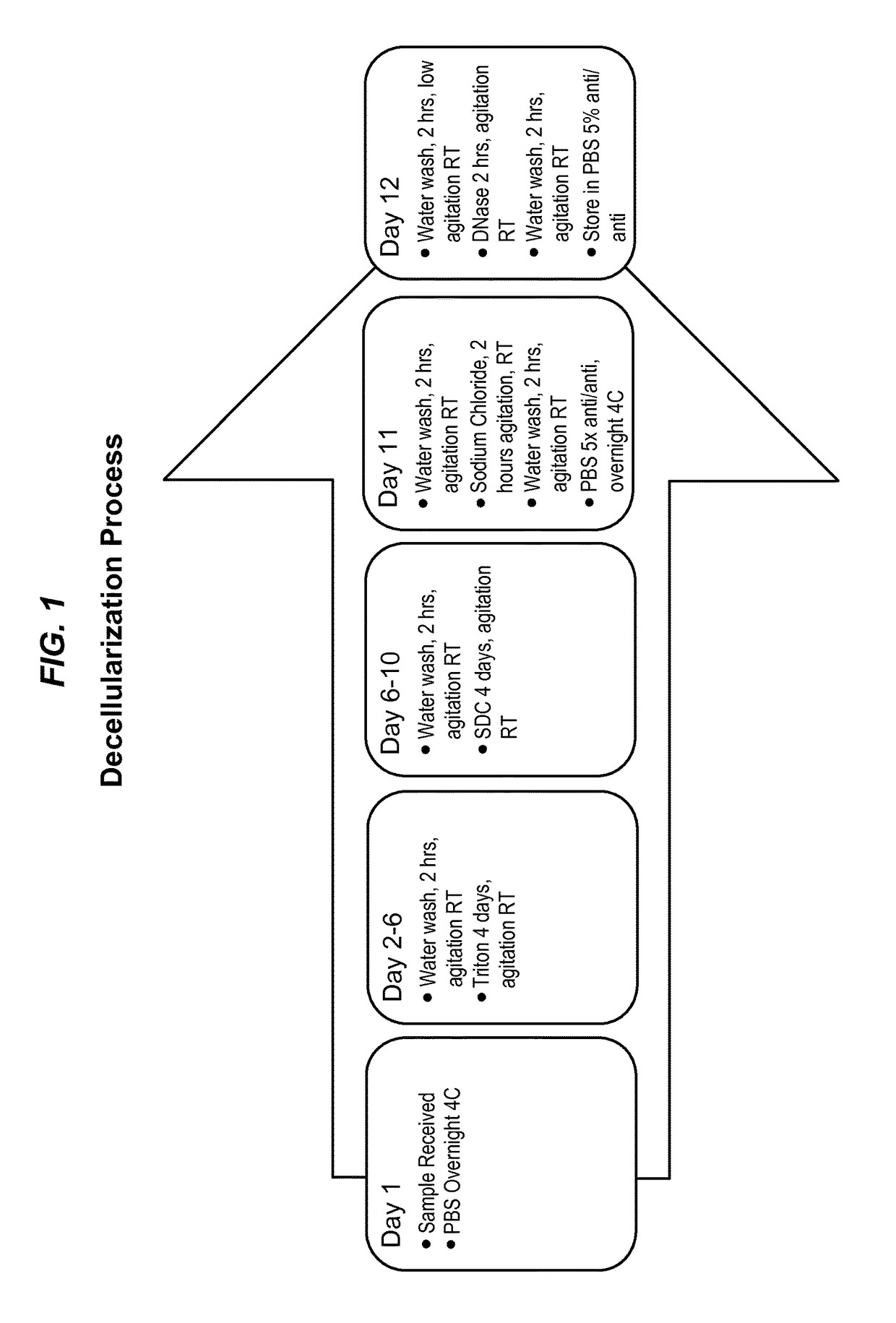
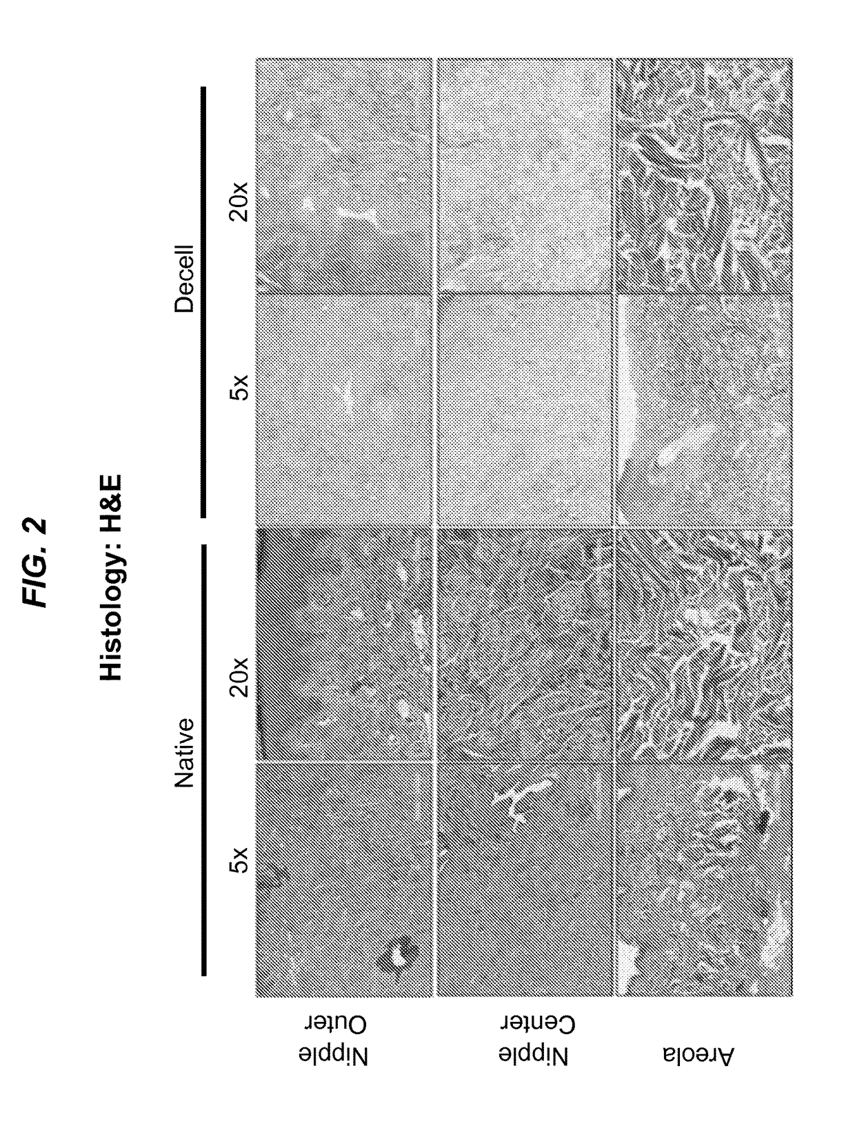
[0073]The process depicted in FIG. 1 used tissue samples collected by trained veterinary technicians at Tulane National Primate Research Center (TNPRC). The technicians collected Rhesus macaque (Macaca mulatta) nipple and areola tissue from animals undergoing routine euthanasia due to chronic diarrhea or self-mutilation, as well as from control animals from various experiments. All animal procedures conformed to the requirements of the Animal Welfare Act and all animal protocols were approved by the Institutional Animal Care and Use Committee (IACUC) of the TNPRC before implementation of experimental protocols. Rhesus macaque tissue samples were decellularized through a modified protocol previously described by Bonvillain et. al., Tissue Eng: Part A, 18(23-24): 2437-52 (2012) (“Bonvillain 2012”), and Scarritt et. al., Tissue Eng: Part A, 20(9-10):1426-43 (2014) (“Scarritt 2014”). In ...

example 2

[0082]This Example sets forth the results of studies conducted in the course of the work reported herein.

Detergent-Based Decellularization Effectively Removes Cells from NAC Tissue

[0083]To determine the efficiency of decellularization to remove DNA from rhesus macaque NAC, gDNA was isolated from native and decellularized NAC tissue (FIG. 7A). There was a significant reduction in DNA content with native NAC containing 2,022.71 (SEM: 751.90) ng DNA per mg lyophilized tissue and decellularized NAC containing 56.51 (SEM: 8.45) ng of DNA per mg of lyophilized tissue (p<0.05). Gel electrophoresis of 1.5 ug of gDNA from native NAC appeared as a heavy, high molecular weight band, whereas DNA from decellularized NAC consisted of small fragments of degraded DNA that did not create a banding pattern (FIG. 7 B).

[0084]To investigate the potential application of decellularization for the generation of tissue-engineering scaffolds for breast reconstruction, NAC tissue was exposed to a series of de...

example 3

[0087]This Example discusses the results set forth in the preceding Example.

[0088]This study demonstrated that a dermal / semi-glandular structure such as the nipple and areola complex can be sufficiently decellularized. Decellularization efficiency was determined based on three key criteria, as outlined by Crapo et al., Biomaterials, 32(12):3233-43 (2011): no visible signs of cells or cell debris, reduction of DNA to less than 50 ng per mg tissue, and degradation of any remaining DNA to less than 200 bp. These criteria are based on the principle that cellular debris, high concentrations of DNA, or the presence of long DNA fragments in a decellularized tissue are potentially immunogenic and, therefore, detrimental to successful incorporation post-transplantation. The concentration of genomic DNA isolated from decellularized NAC was about 56 ng per mg of lyophilized tissue and no visible banding of DNA fragments were found when DNA from decellularized NAC was electrophoresed. The avera...


PUM

PropertyMeasurementUnit
timeaaaaaaaaaa
temperatureaaaaaaaaaa
pressureaaaaaaaaaa

Abstract

The present disclosure relates to surgical grafts for replacing nipples and areolas lost to disease or trauma with surgical grafts of decellularized donor nipples and areolas and to placing and recellularizing such grafts. The disclosure further provides methods for decellularizing epidermis. The decellularized epidermis can be used as a protective cover for skin wounds.

Description

CROSS-REFERENCE TO RELATED APPLICATIONS[0001]This application claims the benefit and priority of U.S. Provisional Patent Application No. 62 / 073,719, filed Oct. 31, 2014, and of U.S. Provisional Patent Application No. 62 / 212,846, filed Sep. 1, 2015. The contents of both applications are hereby incorporated by reference in their entireties.STATEMENT OF FEDERAL FUNDING[0002]Not applicable.PARTIES TO JOINT RESEARCH AGREEMENT[0003]Not applicable.REFERENCE TO SEQUENCE LISTING OR TABLE SUBMITTED ON COMPACT DISC AND INCORPORATION-BY-REFERENCE OF THE MATERIAL[0004]Not applicable.BACKGROUND OF THE INVENTION[0005]In the United States alone, over 40,000 women die from breast cancer each year, and more than 230,000 new cases are diagnosed; and there are approximately 430 deaths and 2,000 new cases expected for men. Additionally, there are more than 2.8 million breast cancer survivors in the United States, many of who have undergone reconstructive surgery. Approximately 36% of patients with early...

Claims


Application Information

Patent Timeline
Patent Type & Authority Applications(United States)
IPC IPC(8): A61L27/36A61L27/22A61L27/38A61F2/00A61L27/24A61F2/52
CPCA61L27/362A61L27/3687A61L27/227A61L27/24A61L27/3813A61L27/3641A61F2/0059A61F2/52A61L2430/04A61F2002/526A61L27/3604A61L27/3666
Inventor PASHOS, NICHOLAS C.BUNNELL, BRUCE A.
Owner TULANE EDUCATIONAL FUND
Learn More