

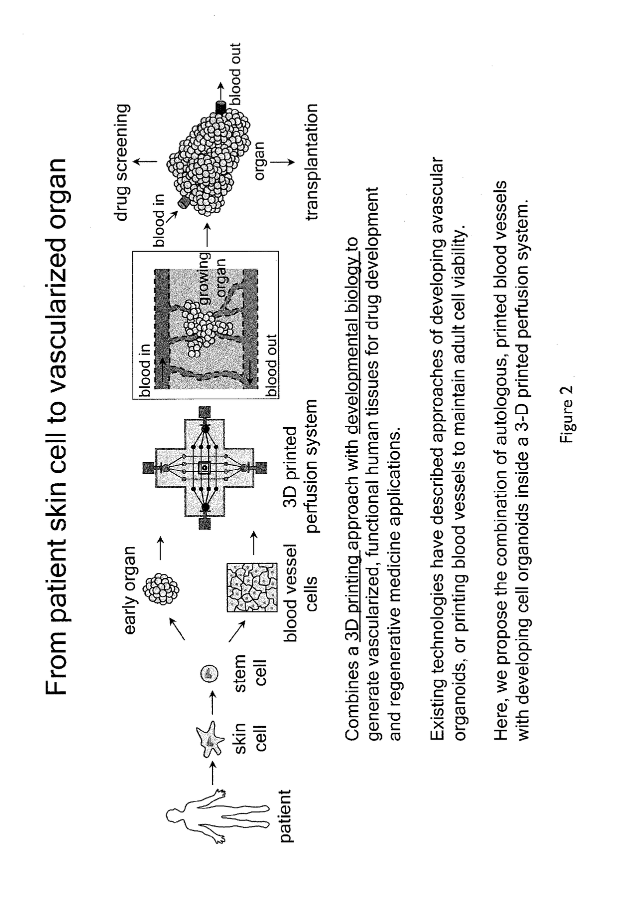
Methods of generating functional human tissue
a human tissue and functional technology, applied in the field of human tissue generation methods, can solve the problems of preventing the development unable to embed vascular networks in tissue constructs, and affecting the progress of 3d tissue engineering for decades, and achieve the effect of enhancing at least one of endothelial sprouting behavior
- Summary
- Abstract
- Description
- Claims
- Application Information
AI Technical Summary
Benefits of technology
Problems solved by technology
Method used
Image

Examples
example 1
Printing Vascularized Tissues
[0199]To create 3D printed vascularized tissues, the following method (illustrated in FIGS. 17 and 18) was used:
[0200]Step 1: PDMS border was 3D printed using SE 1700 from Dow chemicals.
[0201]Step 2: Fugitive ink Pluronic F-127 or gelatin was 3D printed to create vascular channels.
[0202]Step 3: Cell laden ink #1 comprising cells mixed in 10% wt / v gelatin-fibrinogen or gelatin methacrylate (GelMA) hydrogels hydrogels containing a photoinitiator Irgacure-2959 at 0.3% wt / v was 3D printed.
[0203]Step 4: Cell-laden ink #2 I comprising cells mixed 10% wt / v gelatin methacrylate hydrogels containing a photoinitiator Irgacure-2959 at 0.3% wt / v was 3D printed.
[0204]Steps 1-4 above were repeated.
[0205]The resultant fugitive ink and cell-laden filaments were infilled with extracellular matrix comprising gelatin methacrylate at 10% wt / v containing 0.3% wt / v Irgacure 2959 photoinitiator.
[0206]The structure was then exposed to chemically crosslink the gelatin methacryla...
example 2
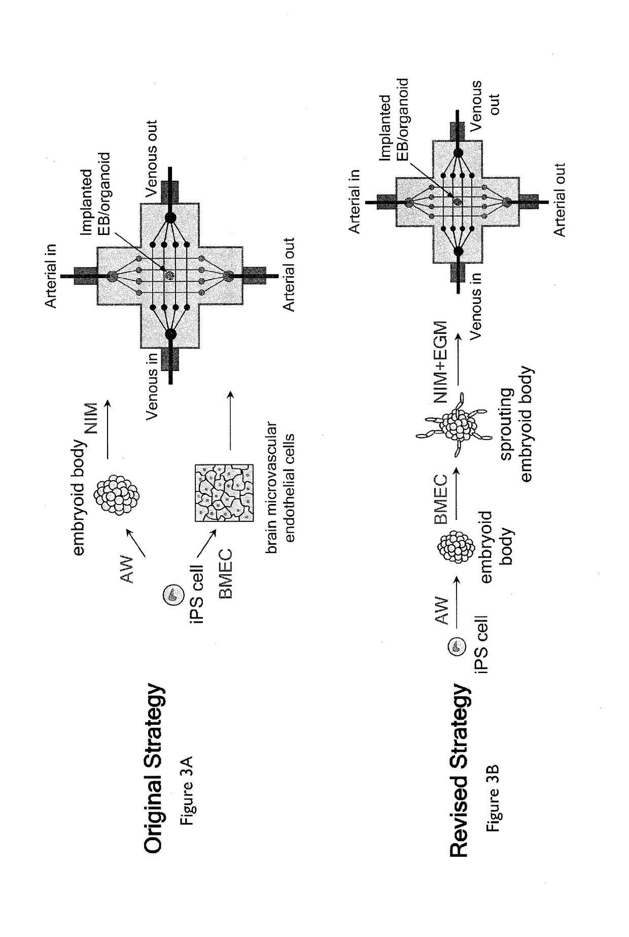
Printing Endothelial Cell-Laden Fugitive Filaments
[0216]The fugitive ink, either Pluronic F-127 or gelatin contained live HUVEC cells, as shown in FIGS. 14A-G.
[0217]Cells were first dispersed in a Pluronic-F127 ink (FIGS. 14A and D), and then a 10% wt / v gelatin methacrylate matrix in DMEM media containing 0.3% wt / v Irgacure 2959 photoinitiator was cast surrounding the cells (FIGS. 14B and E) and crosslinked via UV exposure. Next, the Pluronic F-127 ink was liquefied by cooling to 4° C. and cells suspended in the ink were allowed to settle and stick to the wall (FIGS. 14C and F). After removal of the ink and culturing of the cells for several days, the cells remained adhered to the walls (FIG. 14G).
example 3
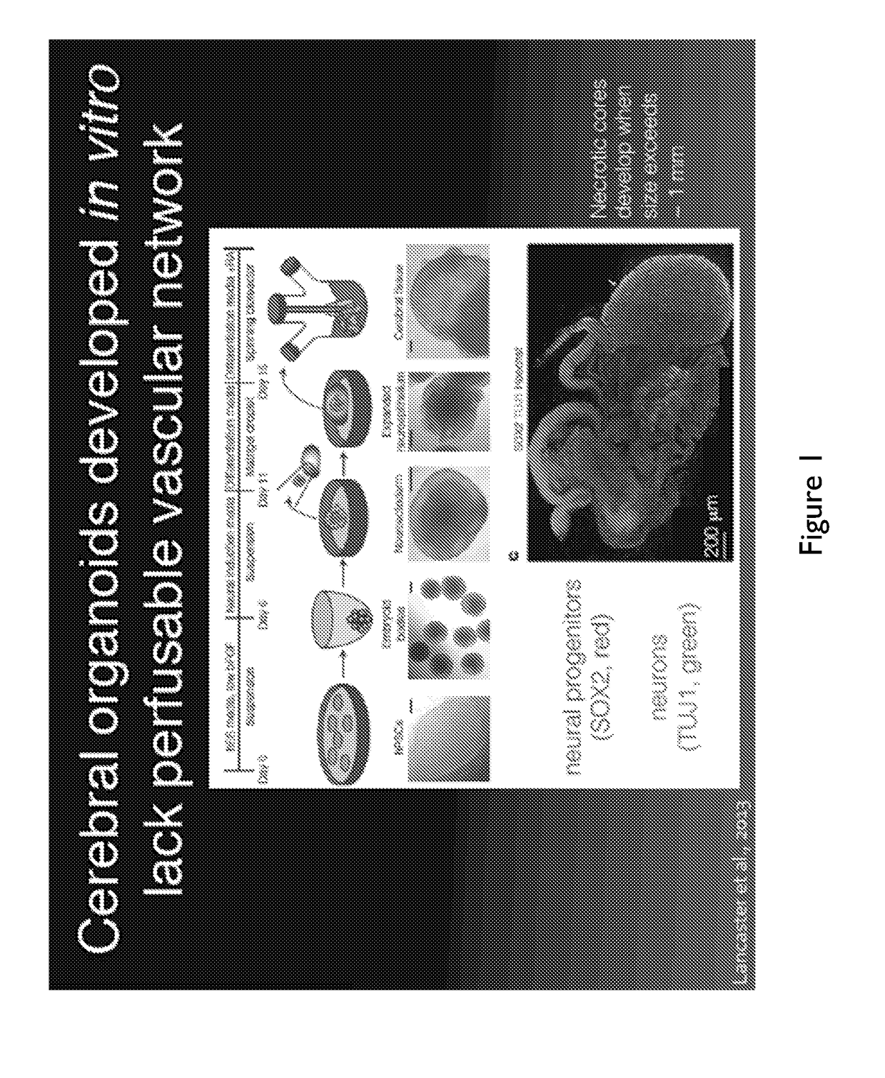
Generating Cerebral Organoids
[0218]To synthesize a cerebral organoid (shown in FIGS. 7A-I) the following method was used.
[0219]6-well plates were coated in matrigel by incubating human ESC-certified growth-factor depleted Matrigel, diluted in DMEM / F12 medium at the manufacturer's (Corning) batch specific recommended concentration, for 1 h at room temperature
[0220]Human iPSCs were maintained in mTeSR medium on the matrigel coated plates and passaged using Accutase when colonies begin to merge.
[0221]At passaging, Human iPSCs were dissociated for 15 minutes using 1 mL Accutase reagent, then diluted in 11 mL of DMEM / F 12 medium, centrifuged at 200 g for 5 minutes, resuspended in 1 mL of AW medium, counted using a cellometer, and seeded at a density of 600,000 cells per well of an Aggrewell™ 400 plate, which corresponds to a per-microwell density of 500 iPSCs (FIG. 7A). The media volume was brought up to 2 mL using AW, containing 10 μM ROCK inhibitor Y-27632. The time point ‘day 0’ corre...
PUM
| Property | Measurement | Unit |
|---|---|---|
| diameter | aaaaa | aaaaa |
| concentration | aaaaa | aaaaa |
| molecular weight | aaaaa | aaaaa |
Abstract
Description
Claims
Application Information