System and method for optimizing an implant position in an anatomical joint

an anatomical joint and implant position technology, applied in the field of system and method for optimizing the implant position in the anatomical joint, can solve the problems of undeveloped positioning of customized implants, inability to optimize other aspects of implant positioning, etc., to achieve less invasive, maintain the position of the cross section area, and optimize the tilt of the implant axis

Active Publication Date: 2018-06-28
EPISURF IP MANAGEMENT
View PDF6 Cites 5 Cited by
  • Summary
  • Abstract
  • Description
  • Claims
  • Application Information

AI Technical Summary

Benefits of technology

[0006]The above described problems are addressed by the claimed system for optimizing a position of an implant having an individually customized implant hat and at least one implant protrusion extending from the implant hat in the direction of an implant axis in an anatomical joint of a patient. The claimed system may comprise a storage media and a processor which is configured to: receive medical image data representing a three dimensional image of the joint from the storage media; obtain a three dimensional virtual model of the joint which is based on the received medical image data; identify damage in the joint based on the received medical image data and/or the three dimensional virtual model of the joint; define an implant area that covers at least a major part of the identified damage; position a virtual implant template having an implant hat corresponding to said implant area in the three dimensional virtual model of the joint; and generate a customized top surface of the implant hat to correspond to a simulated healthy cartilage surface. The processor is configured to position the virtual implant template by: placing the virtual implant

Problems solved by technology

Even though individually customized implants have been used for some time, the positioning of these customized implants has not de

Method used


Image

  • System and method for optimizing an implant position in an anatomical joint
  • System and method for optimizing an implant position in an anatomical joint
  • System and method for optimizing an implant position in an anatomical joint

Examples


use case embodiment

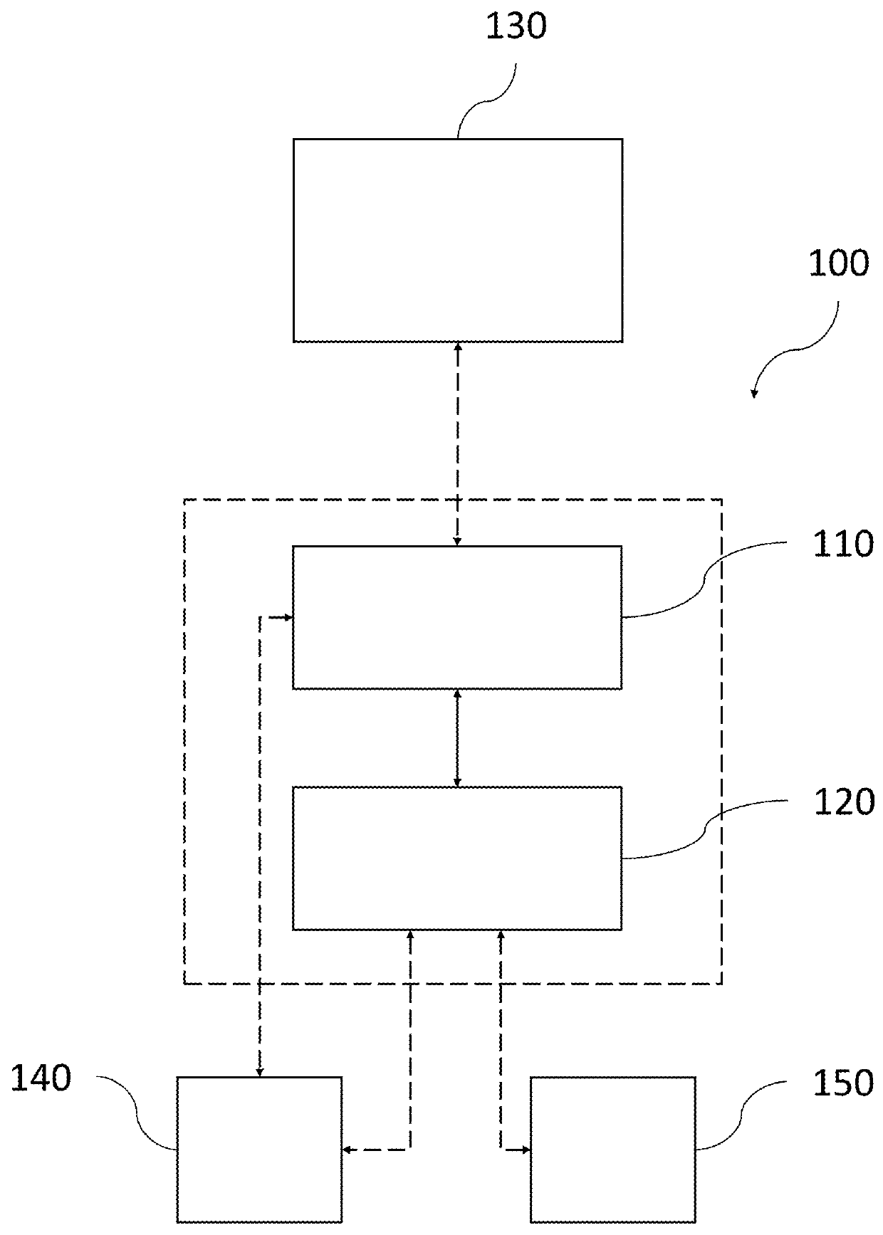
[0110]To set the presently disclosed methods and systems in a larger context, the method of optimizing an implant position in an anatomical joint of a patient according to any of the disclosed embodiments may in use case embodiments be preceded by capturing and / or obtaining medical image data representing an anatomical joint, and may further be followed by the design and production of a medical implant.

[0111]FIG. 11 is a flow diagram exemplifying one such larger context, including receiving radiology / medical image data from an image source, obtaining a three dimensional virtual model of the joint, identifying damage in the joint, defining an implant area based on the identified damage, and positioning a virtual implant template in the virtual model, in accordance with one or more embodiments described herein. In FIG. 11, the various ways of generating the medical image data is marked with dashed lines to clarify that they are optional steps shown in the figure to provide context onl...


PUM


Abstract

In accordance with one or more embodiments herein, a system for optimizing a position of an implant having an individually customized implant hat H and at least one implant protrusion P extending from the implant hat H in the direction of an implant axis A in an anatomical joint of a patient is provided. The system comprises a storage media 110 and a processor 120 which is configured to: receive medical image data representing a three dimensional image of the joint from the storage media 110; obtain a three dimensional virtual model of the joint which is based on the received medical image data; identify damage in the joint based on the received medical image data and/or the three dimensional virtual model of the joint; define an implant area that covers at least a major part of the identified damage; position a virtual implant template having an implant hat H corresponding to said implant area in the three dimensional virtual model of the joint; and generate a customized top surface of the implant hat H to correspond to a simulated healthy cartilage surface. The processor is configured to position the virtual implant template by: placing the virtual implant template so that the cross section area of the implant hat H in a direction perpendicular to the implant axis A covers at least a major part of the damage; and optimizing the tilt of the implant axis A, while maintaining the position of the cross section area of the implant hat H in the joint, by minimizing at least one of the maximum penetration depth Dmax into the bone along the circumference of the implant hat H; the total volume of bone and/or cartilage to be removed for implanting the implant; and/or the surface area of the implant penetration into the bone.

Description

CROSS-REFERENCE TO RELATED APPLICATION[0001]This application is a continuation-in-part of PCT Application No. PCT / EP2016 / 082455, filed Dec. 22, 2016, entitled “SYSTEM AND METHOD FOR OPTIMIZING AN IMPLANT POSITION IN AN ANATOMICAL JOINT,” and is a continuation-in-part of U.S. application Ser. No. 15 / 324,351, filed Jan. 6, 2017, entitled “SURGICAL KIT FOR CARTILAGE REPAIR COMPRISING IMPLANT AND A SET OF TOOLS,” the content of which is incorporated by reference herein.TECHNICAL FIELD[0002]The present disclosure relates generally to systems and methods for optimizing an implant position in an anatomical joint of a patient.BACKGROUND[0003]Surgical implants have since long been used for cartilage repair in anatomical joints. For a long time, a surgeon wishing to use an implant for cartilage repair could only choose from a selection of ready-made implants, and try to fit these in the best possible way, but over the years, various ways of customizing the implants to the individual patient h...

Claims


Application Information

Patent Timeline
IPC IPC(8): A61F2/38A61B5/00A61F2/46A61B34/10A61F2/30
CPCA61B34/10A61B2034/104A61B2034/105A61B2034/108A61F2/30756A61F2002/3013A61F2002/30878A61B2034/102A61B2034/107A61B34/20A61B17/154A61F2/32A61F2/38A61F2/40A61F2/42G06T7/0012G06T19/20G06T2207/30008G06T2219/2004
Inventor KARLSSON, ANDERSLILLIESTR LE, RICHARD
Owner EPISURF IP MANAGEMENT
Why Eureka
Try Eureka