Accessory for external cardiac defibrillation, pacing and monitoring physiological signals/health data in the presence of electromagnetic interference

a technology of electromagnetic interference and accessory, which is applied in the field of biomedical methods and systems for external defibrillation, pacing, and cardioversion, can solve the problems of ic-mri development impeded by defibrillation and pacing, and achieve the effect of reliable wireless data transmission

Active Publication Date: 2018-08-30
SHUSTERMAN VLADIMIR
View PDF0 Cites 0 Cited by
  • Summary
  • Abstract
  • Description
  • Claims
  • Application Information

AI Technical Summary

Benefits of technology

[0018]B. A “defibrillation” mode, in which the defibrillator's current is directed to the defibrillation cables and electrodes, thus enabling: 1) accurate estimation of the electrical impedance of the contact between the patient's skin and the defibrillator's electrodes / pads, and 2) subsequent defibrillation. This mode is useful, e.g., during a test / procedure which does not require high-quality diagnostic MR imaging but may require external defibrillation, e.g., during MR-guided cardiovascular interventions, stress test, or MR-guided EP procedures.
[0024]5. Other operational regimes which optimize the defibrillator's operation for application specifics and minimize the EMI impact on MR images, including the regimes optimized for synchronous (on-demand) pacing with inhibition of pacing during intrinsic cardiac activity, asynchronous pacing, and cardioversion, which involves delivery of electrical discharges at specific time points synchronized with intrinsic cardiac activity.
[0036]B. One or more RF filters to eliminate the RF interference generated by the MRI scanner (e.g., a filter with a stop band at 64 MHz for a 1.5T MRI scanner or at 124 MHz for a 3T MRI scanner);
[0051]D. One or more wireless transmitters for increasing the reliability and speed (throughput) of the wireless data transmission, which is critically important for large volumes of continuous, multichannel data.
[0054]B. Low-amplitude stimulation of skeletal muscles immediately before defibrillation to minimize patient discomfort caused by muscle contraction during defibrillation.
[0058]One important aspect of this invention is mounting (linking / connecting) the (optional) wireless antenna to a movable / wheeled patient table (herein, the terms patient table, bed, and procedure table are used interchangeably) to enable reliable wireless data transmission at different locations of the patient table and during its movement in the course of various procedures, as well as between different procedures and / or rooms. An important aspect of this invention is the positioning of one or more antennas connected to a wireless transmitter and one or more antennas connected to a wireless receiver (herein referred to as the first and second antenna, respectively) to provide a free (unobstructed) plane (space) for substantially continuous wireless communication between the two antennas at various positions of the patient table and during table movement.

Problems solved by technology

Because interventional cardiology procedures (in particular EP studies) often involve arrhythmia induction and termination, an absence of in-bore defibrillation and pacing impedes the development of IC-MRI.

Method used


Image

  • Accessory for external cardiac defibrillation, pacing and monitoring physiological signals/health data in the presence of electromagnetic interference
  • Accessory for external cardiac defibrillation, pacing and monitoring physiological signals/health data in the presence of electromagnetic interference
  • Accessory for external cardiac defibrillation, pacing and monitoring physiological signals/health data in the presence of electromagnetic interference

Examples


example 1

Example 1

An MRI-Guided EP Study

[0104]This hypothetical example describes the application of a medical device of this invention for safe and efficient external cardiac defibrillation during an MRI-guided EP study.

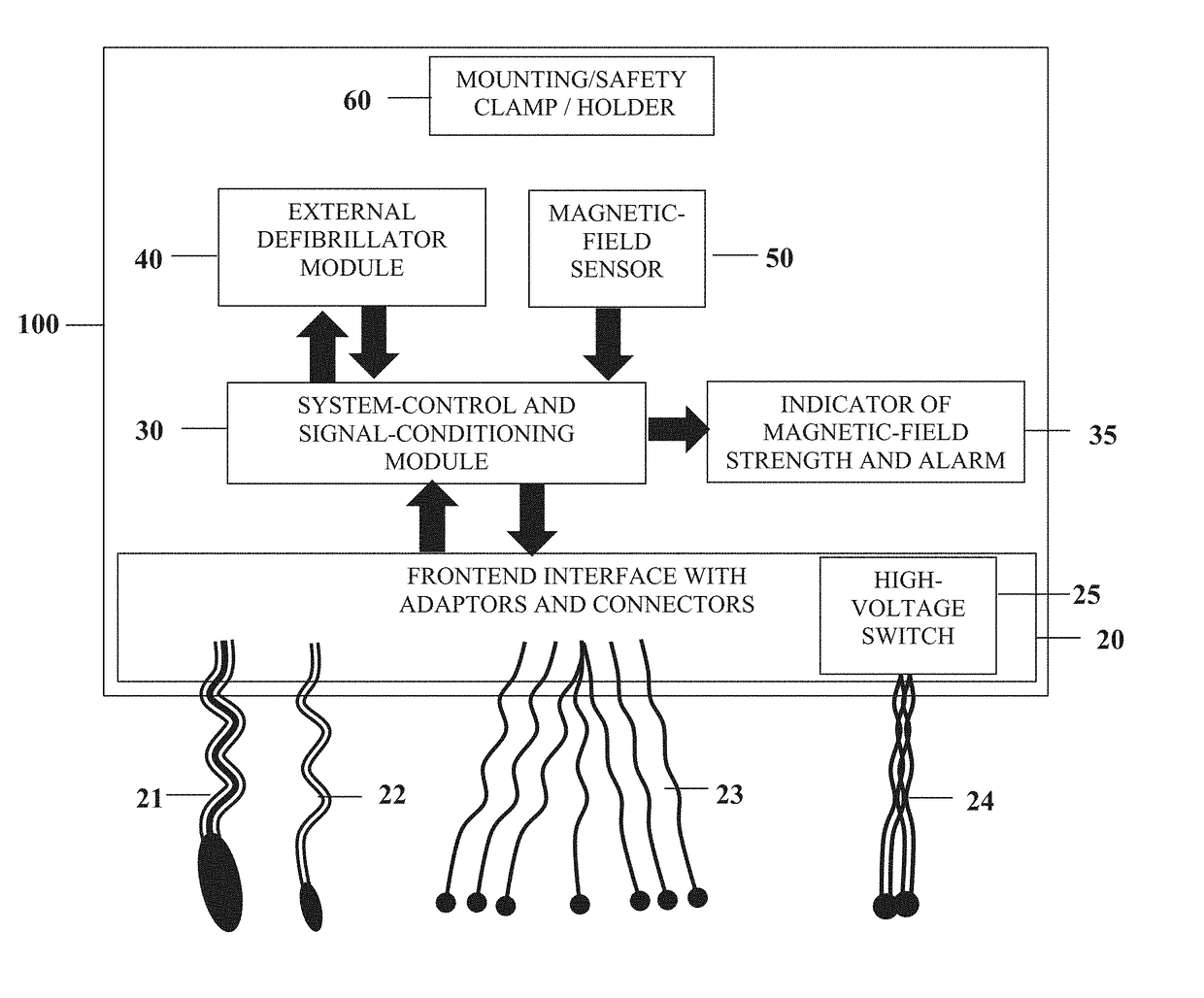
[0105]The MAGNA-DEX device of this invention would be brought into the scanner room and initially positioned 25 cm from the edge of an MRI magnet bore, which would trigger an audio-visual alarm indicating a strong EMF that exceeds the preset threshold of 200 Gauss. The MAGNA-DEX device would then be moved away from the magnet and affixed to the end of a patient table located approximately 1.5 m from the MRI magnet bore.

[0106]Because many defibrillator functions require continuous, high-quality ECG, blood pressure, or pulse-oximetry signals, which may become obscured by the high-level EMI generated by MRI scanners (especially during real-time cardiovascular imaging, which requires the application of steady-state-free-precession pulse sequences with very short duty cycle, time...


PUM


Abstract

This accessory adapts external cardiac defibrillation systems to enable safe defibrillation, pacing, and cardioversion inside the MRI bore with minimal effect on MR image quality. Commercially available external defibrillators are not designed to work in the MRI environment. An MR-compatible defibrillator is needed to safely perform cardiovascular MRI, in particular MR-guided interventional cardiovascular procedures, such as cardiac electrophysiology studies and cardiac catheterization. This accessory includes nonmagnetic defibrillator housing with MRI safety features, provides interface for MRI-compatible physiological monitoring, and optimizes defibrillator operation for the MRI environment. The accessory may include MRI-compatible modules for monitoring / recording electrocardiogram, blood pressure, pulse oximetry, and other physiological signals. It may also include a wireless transmitter and at least one module for electrical energy generation and / or stimulation.

Description

STATEMENT REGARDING FEDERALLY SPONSORED RESEARCH OR DEVELOPMENT[0001]This invention was made with government support under contract HHSN268201500009C awarded by the National Institutes of Health. The government has certain rights in the invention.FIELD OF THE INVENTION[0002]This invention relates to the field of biomedical methods and systems for external defibrillation, pacing, and cardioversion in the presence of strong electromagnetic interference (EMI) and electromagnetic forces (EMF) generated by magnetic resonance imaging (MRI) scanners.BACKGROUND OF THE INVENTION[0003]This application is a continuation-in-part of pending U.S. patent application Ser. No. 15 / 442,705 filed on Feb. 27, 2017, which is a continuation-in-part of U.S. patent application Ser. No. 14 / 470,923 filed on Aug. 27, 2014 (now U.S. Pat. No. 9,610,016), which are incorporated herein in their entirety by reference. This application also claims the benefit of U.S. Provisional Application No. 62,490,031, filed Apr...

Claims


Application Information

Patent Timeline
Patent Type & Authority Applications(United States)
IPC IPC(8): A61N1/39A61B5/00A61N1/36A61N1/362A61N1/37
CPCA61N1/3904A61B5/7217A61N1/36031A61N1/36139A61B5/7221A61N1/3993A61N1/3931A61N1/3625A61N1/3718A61B5/0024A61B5/7203A61B5/7282A61B5/0006A61B5/002A61B5/0205A61B5/021A61B5/0263A61B5/053A61B5/055A61B5/14551A61B5/7225G08C17/02A61B5/318A61B5/369A61B5/389
Inventor SHUSTERMAN, VLADIMIR
Owner SHUSTERMAN VLADIMIR
Why Eureka
Try Eureka