Methods and compositions for identifying or quantifying targets in a biological sample

a biological sample and target technology, applied in the field of biological sample target identification or target quantification, can solve the problems of inability to augment protein information with cytometry, methods that are not well suited to the discovery of novel cell populations, and facs/scrna-seq approaches suffer from relatively low throughpu

Inactive Publication Date: 2018-09-06
NEW YORK GENOME CENT
View PDF2 Cites 71 Cited by
  • Summary
  • Abstract
  • Description
  • Claims
  • Application Information

AI Technical Summary

Benefits of technology

[0016]In still another aspect, the constructs described above are used in a method of batch-barcoding or cell “hashtagging”. An above-described construct, e.g., an antibody or any ligand that binds to a cell, conjugated or associated with an oligonucleotide sequence comprising an Amplification Handle; a Barcode that specifically identifies the ligand, an optional Random Molecular Tag (RMT), or Unique Molecular Identifier (UMI), hereafter referred to as “UMI” that that is positioned adjacent to the Barcode on its 5′ or 3′ end; and an

Problems solved by technology

However, massively parallel approaches based on droplet microfluidics1-3, microwells47,48 or combinatorial indexing20,30 are incompatible with cytometry and therefore cannot be augmented with protein information.
While effective, FACS/scRNA-seq approaches suffer from relatively low throughput and from an experimental bias in that only cell types chosen a priori are sorted and sequenced.
Thus, these methods are not well suited for discovery of novel cell populations or for characterizing complex tissues that require the analysis of tens of thousands of cells.
Although droplet-based advances in single-cell genomics have dramatically changed the scale of scRNA-seq experiments, these methods suffer from a key disadvantage: All drople

Method used


Image

  • Methods and compositions for identifying or quantifying targets in a biological sample
  • Methods and compositions for identifying or quantifying targets in a biological sample
  • Methods and compositions for identifying or quantifying targets in a biological sample

Examples


example 1

Design and Validation of Antibody-oligo Complexes

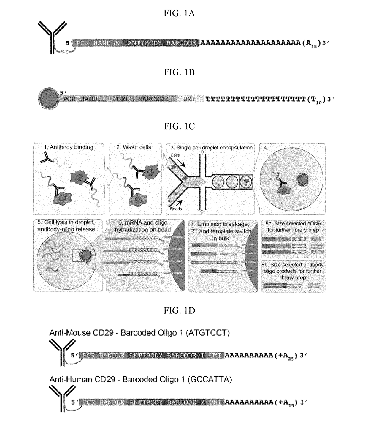
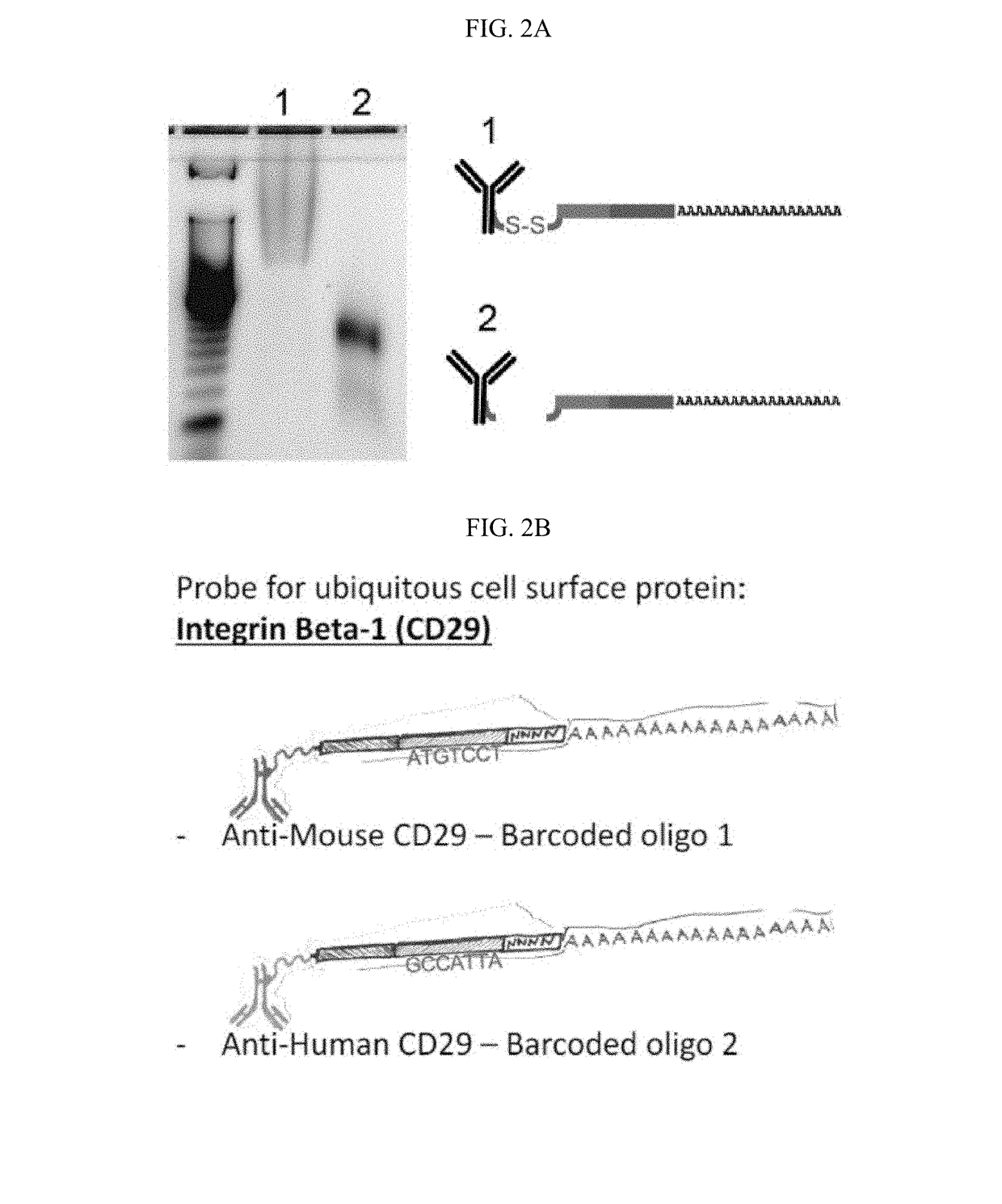
[0391]Antibody-oligos were designed with the following characteristics: a generic Amplification Handle (PCR handle) for next-generation sequencing library preparation, a unique Barcode sequence specific for each antibody, and a polyA stretch at the 3′ end (FIG. 1A). Two antibody-oligos were generated. Anti-Mouse Integrin Beta-1 (CD29) antibodies were linked to Barcoded oligo 1 containing a disulfide bridge, a common sequence (Amplification Handle, PCR handle), a unique antibody identifier Barcode (5′-ATGTCCT-3′) and a UMI containing 4 nt followed by a polyA tail (FIG. 2B, top panel). Anti-human CD29 antibodies were linked to Barcoded oligo 2 containing a disulfide bridge, a common sequence (Amplification Handle, PCR handle), a unique antibody identifier Barcode (5′-GCCATTA-3′) and a UMI containing 4 nt followed by a polyA tail (FIG. 2B, bottom panel).

[0392]For the experiments presented in Examples 1 to 7, the oligos were modified with...

example 2

Methods and Materials

[0393]Conjugation of Antibodies to DNA-barcoding Oligonucleotides. Highly specific, flow-cytometry-tested monoclonal antibodies were conjugated to oligonucleotides containing unique antibody-identifier sequences and a polyA tail. We adopted a commonly used streptavidin-biotin interaction to link oligos to antibodies19. Antibodies were streptavidin labeled using the LYNX Rapid Streptavidin Antibody Conjugation Kit (Bio-Rad, USA) according to manufacturer's instructions with modifications. Specifically, we labeled 15 μg of antibody with 10 μg of streptavidin. At this ratio, an average of two streptavidin tetramers will be conjugated per antibody molecule, which results in an average of eight binding sites for biotin on each antibody. DNA oligonucleotides with a 5′ amine modification were purchased at IDT (USA) and biotinylated using NHS-chemistry according to manufacturer's instructions (EZ Biotin S-S NHS, Thermo Fisher Scientific, USA). The optional disulfide bon...

example 3

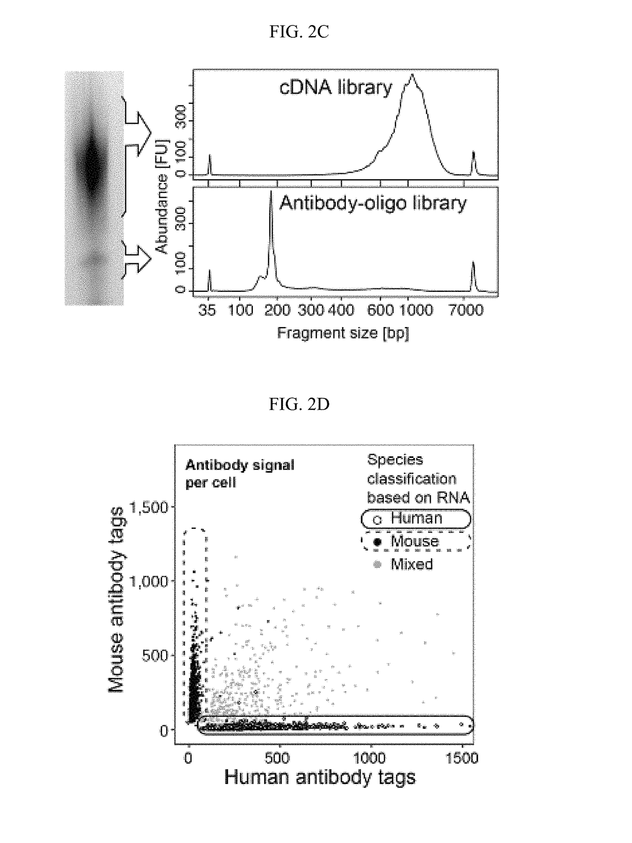
Identification of Different Species in Mixing Sample

[0429]The antibody-oligo complexes described in Example 1 were incubated with cells using conditions established for flow cytometry, such as ref 22. The cells were washed to remove unbound antibodies, then single cells were encapsulated into nanoliter-sized aqueous droplets in a microfluidic apparatus designed to perform Drop-seq1 (FIG. 1C). After cell lysis (which happened immediately in the droplets when the lysis buffer contacted cells), cellular mRNAs annealed to polyT containing Drop-seq beads (FIG. 1B) via their polyA tail (FIG. 1C#6). Oligos from the antibodies also annealed to the Drop-seq beads via their poly-A stretch at the 3′ end. A unique Barcode sequence on the Drop-seq bead indexed the transcriptome of each co-encapsulated cell. After breaking the emulsion and removing the oil, reverse transcription extended the Barcoded oligo to create the first-strand cDNA from both mRNA and antibody-derived oligo templates. The cD...


PUM

PropertyMeasurementUnit
Massaaaaaaaaaa
Lengthaaaaaaaaaa
Lengthaaaaaaaaaa

Abstract

Compositions, kits and methods are described that comprise one or more constructs, each construct comprising a ligand attached or conjugated to a polymer construct, e.g., an oligonucleotide sequence, by a linker, each ligand binding specifically to a single target located in or on the surface of a cell. The polymer construct comprises a) an Amplification Handle; b) a Barcode that specifically identifies a single ligand; c) an optional Unique Molecular Identifier that is positioned adjacent to the Barcode on its 5′ or 3′ end; and d) an Anchor for hybridizing to a complementary sequence, e.g., for generation of a double-stranded oligonucleotide. These compositions are used in methods, including high throughput methods, for detecting one or more targets or epitopes in a biological sample. These compositions are also used in a high throughput method for characterizing a cell by simultaneous detection of one or more epitopes located in or on the cell and its transcriptome.

Description

STATEMENT REGARDING FEDERALLY SPONSORED RESEARCH OR DEVELOPMENT[0001]This invention was made with government support under Grant Nos. R21-HG-009748 awarded by the National Institutes of Health. The government has certain rights in this invention.INCORPORATION-BY-REFERENCE OF MATERIAL SUBMITTED IN ELECTRONIC FORM[0002]Applicant hereby incorporates by reference the Sequence Listing material filed in electronic form herewith. This file is labeled “NYG_LIPP35US_ST25.txt”, dated January 23, 2018 and contains 11kB.BACKGROUND OF THE INVENTION[0003]The ability to characterize individual cells in a heterogeneous population is becoming increasingly important in biological research and clinical diagnostics. The unbiased and high-throughput nature of modern single cell RNA-seq (scRNA-seq) approaches has proven invaluable for describing heterogeneous cell populations1-3. Prior to single-cell genomics, cellular states were routinely described using curated panels of fluorescently labeled antibodi...

Claims


Application Information

Patent Timeline
IPC IPC(8): C12Q1/6844
CPCC12Q1/6844C12N15/1093C12Q1/6804C12Q2531/113C12Q2535/122C12Q2537/143C12Q2563/159C12Q2563/179C12Q2565/519C40B50/06
Inventor STOECKIUS, MARLONSMIBERT, PETERHOUCK-LOOMIS, BRIAN
Owner NEW YORK GENOME CENT
Why Eureka
Try Eureka