Analysis of single cell mechanical phenotyping for metastatic detection

a single cell and mechanical phenotype technology, applied in the field of single cell mechanical phenotype analysis for metastatic detection, can solve the problems of inability to analyze a large number of cells within a limited time, low throughput of most existing methods, and provide an average measurement of the whole cell

Active Publication Date: 2018-10-04
CANON USA
View PDF0 Cites 14 Cited by
  • Summary
  • Abstract
  • Description
  • Claims
  • Application Information

AI Technical Summary

Benefits of technology

[0016]In yet another embodiment, the label-free cytometry used for acquiring the original data set comprises the steps of: illuminating the sample by a light beam along a first direction; collecting a Brillouin scattered light emitted from the sample in response to the illuminating light beam; sending the Brillouin scattered light to a detection unit to generate a spatio-spectral pattern of the Brillouin scattered light, wherein the detection unit is positioned along a second direction; detecting the spatio-spectral pattern of the Brillouin scattering light onto the detection unit, wherein multiple points of the sample along the illuminating light beam are measured simultaneously; calibrating the spectral pattern at each spatial point at the detection unit; and calculating the one or more metric values at each measured sample point based on the detected spatio-spectral pattern to generate the original data set.

Problems solved by technology

However, the existing methods either need physical contact (such as micropipette aspiration and AFM) to deform the cell, or can only provide an average measurement of the whole cell (such as micropipette aspiration, optical tweezer, optical stretcher and deformability cytometry), or is invasive (such as optical tweezer, magnetic twisting cytometry and micro-rheology).
In addition, most of existing methods have very low throughput and thus cannot analyze a large number of cells within limited time.

Method used


Image

  • Analysis of single cell mechanical phenotyping for metastatic detection
  • Analysis of single cell mechanical phenotyping for metastatic detection
  • Analysis of single cell mechanical phenotyping for metastatic detection

Examples


example 1

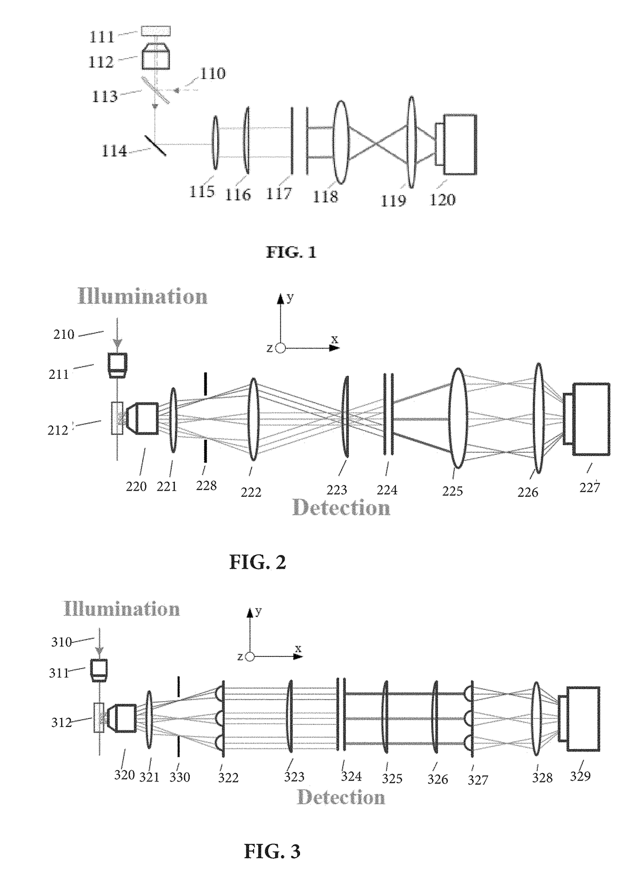
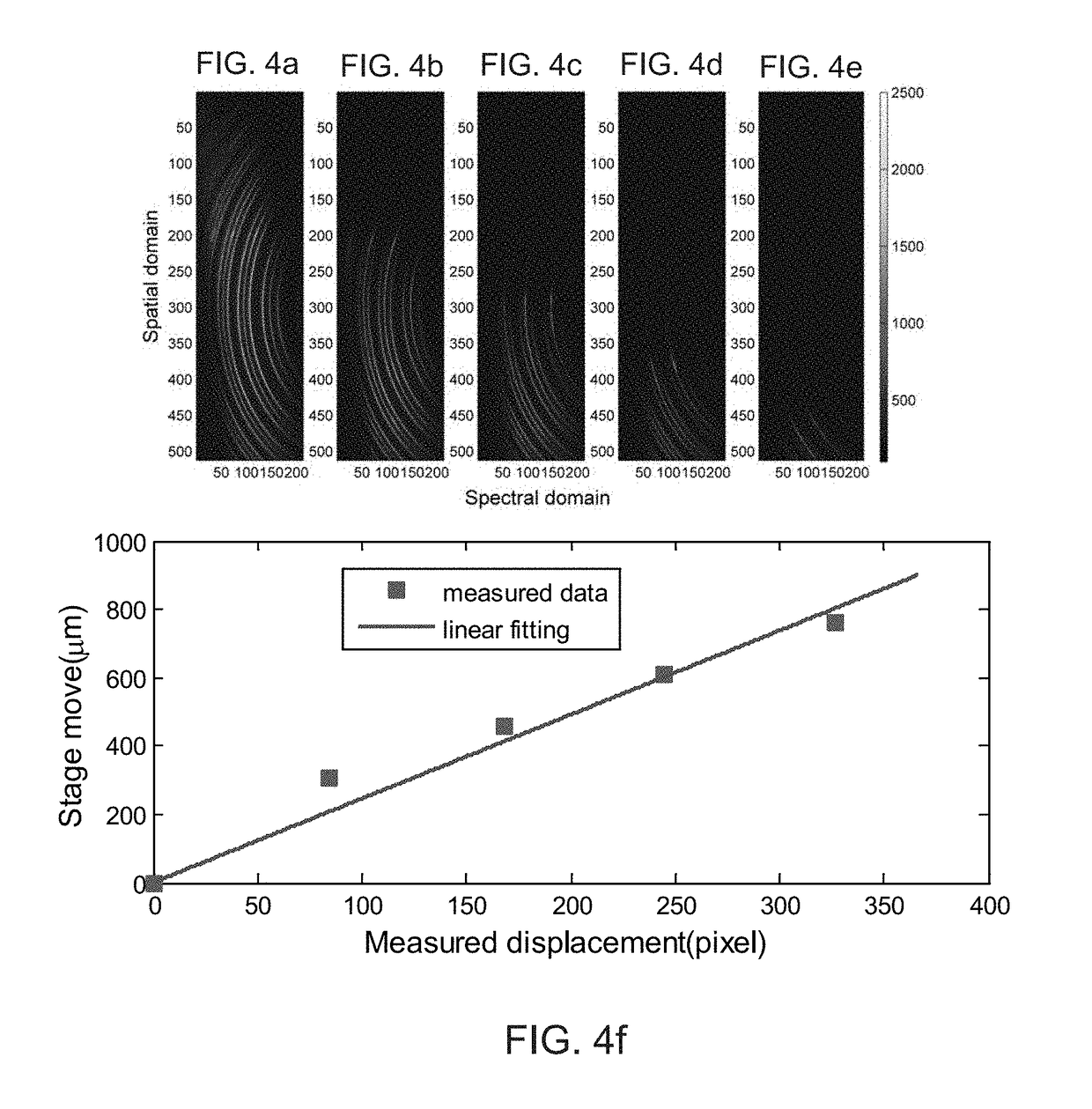
[0105]As an example of validation of the current invention, an experiment was implemented to demonstrate the spatial resolution of the multiplexed Brillouin spectroscopy using the setup of FIG. 2 In this experiment, a single-mode 532 nm continuous-wave laser was used as a light source, a plastic cuvette containing methanol was used as a sample. The objective lens 211 in the illumination path (along y-axis) has a numerical aperture (NA) of 0.0175, and the objective lens 220 in the detection path (along x-axis) has a NA of 0.1. The VIPA 224 has a free spectral range (FSR) of 17 GHz and entrance window of 20 mm. The camera 227 is an electron multiplying coupled charge device (EMCCD). A knife edge was placed between the sample (the plastic cuvette) and the objective lens 220. The knife edge can be moved along y-direction using a translational stage. When moving the knife edge towards y-direction, the scattering signal from the cuvette will be partly blocked. By comparing the displacemen...

example 2

[0109]Example 2 relates to characterization of the multiplexed (line-scanned) Brillouin spectroscopy according to the embodiment of the current invention as shown in FIG. 7. FIG. 7 demonstrates a line-scanned Brillouin spectroscopy setup including a mirror 724, objective lenses 711, 713, and 714, a spherical lens 715 for beam collimation, cylindrical lenses 716, 718, and 719, and a spherical lens 722 for imaging purpose. In one embodiment, a spatial filter or aperture 725 is positioned in an intermediate image plane to reject out-of-focus light coming from the sample.

[0110]The spectral resolution of the spectrometer according to FIG. 7 was characterized in the current example. In one non-limiting embodiment, the light source 710 was a single mode 532-nm cw (continuous-wave) laser (Torus, LaserQuantum). The light from the laser head was focused by the objective lens 711 (NA=0.0175) to generate a line beam for illuminating the sample 712. At 90 degree, the scattering light along the b...

example 3

Two-Dimensional and Three-Dimensional Imaging

[0114]Example 3 relates to characterization of two-dimensional and three-dimensional imaging based on the multiplexed Brillouin spectroscopy according to FIG. 7.

[0115]The Brillouin shift of an aspherical PMMA lens was measured in the setup shown in FIG. 10a. FIG. 10a shows a picture of the PMMA lens taken before filling cuvette with index-matching liquid. The lower is zoomed-in picture of the PMMA lens, the scale bar has a length of 500 μm. The PMMA lens was placed into a plastic cuvette, which was pre-filled with refractive-index-matching liquid to reduce the scattering at the surface of the sample. The cuvette was carried by a vertically placed motorized translation stage, which enabled us to scan the sample in y-direction. In one embodiment, the cuvette (sample 712 in FIG. 7) has a size of 10 mm by 10 mm. To suspend an aspherical Poly methyl methacrylate (PMMA) lens in the central region of the cuvette 712, the margin of the PMMA lens ...


PUM

PropertyMeasurementUnit
sizeaaaaaaaaaa
depthaaaaaaaaaa
widthaaaaaaaaaa

Abstract

The present invention relates to a method and system for analyzing mechanical signatures of a plurality of cells for metastatic detection. Specifically, a data set characterized by at least one metric (such as Brillouin frequency shift and/or Brillouin linewidth) representing a cell mechanical signature is acquired for the plurality of cells by using a label-free Brillouin spectroscopy. A merit function is calculated based on one or more statistical characteristics of the data set, such as sensitivity and specificity. Then, the plurality of cells can be classified to detect metastatic cells based on mechanical signatures provided by the data set and an optimal metric value delivering maximum to the merit function.

Description

CROSS-REFERENCE TO RELATED APPLICATIONS[0001]This application claims the benefit of U.S. Provisional Patent Application Ser. No. 62 / 515,873, filed on Jun. 6, 2017, and is a continuation-in-part of U.S. patent application Ser. No. 15 / 909,699, filed Mar. 1, 2018, which claims the benefit of U.S. Provisional Patent Application Ser. No. 62 / 465,230, filed Mar. 1, 2017, and is a continuation-in-part of U.S. patent application Ser. No. 15 / 388,582, filed Dec. 22, 2016, which claims the benefit of U.S. Provisional Patent Application Ser. No. 62 / 270,982, filed on Dec. 22, 2015; U.S. Provisional Patent Application Ser. No. 62 / 339,512, filed on May 20, 2016; U.S. Provisional Patent Application Ser. No. 62 / 323,176, filed on Apr. 15, 2016; and U.S. Provisional Patent Application Ser. No. 62 / 425,070, filed on Nov. 21, 2016. All of the above listed applications are incorporated herein by reference in their entirety.FIELD OF THE INVENTION[0002]The present invention relates to a method and system for...

Claims


Application Information

Patent Timeline
Patent Type & Authority Applications(United States)
IPC IPC(8): G01N15/14G01N21/64G06T7/00G01N21/53G01N21/63
CPCG01N15/1429G01N15/1434G01N15/147G01N15/1475G01N2015/1006G01N2015/1445G01N2015/1488G01N2021/638C12Q1/00G01J3/44G01N21/63G01N21/64G16B20/00
Inventor SCARCELLI, GIULIANOZHANG, JITAO
Owner CANON USA
Why Eureka
Try Eureka