Convolutional deep learning analysis of temporal cardiac images

Active Publication Date: 2018-11-22
KONINKLJIJKE PHILIPS NV
View PDF0 Cites 14 Cited by
  • Summary
  • Abstract
  • Description
  • Claims
  • Application Information

AI Technical Summary

Benefits of technology

The present patent text describes a system and method for using a deep learning approach to diagnose cardiovascular disease (CVD) based on echocardiograms. The system includes an ultrasound device and an echocardiogram controller for generating and analyzing echocardiograms. The system can also combine electrocardiograms and echocardiograms to improve the diagnosis of CVD. The technical effects of this system include standardizing the interpretation of echocardiograms, reducing inter-observer and intra-observer variability, and improving the diagnosis of CVD using a combination of echocardiograms and electrocardiograms.

Problems solved by technology

One of the major problems of the aforementioned Echo procedures is the diagnosis of CVD based on motion of the cardiac wall is done in a completely subjective manner.
The other difficulty with interpretation of Echo is that it requires highly trained professionals needed for interpretation of echo images.

Method used


Image

  • Convolutional deep learning analysis of temporal cardiac images
  • Convolutional deep learning analysis of temporal cardiac images
  • Convolutional deep learning analysis of temporal cardiac images

Examples


second embodiment

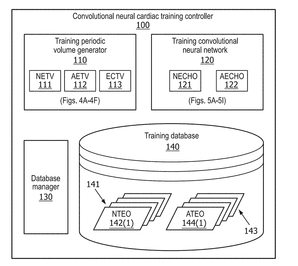
[0121]Referring back to FIG. 2A, in a second embodiment, training CNN 120 executes a multiple stream CNN involving an execution of a spatial-temporal CNN for each echocardiogram slice of a normal echocardiogram training volume 111 or an abnormal echocardiogram training volume 112 (i.e., a spatial stream CNN) and an execution of spatial-temporal CNN for a motion flow of normal echocardiogram training volume 111 or abnormal echocardiogram training volume 112 (i.e., a temporal stream CNN). The multiple (dual) streams are combined by a late fusion of scores (e.g., an averaging a linear SVM, another neural network). The information from the multiple (dual) streams can also be combines by using shared convolutional kernels between different streams.

[0122]For example, FIG. 5D illustrates a training CNN 180a for executing a spatial stream CNN 182a for each eco echocardiogram slice 181a of a segmental view of a normal echocardiogram training volume 111 or of an abnormal echocardiogram traini...

third embodiment

[0125]Referring back to FIG. 2A, in a third embodiment, training CNN 120 executes a memory recurrent CNN involving an execution of a spatial-temporal CNN for each echocardiogram slice or a sliced 3D volume of a normal echocardiogram training volume 111 or an abnormal echocardiogram training volume 112, a mean polling of the outputs of the spatial temporal CNNs, and an execution of a recurrent neural network (RNN) of the mean polling to obtain a scoring output.

[0126]For example, FIG. 5G illustrates a memory recurrent CNN 190a involving an execution of a mean polling 192a of a spatial-temporal CNN 191a for each echocardiogram slice of a segmental view of a normal echocardiogram training volume 111 or an abnormal echocardiogram training volume 112, followed by an execution of a Long Short Term Memory (LSTM) RNN 193a and LSTM RNN 194a to obtain a scoring output 195a. In practice, a particular setup of training CNN 190a in terms of a complexity of spatial-temporal CNN 191a, LSTM RNN 193a...


PUM


Abstract

A convolutional neural cardiac diagnostic system employs an echocardiogram diagnostic controller for controlling a diagnosis of an echocardiogram including a temporal sequence of echocardiac cycles. The echocardiogram diagnostic controller includes a diagnostic periodic volume generator generating an echocardiogram diagnostic volume including a periodic stacking of the temporal sequence of echocardiac cycles, and further includes a diagnostic convolutional neural network classifying the echocardiogram as either a normal echocardiogram or an abnormal echocardiogram based on a convolutional neural analysis of the echocardiogram diagnostic volume. The diagnostic periodic volume generator may further generate an electrocardiogram diagnostic volume including a periodic stacking of the temporal sequence of electrocardiac waves, and the convolutional neural network may classify the echocardiogram as either a normal echocardiogram or an abnormal echocardiogram based on a convolutional neural analysis of both the echocardiogram diagnostic volume and the electrocardiogram diagnostic volume.

Description

CROSS REFERENCE TO RELATED APPLICATION[0001]The present application claims the benefit of U.S. Provisional Application No. 62 / 508,087, filed May 18, 2017. These applications are hereby icorporated by reference herein.TECHNICAL FIELD[0002]The present disclosure generally relates to image processing. More specifically, but not exclusively, the present disclosure relates to classifying a cardiac image as illustrating normal cardiac functions or abnormal cardiac functions.BACKGROUND[0003]Many medical imaging modalities (e.g., ultrasound US, magnetic resonance imaging MRI, CT, positron emission tomography PET, etc.) provide temporal data describing functions of various body organs. One of the main application of the temporal functional imaging is the diagnosis and monitoring of heart disease. Because the heart is in constant periodic motion, the temporal imaging is extensively used to characterize cardiac function by analysis of the cardiac deformation.[0004]Echocardiography (echo) as kn...

Claims


Application Information

Patent Timeline
IPC IPC(8): A61B5/00G06N5/04A61B5/0468G06T7/00A61B5/364
CPCA61B5/7267G06N5/046A61B5/0468G06T7/0012G06T2207/30048G06T2207/10132G06T2207/10081G06T2207/10088G06T2207/10121G06T2207/10104G06T2207/10108G06N3/08G06T2207/20084G06N3/044G06N3/045A61B5/364
Inventor SITEK, ARKADIUSZ
Owner KONINKLJIJKE PHILIPS NV
Why Eureka
Try Eureka