
Method to measure the shortest telomeres

a telomere and telomere technology, applied in the field of cell biology, molecular biology, diagnostics, and medicine, can solve the problems of inability to quantify tl for cancer studies, inability to analyze non-dividing cells such as senescent cells or resting lymphocytes, and inability to quantify tl. sensitivity, efficiency and specificity increase

Active Publication Date: 2018-11-29
BOARD OF RGT THE UNIV OF TEXAS SYST
View PDF0 Cites 1 Cited by
  • Summary
  • Abstract
  • Description
  • Claims
  • Application Information

AI Technical Summary

Benefits of technology

The patent describes a new method called TeSLA for measuring the length of telomeres, which are important parts of chromosomes. This method allows for more sensitive and accurate detection of telomere length compared to other methods. The inventors used TeSLA in combination with an image-processing program to measure telomere length after Southern blot analysis. They were able to detect changes in telomere length in normal aging, cancer progression, and related disorders. The TeSLA method was also applied to different animal species, including humans. Overall, this improved technology provides a better way to measure telomere length and study its dynamic behavior.

Problems solved by technology

The TRF technique not only requires a large amount of genomic DNA, but due to the lower hybridization signal of the shortest telomeres, TRF underestimates information about the abundance of the shortest telomeres.
In addition, the qPCR method is not suitable to quantify TL for cancer studies since most of cancer cells are aneuploidy (Holland and Cleveland, 2009).
Although metaphase Q-FISH (Lansdorp et al., 1996) can detect TL from each chromosome, this method does not permit analyses on non-dividing cells, such as senescent cells or resting lymphocytes.
While these approaches are an improvement over Q-PCR, one disadvantage of these techniques is the probe not only binds to telomeric repeats but also interacts with non-specific components in the cytoplasm (Aubert et al., 2012; Wieser et al., 2006).
Importantly, probe hybridization kinetics does not permit robust quantitation of the shortest telomeres (<2-3 Kb).
This is one major limitation of STELA.
It is not sufficient to suppress the amplification of larger genomic DNA fragments.
In addition, U-STELA is not efficient to detect TL over 8 kb (Bendix et al., 2010) which could affect the detection of accuracy of TL distribution.

Method used


Image

  • Method to measure the shortest telomeres
  • Method to measure the shortest telomeres
  • Method to measure the shortest telomeres

Examples


example 1

TeSLA: A Method for Measuring the Distribution of the Shortest Telomeres in Cells and Tissues

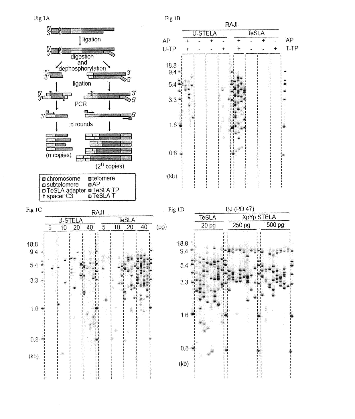
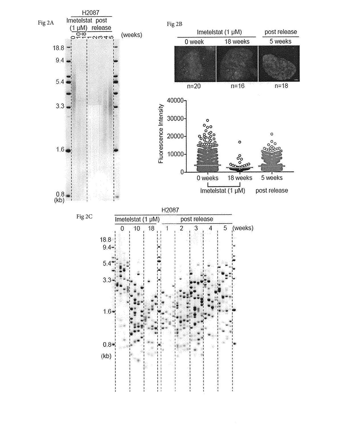
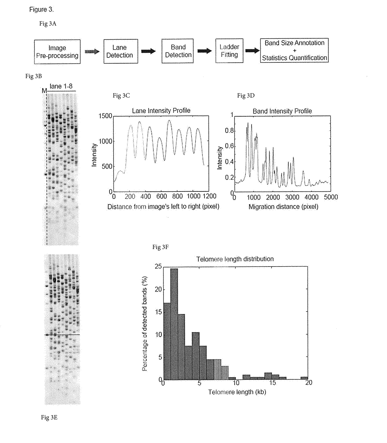
[0050]Methods encompassed by the disclosure allow for more sensitive, efficient and specific for telomere length (TL) detection when directly compared to other methods for TL measurement. The methods may be used in combination with any other method, and in some cases may be used in combination with any imaging method, whether it is automated or not. As shown herein, one can detect telomere dynamics, especially for the shortest telomeres, in normal aging process, cancer, and telomere-related disorders in humans, for example. Also, one can apply the TeSLA method to different mammals such as mice, elephants, and bowhead whales to demonstrate the effectiveness of this improved technology.

Results

The Principle of TeSLA

[0051]A schematic presentation of the TeSLA method is shown in FIG. 1A. TeSLA is based on a ligation and PCR approach to detect amplified terminal restriction fragments which contain...


PUM

PropertyMeasurementUnit
lengthaaaaaaaaaa
genomic stabilityaaaaaaaaaa
size distributionaaaaaaaaaa

Abstract

The present disclosure encompasses methods and compositions for analyzing telomeres of genomic DNA, including the length of one or more telomeres. In specific embodiments, methods and compositions are employed for identification of the length of the shortest telomere in a collection of genomic DNA. In particular cases, the method greatly suppresses amplification of intra-genomic DNA fragments in comparison to telomeres by hindering their amplification using particular adaptors.

Description

[0001]This application claims priority to U.S. Provisional Patent Application No. 62 / 504,009, filed May 10, 2018, which is incorporated by reference herein in its entirety.TECHNICAL FIELD[0002]Embodiments of the disclosure include at least the fields of cell biology, molecular biology, diagnostics, and medicine, including at least cancer medicine.BACKGROUND[0003]In vertebrates, telomeres consisting of repeat arrays of the canonical TTAGGG DNA sequence and their associated shelterin proteins reside at the ends of each chromosome to maintain genomic stability by preventing the ends from being recognized as DNA double strand breaks (Palm and de Lange, 2008). In most adult human somatic cells telomerase, a cellular reverse transcriptase that adds the repetitive DNA to telomeres is not detected. Therefore, telomeres progressively shorten with each cell division due to the “end replication problem” (Olovnikov, 1973; Watson, 1972). In humans, telomere shortening has been implicated as a ri...

Claims


Application Information

Patent Timeline
Patent Type & Authority Applications(United States)
IPC IPC(8): C12Q1/683C12Q1/6855C12Q1/6851C12Q1/6858C12Q1/686G06F19/22G16B30/00
CPCC12Q1/683C12Q1/6855C12Q1/6851C12Q1/6858C12Q1/686G06F19/22C12Q1/6886G16B30/00C12Q2521/313
Inventor SHAY, JERRY W.LAI, TSUNG-PO
Owner BOARD OF RGT THE UNIV OF TEXAS SYST
Learn More