
Transcutaneous device for removal of fluid from a body

a technology of transcutaneous device and body, which is applied in the field of transcutaneous device for removal of fluid from the body, can solve the problems of nt failure, prone to blockage of cannula used in current nt's, and insufficient complications or indeed inadequate, so as to improve the function of the device, improve the utility, and reduce the effect of nt failur

Pending Publication Date: 2019-01-03
SAFEGUARD MEDICAL HOLDCO LLC
View PDF7 Cites 5 Cited by
  • Summary
  • Abstract
  • Description
  • Claims
  • Application Information

AI Technical Summary

Benefits of technology

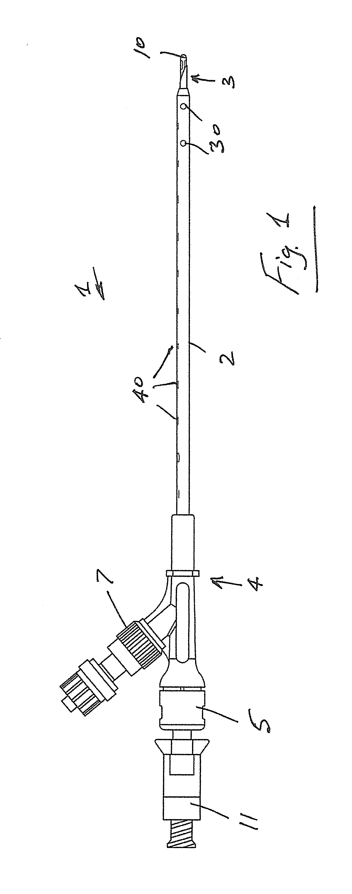
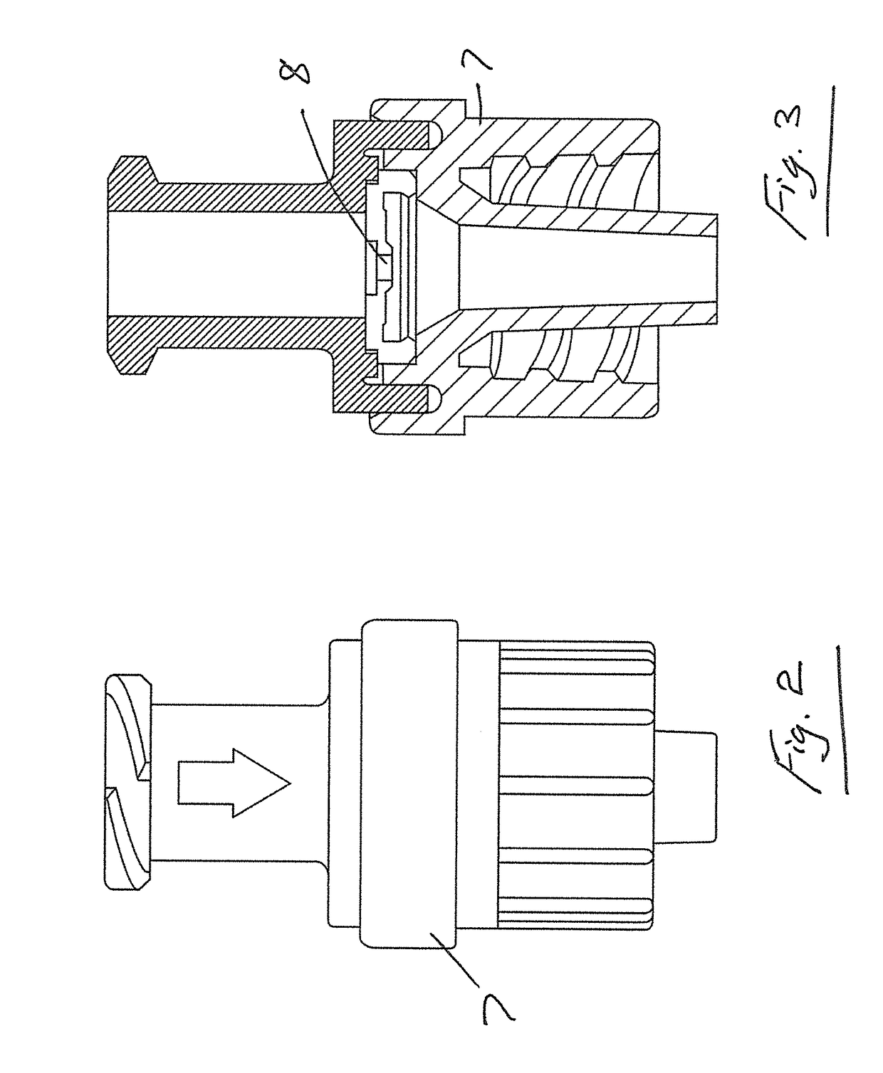
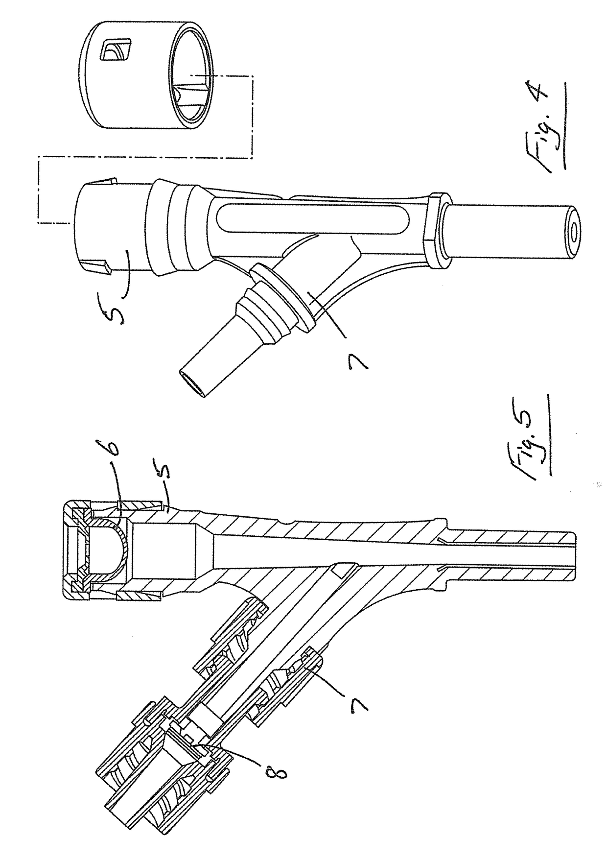
The invention is a device that can manage pneumothoraxes (when air bubbles accumulate in the chest) and effusions (when there is excess liquid in the chest) for both prehospital and hospital medical professionals. It has multiple functions in one device, including needle blunting and medical drainage. It is easy to use, can be inserted in patients at various locations, and is resistant to kinking and skin puncture. It also has a valve to prevent air re-occurrence after use.

Problems solved by technology

Much evidence is now available that indicates using an inappropriate product to perform this procedure may lead to complications or indeed simply be inadequate.
In either scenario, this can lead to an NT failure.
The cannulas used in current NT's are also prone to blockage through kinking of the cannula.
The three cases discussed showed that there is a potential for failure of the NT due to the cannula.
While it is not always possible to exactly determine the cause of a haemothorax it is widely considered a significant issue that most NT products or improvised cannulas have sharp unprotected ends that can cause bleeding through laceration of blood vascularisation in the areas of insertion.
Even more significantly if misdiagnosis of a pneumothorax occurs there is a strong possibility of lung laceration with the needle if no pneumothorax is present.
However, it has an unprotected sharp end which could cause lung laceration or laceration of blood vascularisation and the cannula also kinks easily which could lead to cannula occlusion and the effects that can cause.
The length of the product also means that it may also be too short to reach the pleural space in some individuals.
However, the cannula kinks easily and the device cannot be inserted in a single operation.
The device requires that a scalpel be used to cut skin before it can be used which limits its use in the prehospital environment where paramedic staff do not always carry scalpels and are usually precluded from using scalpels in some territories.

Method used


Image

  • Transcutaneous device for removal of fluid from a body
  • Transcutaneous device for removal of fluid from a body
  • Transcutaneous device for removal of fluid from a body

Examples


example 1

Tissue Penetration

[0168]The device of the invention successfully penetrated skin / muscle penetration using an uncooked pork belly sample. Uncooked pork belly provides a good human skin / muscle substitute due to its outer tough skin and multiple tissue layers.

example 2

hawed Cadaver Pleural Cavity Penetration

[0169]A cadaver study was performed with the invention. The device was tested for insertion into a freshly thawed non-embalmed cadaver. The study mimicked the placement of the device for the management of a tension pneumothorax. The device insertion site was located and insertion with the device was as described above. The device successfully penetrated the skin surface.

example 3

Cadaver Fluid Expiration Test

[0170]Example 2 was repeated using an embalmed cadaver. After insertion, embalming fluid escaped through the Veress needle on entry and after Veress needle withdrawal the fluid escaped through the side port one way crack valve but not through the self-sealing bung. This was representative of the in vivo fluid / gas functionality of the device.


PUM


Abstract

A single step body insertion device comprises a cannula and a Veress needle which penetrates the skin surface and relevant tissue layers to reach fluid and / or gases that need to be removed from the body. The cannula shaft and tapered tip are of a polymeric material which is flexible and kink resistant. The Veress needle has an engagement feature for engagement with the tapered tip of the cannula for delivery of the cannula into the body as the Veress needle is inserted into the body. The device is used for the management of conditions such as pneumothoraxes and pleural effusions as well as other conditions that require release of fluid and / or gas from the body.

Description

CROSS REFERENCE TO RELATED APPLICATIONS[0001]This application is a divisional of application Ser. No. 14 / 844,533 filed on Sep. 3, 2015, which is incorporated herein in it's entirety by reference.FIELD OF INVENTION[0002]The invention relates to a device used to treat and manage pneumothoraxes' and effusions through a controlled drainage system while maximising patient safety and minimising the steps required and complexity for the practitioner.BACKGROUND OF THE INVENTION[0003]US2010 / 0268156A describes a medical device for supplying and / or removing fluid into and / or from body cavities which comprises a catheter with a valve at the proximal end through which a Veress needle is inserted.[0004]EP1358904A describes a thoracic vent kit.[0005]WO2006 / 090148 describes an apparatus for the aspiration of a fluid from a body including a catheter and a needle.[0006]Needle thoracocentesis (NT) is the insertion of a needle into the pleural space and the drainage of air that has accumulated. This ca...

Claims


Application Information

Patent Timeline
Patent Type & Authority Applications(United States)
IPC IPC(8): A61M1/00A61B17/34A61M25/06A61M25/01A61M25/00A61B17/00A61M39/10
CPCA61M2025/0008A61M25/065A61M25/0041A61M2039/1044A61B2017/00915A61B2017/00477A61M25/0606A61M25/0105A61M25/0102A61M25/0097A61M25/0069A61B17/3474A61B17/3415A61M1/008A61M1/0019A61B2017/3454A61B17/3498A61B17/3421A61M2210/1039A61M25/008A61B17/3462A61M1/84A61M1/69
Inventor REAL, KEITH JOSEPHRUSSELL, MALCOLM QUENTIN
Owner SAFEGUARD MEDICAL HOLDCO LLC
Learn More