Non-invasive ocular drug delivery insert technology

a technology of ocular drug delivery and insert, which is applied in the field of non-invasive ocular drug delivery insert, can solve the problems of high cost and achieve the effect of inhibiting the risk of displacement or expulsion

Active Publication Date: 2019-02-14
UNIVE DE COIMBRA
View PDF0 Cites 10 Cited by
  • Summary
  • Abstract
  • Description
  • Claims
  • Application Information

AI Technical Summary

Benefits of technology

[0117]The insert can be prepared with different shapes (spherical or hemispherical) and/or architectures (monolithic/layered either with or without a drug core), allowing the incorporation of at least on

Problems solved by technology

The currently available technologies for ocular drug delivery present several limitations which include at least one of the listed below.Short residence time of the drugs in the eye;Fluctuations of the drug concentration (namely with eye drops);Low compliance of the patient to the therapeutics;Short release periods (from

Method used


Image

  • Non-invasive ocular drug delivery insert technology
  • Non-invasive ocular drug delivery insert technology
  • Non-invasive ocular drug delivery insert technology

Examples


example 1

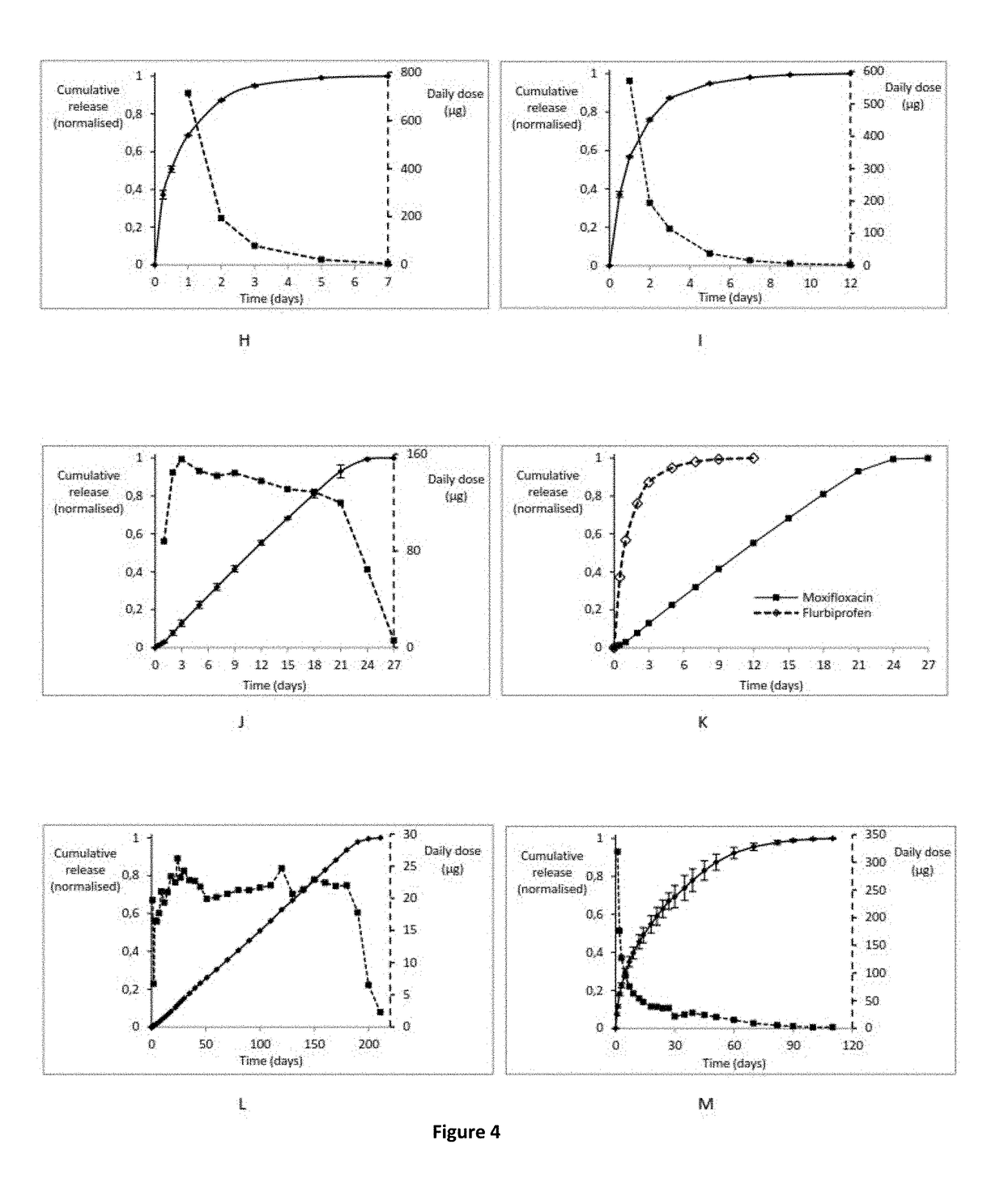
[0185]A formulation of the ocular insert embodying the invention, Formulation 1, was prepared as follows: The copolymer used in this formulation was a low molecular weight polycaprolactone / polyethelyneglycol in a ratio of 75 / 25 (PCL / PEG; mass / mass). The drug moxifloxacin was mixed with the copolymer in a beaker under magnetic stirring at 70° C. for a few minutes. The drug percentage regarding the total mixture weight was 2% (mass / mass).

[0186]The ocular inserts embodying the invention were then prepared from that drug / polymer mixture with asymmetrical shape as represented in scheme B of FIG. 2. The average length of the large diameter was 5.8 mm while the small diameter measured 2.7 mm. The average weight of the ocular inserts of Formulation A was approximately 50 mg with approximately 1 mg of drug dispersed in the polymeric matrix.

[0187]Each insert of Formulation 1 was placed in phosphate-buffered saline solution (PBS) at 37° C. until all the drug was released to the aqueous media. ...

example 2

[0188]A formulation of the ocular insert embodying the invention, Formulation 2, is similar to Formulation 1 except that the drug entrapped in the polymer matrix was flurbiprofen instead of moxifloxacin.

[0189]The architecture, size and shape are the same applied on formulation 1. Therefore the average weight of the ocular inserts of Formulation 2 was also approximately 50 mg with approximately 1 mg of drug dispersed in the polymeric matrix. Each insert of Formulation 2 was placed in phosphate-buffered saline solution (PBS) at 37° C. until all the drug was released to the aqueous media. The flurbiprofen dissolved was quantified by an UV-Vis spectrophotometric method and the results shown a “Fickian” release where approximately 50% of the drug was released in the initial 24 hours and almost all of the entrapped drug was dissolved at day 5 of the dissolution study (FIG. 4I).

example 3

[0190]A formulation of the ocular insert embodying the invention, Formulation 3, was prepared as follows: The copolymer used in this formulation was a low molecular weight polycaprolactone / polyethelyneglycol in a ratio of 75 / 25 (PCL / PEG; mass / mass). 100 mg of Moxifloxacin was slightly compressed under 300 mbar for half a minute to form a tablet from which small portions of 3 mg were obtained. Each of these small tablets were then covered with melted copolymer to form the final ocular insert devices embodying the invention.

[0191]These ocular inserts embodying the invention in Formulation 3 were of an assymetrial shape as represented in scheme B of FIG. 2. The average length of the large diameter was 5.8 mm while the small diameter measured 2.7 mm. The average weight of the ocular inserts of Formulation 3 was 50 mg with approximately 3 mg of drug in a central core.

[0192]Each insert of Formulation 3 was placed in phosphate-buffered saline solution (PBS) at 37° C. until all the drug was...


PUM

PropertyMeasurementUnit
Fractionaaaaaaaaaa
Fractionaaaaaaaaaa
Fractionaaaaaaaaaa

Abstract

Non-invasive Ocular Drug Delivery Insert Technology. The invention concerns an ocular insert which is a new biocompatible polymer-based controlled drug delivery system (CDDS) applicable to a variety of drugs and other compounds for the treatment of different ocular pathologies. This ocular insert allows releasing of at least one drug under suitable concentration levels during suitable periods of time. The device may be inserted in the lower or upper fornix conjunctiva, in a non-invasive way, meaning that the patient will be able to place the device himself, without intervention of medical specialized staff. The insert of the invention will release the drug in such a controlled rate that will allow the drug release up to 300 days by either a “Fickian” or a linear profile according to the intend purpose or pathology. The insert can be prepared with different shapes (spherical or spherical dome) and/or architectures (monolithic/layered either with or without a drug core) allowing the incorporation of at least one drug which can be released at different rates. The size, shape and design of the insert is adjusted in order to tune the drug(s) delivery profile(s) and to inhibit the risk of displacement or expulsion.

Description

FIELD OF THE INVENTION[0001]The present invention is related to a biocompatible ocular insert with polymeric composition, designed for “in vivo” sustained drug release to be applied in the treatment of different ocular pathologies.BACKGROUND OF THE INVENTION[0002]In the area of ophthalmology, the conventional method of drops topical application involves repeated administrations of the drugs solutions and the drugs concentration profiles are characterized by extreme fluctuations with an excessive concentration stage, followed by a brief period of correct dose and ending in a long underdosing period. Moreover, it is necessary to take into consideration the drainage of the lacrimal film and the small corneal permeability which require the use of high doses of drug in order to secure the intraocular therapeutic level. Since only 1 to 3% of the drug penetrates the cornea [1, 2] and the remainder is absorbed by systemic routes, high concentrations can bring toxic side effects at a systemi...

Claims


Application Information

Patent Timeline
IPC IPC(8): A61K9/00A61K47/34A61K31/4709A61K31/192A61K31/165B29D11/00
CPCA61K9/0051A61K47/34A61K31/4709A61K31/192A61K31/165B29D11/00096B29K2071/02B29K2067/046B29K2105/0035B29K2105/0085A61K45/06B29D11/023A61P27/02A61K2300/00A61F9/00A61K9/00
Inventor MOTA LEITE MACHADO MARIZ, MARCOS JOAONUNES FERREIRA CALVINHO, PAULA CRISTINAMENDES GIL, MARIA HELENANETO MURTA, JOAQUIM CARLOS
Owner UNIVE DE COIMBRA
Why Eureka
Try Eureka