Bioscaffold, method for producing the same, and uses thereof

a bioscaffold and cell-free technology, applied in the field of tissue engineering, can solve the problems of limiting the application of thus produced biological scaffolds in tissue regeneration, and none of the above-mentioned methods alone is capable of producing a cell-free bioscaffold, so as to reduce the fluidity of the digestion buffer, restrict digestion, and increase the viscosity

Inactive Publication Date: 2019-05-02
NAT CHENG KUNG UNIV
View PDF0 Cites 6 Cited by
  • Summary
  • Abstract
  • Description
  • Claims
  • Application Information

AI Technical Summary

Benefits of technology

[0010]According to some preferred embodiments of the present disclosure, in the step (a), the organ or the vessel is treated with the digestion buffer comprising a thickening agent and a digesting enzyme. Preferably, the digestion buffer is applied on the outer surface of the organ or the vessel. The purpose of adding thickening agent in the digestion buffer is to increase the viscosity (i.e., reduce the fluidity) of the digestion buffer, restricting the digestion to the outer surface of the organ or the vessel. That is, the lining tissue can thus be removed without damaging the underlying tissue / organ. According to the embodiments of the present disclosure, the digestion buffer has a viscosity suffice enough to prevent free-flow of the digestion buffer so that the digestion buffer remains on the outer surface of the organ or the vessel. According to one working example of the present disclosure, the viscosity of the digestion buffer is 8 centipoise (cP).

Problems solved by technology

However, none of above-mentioned methods alone is capable of producing a completely cell-free biological scaffold.
In addition, some decellularization treatments may damage the structure of ECM that further limits the application of thus produced biological scaffolds in tissue regeneration and / or remodeling.

Method used


Image

  • Bioscaffold, method for producing the same, and uses thereof
  • Bioscaffold, method for producing the same, and uses thereof
  • Bioscaffold, method for producing the same, and uses thereof

Examples


example 1 preparation

and Characterization of Decellularized HUAs

[0100]1.1 Preparation of Adventitia-Free HUAs

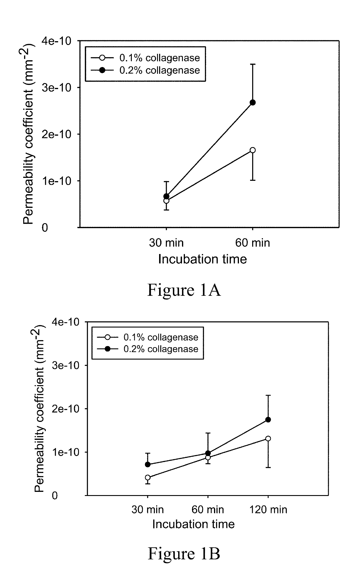
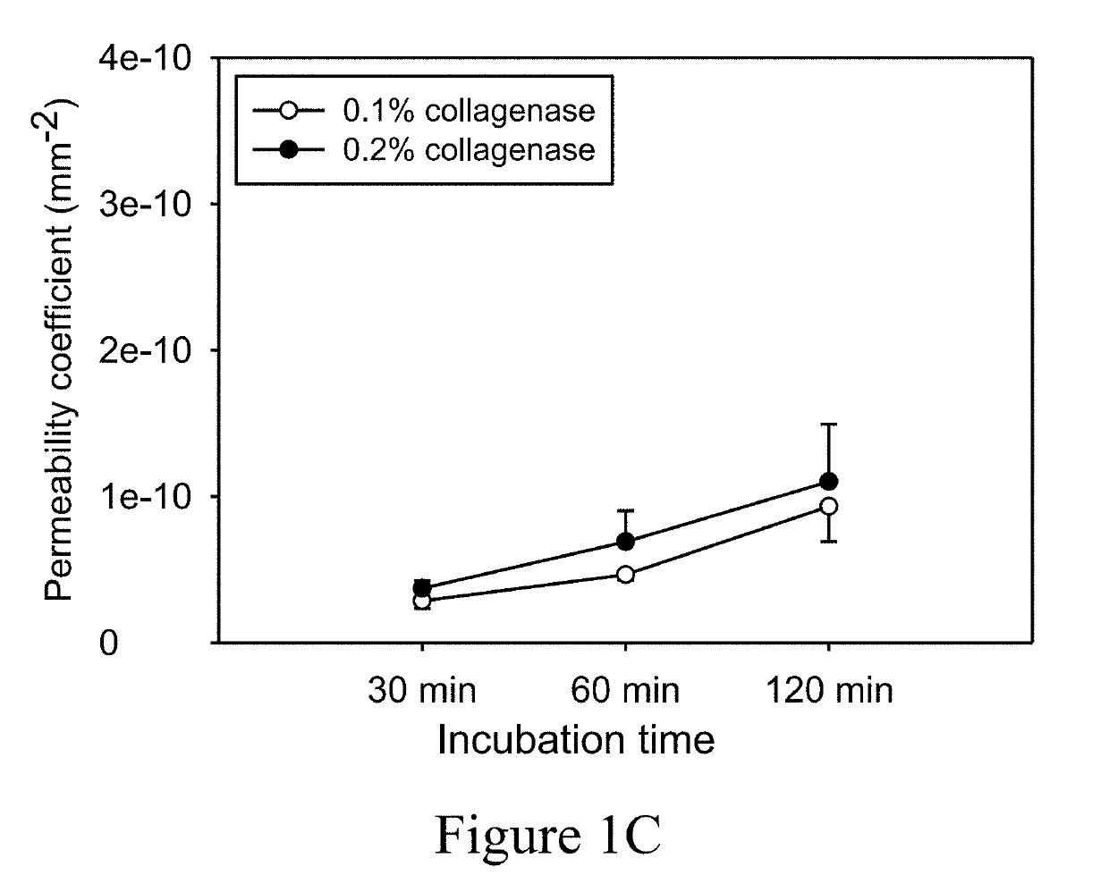
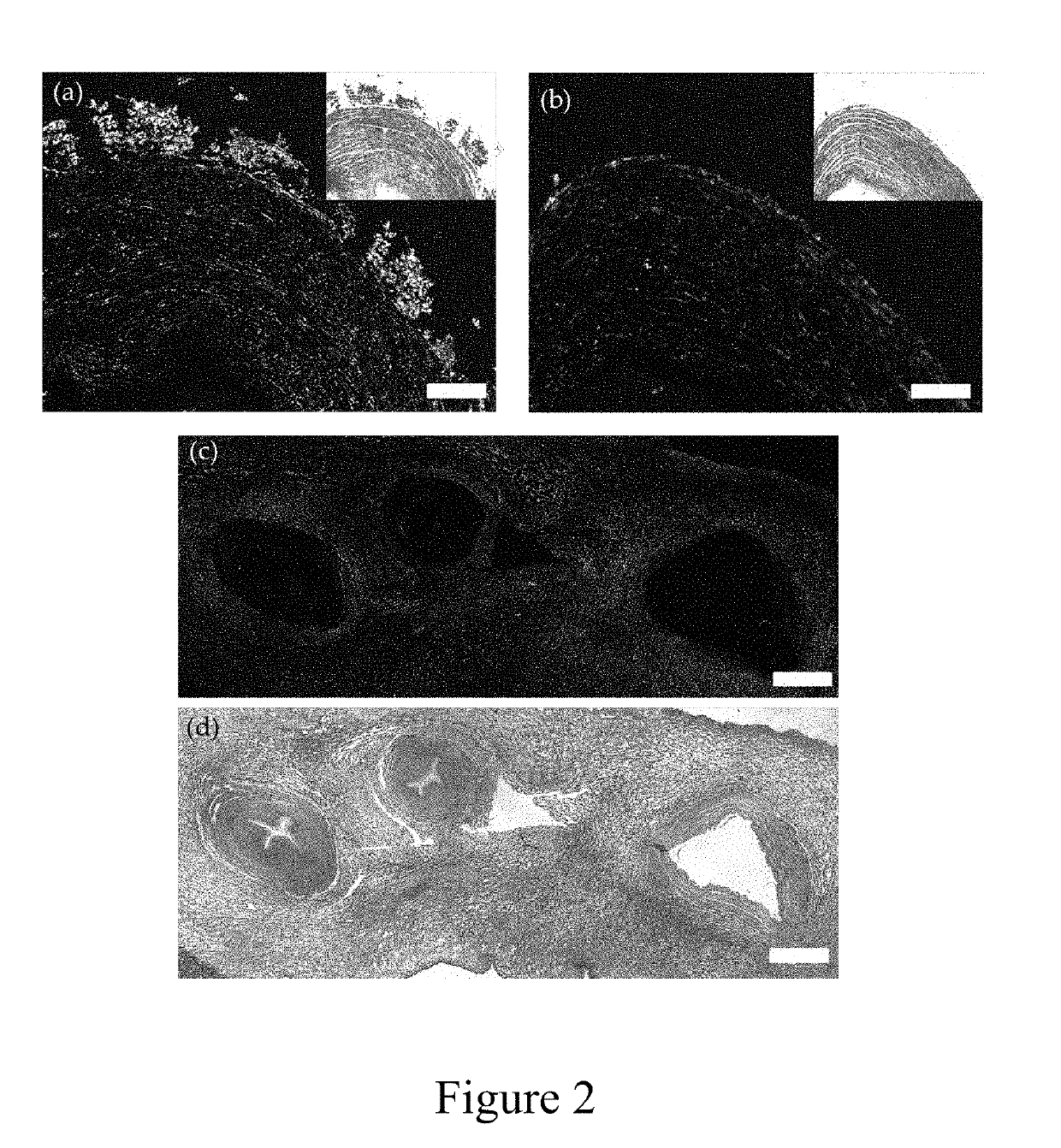
[0101]To remove the lining tissue of HUAs, the HUAs were subjected to the treatment of collagenase solution of low, moderate or high viscosity for 30, 60 or 120 minutes as descried in “Materials and Methods” section, and the effect of the treatment was evaluated by measuring the permeability of the collagenase-treated HUAs. Results were illustrated in FIGS. 1A-1C.

[0102]It was found that immersing the HUAs in 0.1% or 0.2% (w / v) collagenase solution of low viscosity significantly damaged the structure of the vessel wall, rendering the vessel highly water permeable (FIG. 1A). For the HUAs treated with 0.1% or 0.2% (w / v) collagenase solution of moderate viscosity, the value of permeability increased with an increase in the collagenase concentration or treatment time (FIG. 1B). Nevertheless, treating the HUAs with 0.2% (w / v) collagenase solution of high viscosity for 60 minutes more consistently produ...

example 2

In Vivo Cell Infiltration

[0117]To investigate the efficacy of the decellularized HUAs of example 1 as a bioscaffold, the HUAs were implanted subcutaneously or intraperitoneally in animals as described in the “Materials and Methods” section. Short segments of the HUAs with the intact adventitia that were decellularized by immersing in 1% SDS solution with simple agitation served as the control. Results were illustrated in FIGS. 8 to 11.

[0118]FIGS. 8A and 8B are photographs of the histological staining of the decellularized HUAs taken after being implanted for 1 week (panels a and b), 2 week (panels c and d), or 4 weeks (panels e and f) in the subcutaneous space. Cells were found to stay in the outer area of the vessel in the sections of the decellularized HUAs having the intact adventitia, and tissue degradation was found at 4 weeks post-implantation. For the sections of the decellularized HUAs having the adventitia removed, cells were found in the exterior of the vessel at first wee...


PUM

PropertyMeasurementUnit
viscosityaaaaaaaaaa
kinematic viscosityaaaaaaaaaa
absolute viscosityaaaaaaaaaa

Abstract

Disclosed herein is a method of producing a decellularized bioscaffold from an organ or a vessel covered by a lining tissue. The method comprises subjecting the organ or the vessel to a digestion buffer thereby removing the lining tissue from the tissue or organ; and then treating the tissue or organ with a decellularization buffer. The present decellularized bioscaffold is useful in promoting tissue regeneration and/or remodeling.

Description

BACKGROUND OF THE INVENTION1. Field of the Invention[0001]The present disclosure in general relates to the field of tissue engineering. More particularly, the present disclosure relates to a decellularized bioscaffold, the manufacture and uses thereof.2. Description of Related Art[0002]Decellularization is a process that removes cellular contents from a tissue or an organ, while minimizing adverse effects on the composition, biological activity and mechanical integrity of the extracellular matrix (ECM). Biological scaffolds derived from decellularized tissues or organs have been commonly and successfully used in both animal studies and human clinical applications. Compared with man-made scaffolds, decellularized biological scaffolds possess several advantages, including high biocompatibility, low immunogenicity and intrinsic mechanical competence.[0003]In general, the cellular contents of a tissue or an organ may be removed by physical, chemical or enzymatic methods. The physical me...

Claims


Application Information

Patent Timeline
Patent Type & Authority Applications(United States)
IPC IPC(8): A61L27/36A61L27/38
CPCA61L27/3687A61L27/3604A61L27/38A61L2430/40A61L27/3804
Inventor TUAN-MU, HO-YICHANG, YI-HAOHU, JIN-JIA
Owner NAT CHENG KUNG UNIV
Why Eureka
Try Eureka