
Isolation of oogonial stem cells

a technology of oogonial stem cells and isolating cells, which is applied in the field of isolating oogonial stem cells, can solve the problems of time-consuming methods, low efficiency, and inability to achieve isolating these cells

Inactive Publication Date: 2019-06-13
ROYAN INST
View PDF0 Cites 4 Cited by
  • Summary
  • Abstract
  • Description
  • Claims
  • Application Information

AI Technical Summary

Benefits of technology

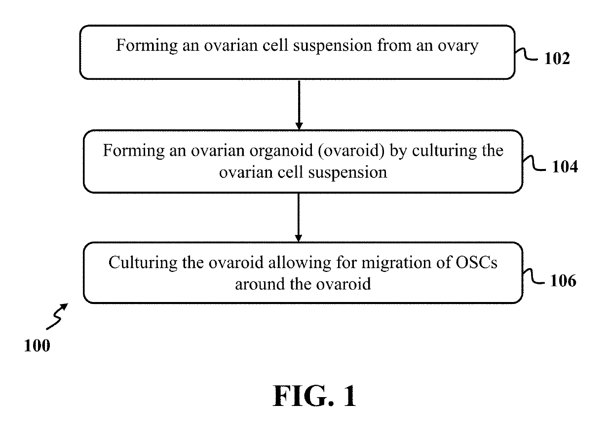
The present disclosure describes an exemplary method for isolating oogonial stem cells (OSCs) from an ovary. The method involves forming an ovarian cell suspension from the ovary and culturing it in a three-dimensional culture to allow the OSCs to migrate around an ovaroid. The OSCs can then be isolated by culturing the ovaroid on a mouse embryonic fibroblast (MEF)-coated plate. The method may include using an agarose-coated plate, a hanging drop, or a microwell for culturing the ovarian cell suspension. The ovarian cell suspension may be cultured for a period of time between about 2 days and about 3 days. The ovaroid may have a diameter between about 50 μm and about 100 μm and may have a round or oval shape. The OSCs may have a genii cell-specific marker and may include mouse or human cells. The technical effect of this method is the efficient isolation of OSCs, which can be useful for research and potential therapeutic applications.

Problems solved by technology

Despite the crucial importance of OSCs in reproductive medicine, a feasible method for isolating these cells has remained elusive due to the relative rarity of OSCs.
However, these methods are time-consuming and have various shortcomings such as low efficiency, low reproducibility, low expansion ability, and high technical difficulty.
However, a feasible method for isolation of OSCs has not been within grasp.
The ovaroid provides us with an invaluable source of functional OSCs with high potentials for in-vitro expansion, while OSCs that may be acquired from fluorescent-and magnetic-based methods are not expandable or show poor expansion ability in-vitro.
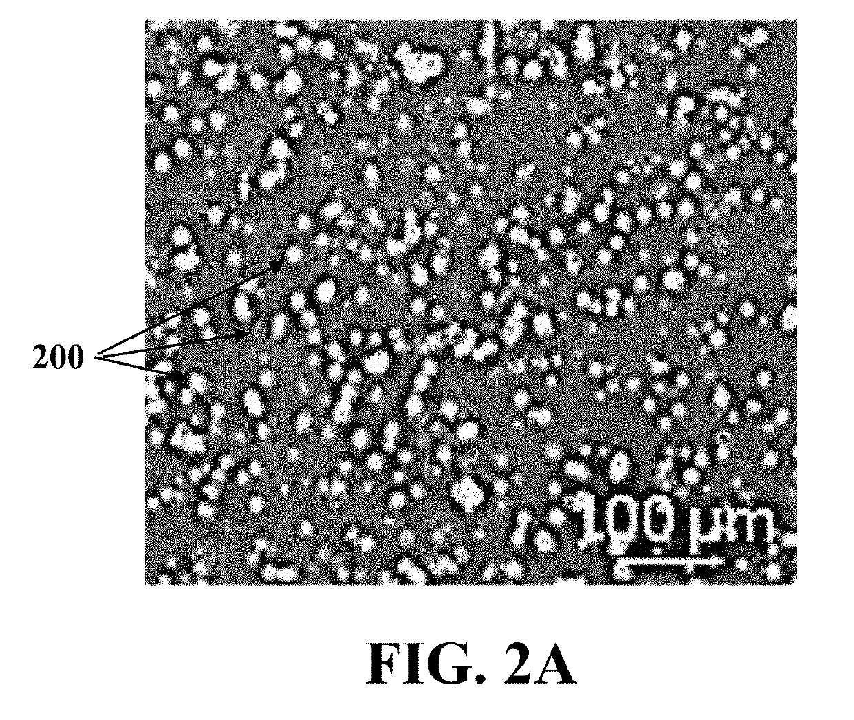
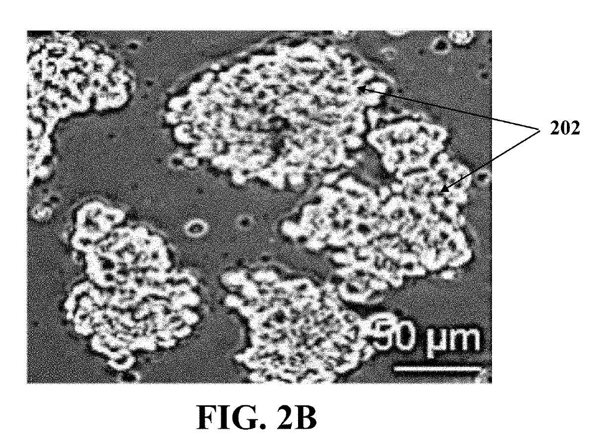
Furthermore, aggregate formation with higher cellular density lead to the aggregates joining together in 24 hours which led to the formation of dense structures.
Furthermore, aggregate formation with higher cellular density joined the aggregates together in 24 hours which led to the formation of dense structures.

Method used


Image

  • Isolation of oogonial stem cells
  • Isolation of oogonial stem cells
  • Isolation of oogonial stem cells

Examples


example 1

Of Mouse Oogonial Stem Cells

[0075]In this example, mOSCs were isolated through steps of forming an ovarian cell suspension using an adult mouse ovary, forming ovaroids by culturing the ovarian cell suspension and migrating mOSCs around the ovaroids by culturing the ovaroids.

[0076]Adult 6-8 week-old female NMRI mice were used for ovarian cells isolation from ovarian tissues. Also, green fluorescent protein (GFP)-positive OSCs were isolated from ovarian tissues of transgenic mice which expressed GFP under chicken beta-actin promoter and cytomegalovirus enhancer (pCAG). All animals were maintained in a 12-hour light-dark cycle with access to water and standard chow ad libitum.

[0077]At first, the ovarian cell suspension was formed using the adult mouse ovary through washing, dissecting, and digesting the ovary. In each experiment, ovarian tissues of 4 mice were collected and fat pad, bursa, and oviduct were removed from each of the mice ovarian tissues carefully. The ovaries were dissec...

example 2

Of Human Oogonial Stem Cells

[0109]In this example, hOSCs were isolated through the steps of forming an ovarian cell suspension using an adult human ovary, forming ovaroids by culturing the ovarian cell suspension in a three-dimensional culture, and migration of hOSCs around the ovaroids by culturing the ovaroids on a mouse embryonic fibroblast (MEF)-coated plate.

[0110]Human ovarian tissue samples were obtained after written informed consent from 8 women in reproductive age, between about 35 and about 45 years old. These women were a candidate for myomectomy, hysterectomy, or tubectomy due to different reasons including uterine fibroids that cause pain, bleeding, or blocked fallopian tubes. All of them signed informed consents, and recorded sex, age, medical history, and relatedness were obtained through a questionnaire at enrollment.

[0111]At first, the ovarian cell suspension was formed using the adult human ovary. Biopsies of human ovarian tissues with the size of about 8 cm3 were ...

example 3

Differentiation of OSCs into Oocyte-Like Cells (OLCS)

[0132]In this example, mouse and human OSCs isolated in EXAMPLE 1 and EXAMPLE 2 were differentiated into oocyte-like cells (OLCs) in-vitro. During in-vitro expansion of OSCs, spherical OLCs formed in the culture through spontaneous differentiation. However, in order to conduct directed oocyte differentiation, the OSCs were cultured in a differentiation medium. Also, every 2 or 3 days, half of the medium was replaced with a fresh medium.

[0133]The differentiation medium contained α-MEM supplemented with 10% FBS, 1 mM sodium pyruvate, 1 mM nonessential amino acids, 2 mM L-glutamine, 1× concentrated penicillin-streptomycin, 0.1 mM β-mercaptoethanol, 1× concentrated N2 supplement, 10 ng / ml rhEGF, a solution of insulin, transferrin, and selenium (ITS) with a concentration of about 5 μl / ml, 0.05 IU follicle-stimulating hormone (FSH), and 0.03 IU luteinizing hormone (LH).

[0134]Characteristics of OLCs generated from mOSCs were determined. ...


PUM

PropertyMeasurementUnit
timeaaaaaaaaaa
timeaaaaaaaaaa
diameteraaaaaaaaaa

Abstract

A method for isolating oogonial stem cells (OSCs) including forming an ovarian cell suspension from an ovary, forming ovaroids by culturing the ovarian cell suspension in a three-dimensional culture, and migrating the OSCs around the ovaroids by culturing the ovaroids on a mouse embryonic fibroblast (MEF)-coated plate.

Description

CROSS-REFERENCE TO RELATED APPLICATION[0001]This application claims the benefit of priority from pending U.S. Provisional Patent Application Ser. No. 62 / 597,446, filed on Dec. 12, 2017, and entitled “OOGONIAL CELL ISOLATION VIA ORGANOID APPROACH,” which is incorporated herein by reference in its entirety.SPONSORSHIP STATEMENT[0002]This application has been sponsored by Iran Patent Center, which does not have any rights in this application.TECHNICAL FIELD[0003]The present disclosure generally relates to oogonial stem cells (OSCs), particularly to a method for isolating OSCs, and more particularly to a method for isolating OSCs via forming ovarian organoids (ovaroids).BACKGROUND[0004]Discovery of oogonial stem cells (OSCs) in adult ovaries and their contribution to forming new offspring has opened new ways to study female reproductive biology and develop advanced therapies for ovarian diseases. Despite the crucial importance of OSCs in reproductive medicine, a feasible method for isol...

Claims


Application Information

Patent Timeline
Patent Type & Authority Applications(United States)
IPC IPC(8): C12N5/075
CPCC12N5/0609C12N2501/11C12N2501/115C12N2501/13C12N2501/235C12N2513/00C12N2533/00C12N2533/76
Inventor BAHARVAND, HOSSEINSABER, MARYAMESFANDIARI NEZHAD, FERESHTEH
Owner ROYAN INST
Learn More