Method for Identification of Tissue Objects in IHC Without Specific Staining

a tissue object and ihc technology, applied in the field of image analysis methods for the assessment of tissue samples, can solve the problems of limited information transfer scope, inconsistent method, and inconsistent alignment of alignments

Inactive Publication Date: 2019-06-13
FLAGSHIP BIOSCI
View PDF3 Cites 2 Cited by
  • Summary
  • Abstract
  • Description
  • Claims
  • Application Information

AI Technical Summary

Problems solved by technology

Rigid registration is not favored in the art as it typically does not provide the same quality of alignment compared to an elastic alignment for consecutive tissue sections.
However, in the industry, as alignment is typically used for images of multiple tissue sections, the transfer of information is limited in scope, as the distance between tissue sections is such that at most a small fraction of cells will show in both sections.
This lack of correspondences can make this method very inaccurate for consecutive sections, and useless if the distance between sections increases.

Method used


Image

  • Method for Identification of Tissue Objects in IHC Without Specific Staining
  • Method for Identification of Tissue Objects in IHC Without Specific Staining

Examples


Embodiment Construction

[0015]In the following description, for purposes of explanation and not limitation, details and descriptions are set forth in order to provide a thorough understanding of the present invention. However, it will be apparent to those skilled in the art that the present invention may be practiced in other embodiments that depart from these details and descriptions without departing from the spirit and scope of the invention.

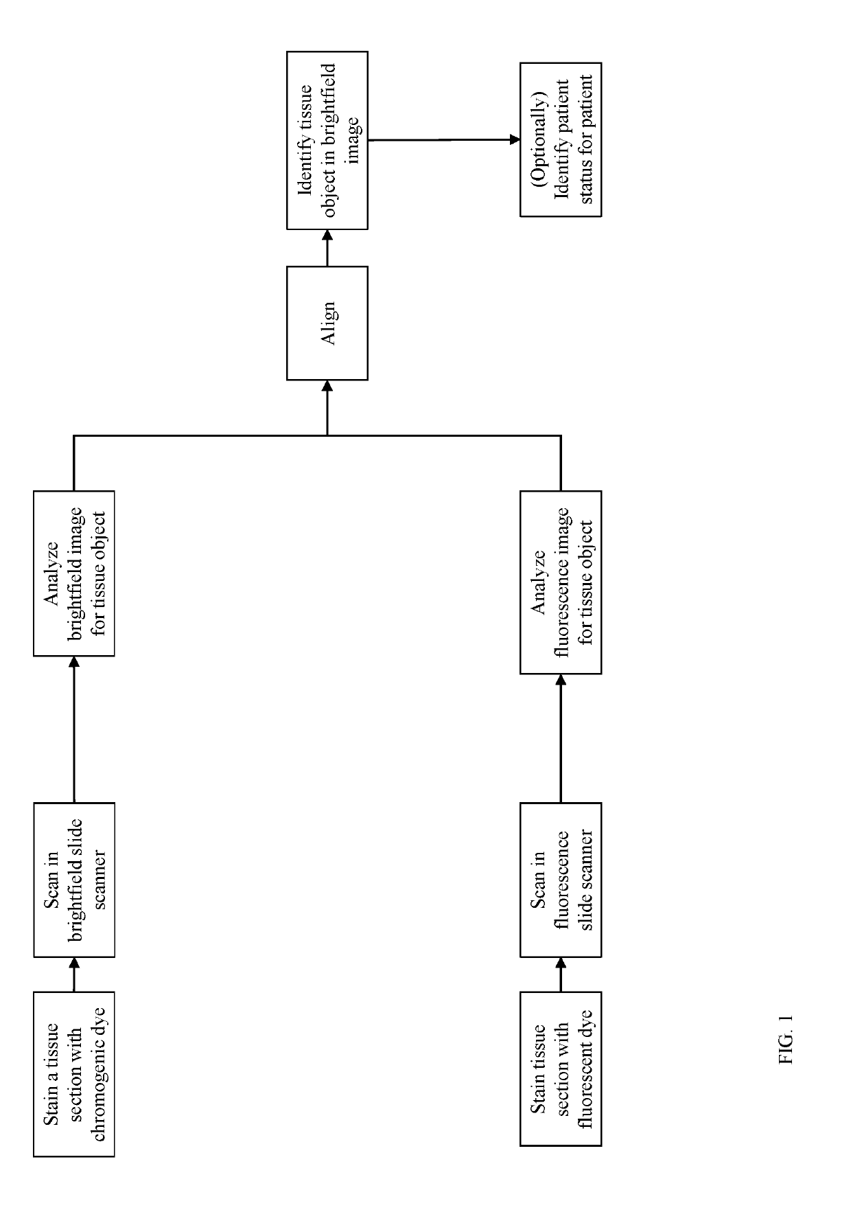
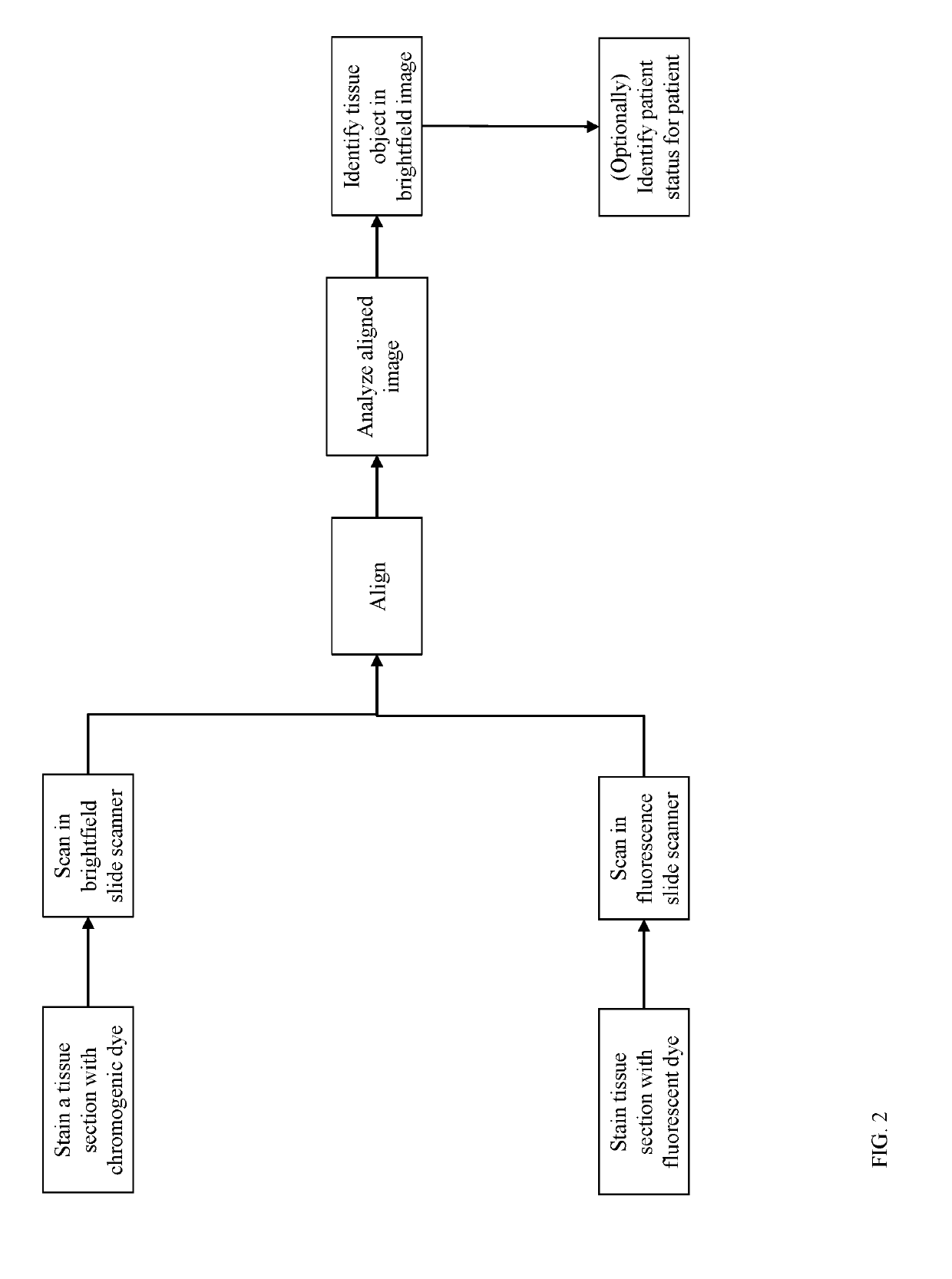
[0016]For purpose of definition, a tissue object is one or more of a cell (e.g., immune cell), cell sub-compartment (e.g., nucleus, cytoplasm, membrane, organelle), cell neighborhood, tissue compartment (e.g., tumor, tumor microenvironment (TME), stroma, lymphoid follicle, healthy tissue), blood vessel, and lymphatic vessel. Tissue objects are visualized by histologic stains which highlight the presence and localization of the tissue object. Tissue objects can be identified directly by stains specifically applied to highlight that tissue object (e.g., hematoxylin to...


PUM

PropertyMeasurementUnit
fluorescence digital imageaaaaaaaaaa
fluorescenceaaaaaaaaaa
fluorescent stainingaaaaaaaaaa

Abstract

The present invention concerns detection of specific tissue objects within thin sections of tissue samples as imaged in a brightfield microscope, such as a predetermined type of immune cells, without using a chromogenic stain that is specific to those tissue objects. The invention uses fluorescent stain and fluorescence imaging to detect these tissue objects. By combining a brightfield and a fluorescence image of the same tissue sample, it is possible to automatically identify objects in the brightfield image that have been stained for in the fluorescence image. The fluorescence stain does not affect the appearance of the tissue sample under the brightfield microscope. Therefore, the invention is ideal to automate the collection of training data for machine learning systems that are to be trained to detect these specific tissue objects in brightfield images of tissue that has not been stained to specifically highlight these tissue objects.

Description

CROSS-REFERENCE TO RELATED APPLICATIONS[0001]This application is a continuation-in-part (CIP) of U.S. Ser. No. 15 / 396,552, filed Dec. 31, 2016, and titled “METHODS FOR DETECTING AND QUANTIFYING MULTIPLE STAINS ON TISSUE SECTIONS”;[0002]the contents of which are hereby incorporated by reference.BACKGROUNDField of the Invention[0003]The present invention relates generally to image analysis methods for the assessment of tissue samples. More specifically, the present invention relates to image analysis methods for the evaluation of tissue objects within a tissue sample without directly staining for those tissue objects.[0004]Tissue samples are generally preserved, embedded in a block of paraffin, cut into thin sections, and one section placed on a glass slide. This section is then further prepared and stained for viewing. Stains can be either chromogenic or fluorescent, which are visible in a brightfield and a fluorescence microscope, respectively. The staining aids in viewing the tissu...

Claims


Application Information

Patent Timeline
Patent Type & Authority Applications(United States)
IPC IPC(8): G01N33/483G01N1/30G01N33/52G01N21/64
CPCG01N33/4833G01N1/30G01N33/52G01N21/6428G01N2021/6441G01N21/6456G06V20/698
Inventor LUENGO HENDRIKS, CRIS LGIANANI, ROBERTO
Owner FLAGSHIP BIOSCI
Why Eureka
Try Eureka