Cell-permeable imaging sensors and uses thereof
- Summary
- Abstract
- Description
- Claims
- Application Information
AI Technical Summary
Benefits of technology
Problems solved by technology
Method used
Image
Examples
example 1
Sensing Intracellular Calcium Ions Using a Manganese-Based MRI Contrast Agent Building Block for Intracellular MRI Calcium Sensors
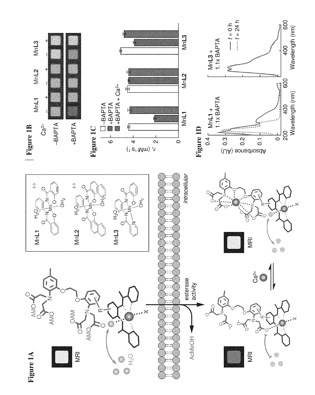
[0068]To examine the potential of Mn-PDA contrast agents to function as building blocks for the sensor design of FIG. 1A, MRI measurements were performed to assess the potential of BAPTA to interact with three paramagnetic chelates (FIG. 1A, inset). Addition of 1.1 equivalents of BAPTA to two of the Mn-PDA complexes, MnL1 and MnL3, produced significant effects on the T1-weighted MRI contrast induced by these compounds in buffer. These BAPTA-dependent contrast changes were reversed by addition of equimolar Ca2+ (FIG. 1B). This was consistent with Mn-PDA moieties competing with Ca2+ for binding to BAPTA molecules. The changes were quantified in terms of T1 relaxivity (r1) values, which were defined as the slope of the T1 relaxation rate (1 / T1=R1) vs. concentration of each paramagnetic complex. Calcium-dependent changes in r1 observed for MnL1 and MnL3 in th...
example 2
Methods and Materials
[0078]Most MRI data were acquired on a 7 T Bruker Biospec system using a T1-weighted 2D gradient echo sequence (echo time, TE=5 ms, repetition time, TR=100 ms; flip angle, FA=65 degrees (°)). Longitudinal relaxivity (r1) measurements were acquired using a 2D spin echo sequence (TE=11 ms, TR=125, 200, 400, 800, 1500, 3000, and 5000 ms), with in-plane resolution of 200×200 microns squared ((μm)2) and 2 mm slice thickness. R1 maps and values were generated using an MRI Analysis Calculator Plugin for ImageJ (National Institutes of Health, Bethesda, Md.) or Matlab scripts (Mathworks, Natick, Mass.). Stimulus-dependent R1 changes in cells were calculated as ΔR1 / R1=[R1(incubated cells)−R1(naive cells)] / R1(naive cells). Statistical comparisons between paired conditions were performed using Student's t-test, and all error bars denote the standard error of the mean from multiple measurements, unless otherwise noted.
In Vitro Characterization of Ma...
example 3
ManICS1-AM Labels Cells and Enables Intracellular Calcium-Sensitive MRI
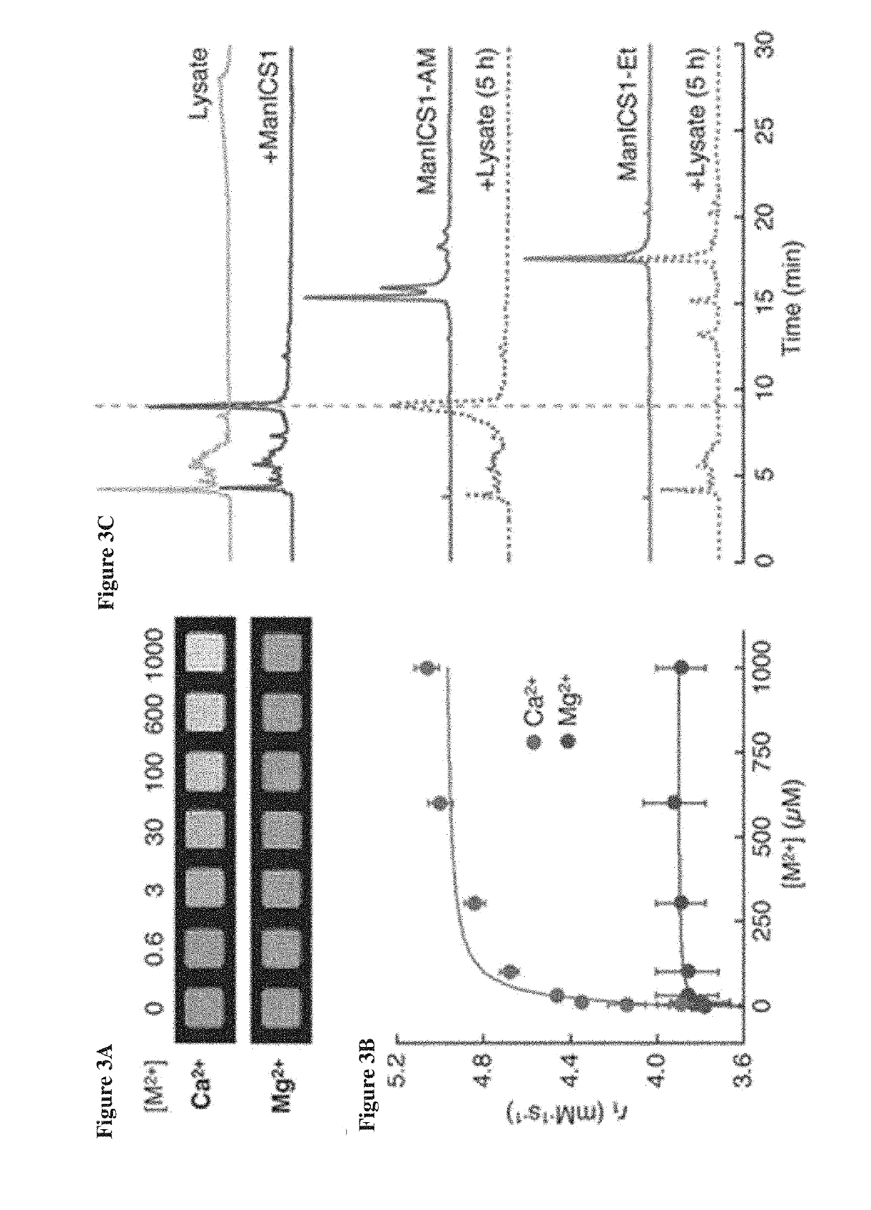
[0083]To test the ability of ManICS1-AM to enable readouts of intracellular calcium by T1-weighted MRI, its propensity was first examined to accumulate within cells. Cultured HEK293 cells were incubated with 10 μM ManICS1-AM or ManICS1 for 30 minutes, followed by washing and MRI analysis (FIG. 7A). It was found that cells incubated with the cell permeable ManICS1-AM underwent a substantial increase in R1 that persisted above basal levels for up to 24 h, while cells labeled with ManICS1 experienced a somewhat lesser increase in R1 that returned to baseline within 5 h. To assess the subcellular localization of manganese following ManICS1-AM or ManICS1 labeling, cytosolic fractions were isolated from cell lysates and the samples were analyzed by inductively coupled plasma mass spectrometry (ICP-MS). The ICP-MS results indicated that only ManICS1-AM labeling produced cytosolic elevations in manganese content (FIG. 7A...
PUM
 Abstract
Description
Claims
Application Information
 - R&D
- Intellectual Property
- Life Sciences
- Materials
- Tech Scout
- Unparalleled Data Quality
- Higher Quality Content
- 60% Fewer Hallucinations
Browse by: Latest US Patents, China's latest patents, Technical Efficacy Thesaurus, Application Domain, Technology Topic, Popular Technical Reports.
© 2025 PatSnap. All rights reserved.Legal|Privacy policy|Modern Slavery Act Transparency Statement|Sitemap|About US| Contact US: help@patsnap.com


