Dectection of exosomes and exosomal biomarkers for the diagnosis and prognosis of diseases and disorders

Inactive Publication Date: 2019-07-18
NANOSOMIX
View PDF0 Cites 10 Cited by
  • Summary
  • Abstract
  • Description
  • Claims
  • Application Information

AI Technical Summary

Benefits of technology

The present invention provides methods, compositions, and kits for detecting and quantitating vesicles (e.g., exosomes) and their biomarkers. The methods involve capturing vesicles on a solid support and then lysing or permeabilizing them while maintaining contact with the solid support. The vesicles are then isolated and the biomarkers are detected using various techniques such as immunoassay, Western blot, Northern blot, chromatography, mass-spectrometry, sequencing, and reaction with nucleic acid dye. The invention can be used for detecting biomarkers in various biological samples such as whole blood, serum, plasma, urine, interstitial fluid, and saliva.

Problems solved by technology

However, even though exosomes are recognized as valuable resources for diagnostics, current analytical methods of analyzing exosomes are too complicated and expensive for routine use in diagnostic testing.
Doctors use brain imaging, evaluation of behavior, psychiatric tests, and other means to diagnose the disease in the patients suspected of having Alzheimer's disease, but none are highly accurate, and many are costly or not practical.

Method used


Image

  • Dectection of exosomes and exosomal biomarkers for the diagnosis and prognosis of diseases and disorders
  • Dectection of exosomes and exosomal biomarkers for the diagnosis and prognosis of diseases and disorders
  • Dectection of exosomes and exosomal biomarkers for the diagnosis and prognosis of diseases and disorders

Examples


example 1

of Inner Membrane-Bound Biomarkers from Exosomes

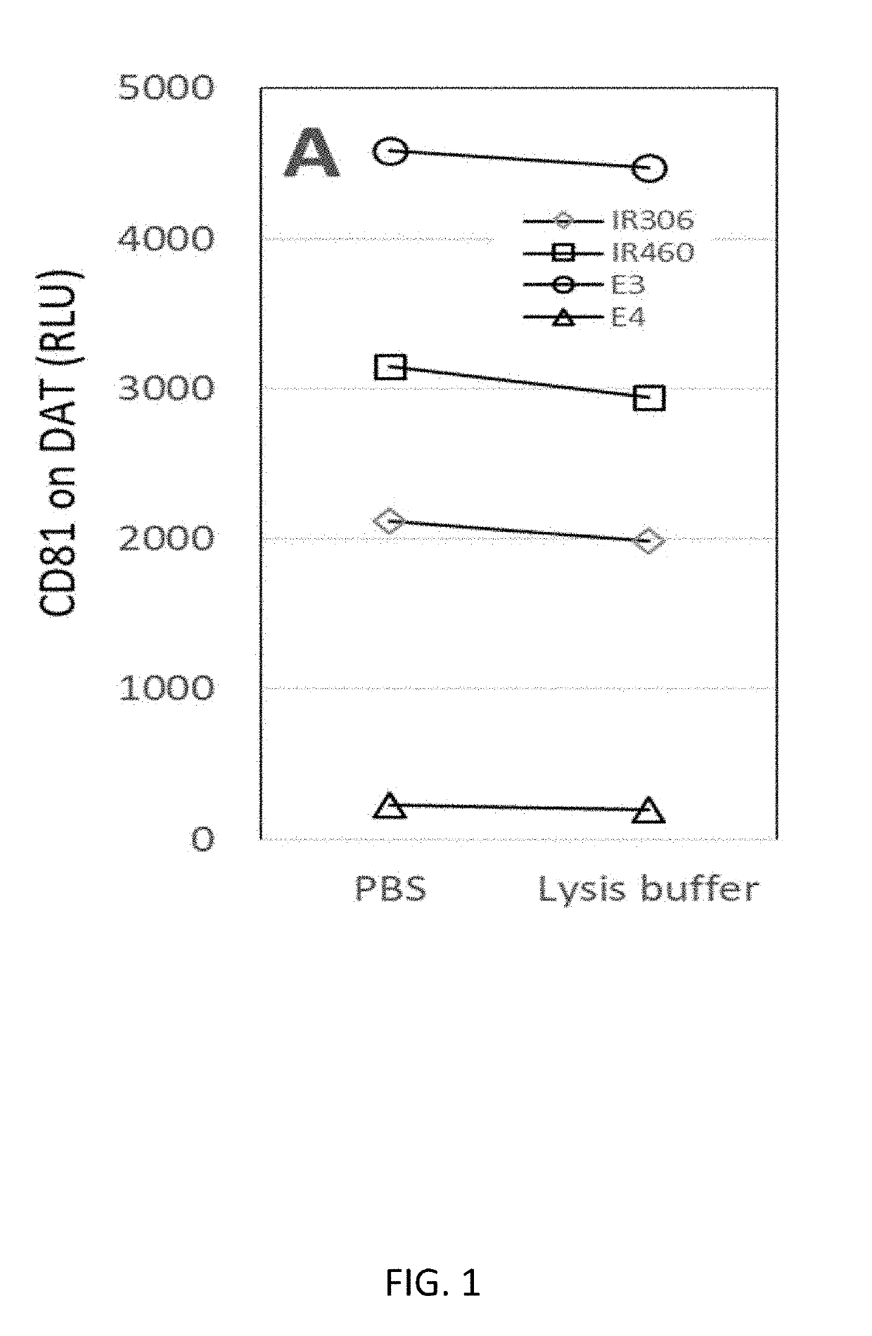
[0118]Inner membrane-bound biomarkers from exosomes were detected as follows. To capture exosomes, four plasma samples were added to duplicate wells on an anti-DAT-immobilized (dopamine transporter) ELISA plate. Prior to reacting with biotinylated anti-CD81 probes, one well for each sample was exposed to a lysis buffer and the other well for the sample was suspended in PBS buffer. Incubation of all wells was continued for 1 hour. Next, biotinylated anti-CD81 antibodies was added to all wells to react with exosomes followed by streptavidin-horseradish peroxidase reaction for chemiluminescent ELISA.

[0119]As shown in FIG. 1, CD81 signal was detected in wells treated with lysis buffer, demonstrating that exosomes were maintained on the ELISA plate even lysis buffer was applied.

[0120]In another series of experiments, plasma samples were added to wells on an anti-SNAP25-immobilized ELISA plate to capture exosomes. After exosomes were capture...

example 2

of Cytosolic Biomarkers from Exosomes

[0124]Cytosolic biomarkers were detected from exosomes as follows. Plasma samples were applied to anti-SNAP25-immobilized ELISA plates to capture exosomes. After exosomes were captured on the anti-SNAP25-immobilized ELISA plates, each well was washed with elution solutions with varying pH levels (from pH 7 to pH 1.8), followed by reaction with labeled anti-CD81 antibody. Results were expressed as % Control with the values at pH 7 as 100% and no plasma control as 0%. As shown in FIG. 4, ELISA signals were decreased by lowering pH, but even at pH1.8, approximately 40% of signals remained, suggesting the difficulty of eluting exosomes from the solid support.

[0125]In another series of experiments, plasma samples were incubated with various volumes of magnetic beads with or without anti-SNAP25 antibody immobilization. Next, supernatants were applied to anti-SNAP25-immobilized ELISA plates followed by the reaction with labeled anti-CD81 antibody. As sh...

example 3

of miRNA from Exosomes

[0129]miRNA was detected from exosomes as follows. Plasma samples were incubated with anti-SNAP25-antibody magnetic beads and control antibody-free magnetic beads. After washing extensively to remove any non-specifically bound materials, magnetic beads were exposed to Trizol. The resultant lysates were used for RNA gel electrophoresis. miRNA was detected in plasma samples applied to anti-SNAP25-bound magnetic beads, but not from control antibody-free magnetic beads. These results showed that the methods of the present invention are useful for detecting miRNA from exosomes. These results further showed that methods of the present invention are useful for selectively capturing exosomes on a solid support, lysing or permeabilizing the exosomes while maintaining contact between the exosome membrane and the solid support, and detecting miRNA from the captured exosomes.


PUM


Abstract

The present invention relates to methods, compositions, and kits for detecting and quantitating exosomes and exosomal biomarkers and the use of exosomes and exosomal biomarkers in diagnostic and prognostic methods for various diseases and disorders. Disease and disorders of the present invention include neurological disorders, immunological disorders, placental diseases, cancer, hematological disorders, kidney disease, gastrointestinal diseases, liver diseases, and musculoskeletal diseases

Description

RELATED APPLICATIONS[0001]This application claims priority to U.S. Provisional Patent Application Ser. No. 62 / 619,080, filed on Jan. 18, 2018, which is hereby incorporated by reference herein in its entirety.FIELD OF THE INVENTION[0002]The present invention relates to methods, compositions, and kits for detecting and quantitating exosomes and exosomal biomarkers and the use of exosomes and exosomal biomarkers in diagnostic and prognostic methods for various diseases and disorders. Disease and disorders of the present invention include neurological disorders, immunological disorders, placental diseases, cancer, hematological disorders, kidney disease, gastrointestinal diseases, liver diseases, and musculoskeletal diseases.BACKGROUND OF THE INVENTION[0003]Exosomes in biological fluids are potentially useful diagnostically for various diseases, because exosomes carry physiological and pathological materials (proteins, metabolites, RNAs, small molecules, etc.) of the mother cells from w...

Claims


Application Information

Patent Timeline
IPC IPC(8): G01N33/574G01N33/543G01N33/68G01N33/552G01N33/553
CPCG01N33/574G01N33/54326G01N33/6896G01N33/54306G01N33/5432G01N33/54346G01N33/552G01N33/553G01N33/543G01N33/57484
Inventor MITSUHASHI, MASATO
Owner NANOSOMIX
Why Eureka
Try Eureka