Glaucoma diagnosis method using fundus image and apparatus for the same

Active Publication Date: 2019-07-18
ELECTRONICS & TELECOMM RES INST +1
View PDF0 Cites 8 Cited by
  • Summary
  • Abstract
  • Description
  • Claims
  • Application Information

AI Technical Summary

Benefits of technology

[0013]Accordingly, the present invention has been made keeping in mind the above problems occurring in the prior art, and an object of the present invention is to provide a glaucoma diagnosis

Problems solved by technology

Glaucoma is a disease by which a retinal ganglion cell (RGC) and an axon thereof are damaged, thus causing a patient to become blind.
However, there are many cases where subjective symptoms that can be clearly felt by patients, such as visual field defects or the deterioration of eyesight, do not appear until the last stage of glaucoma due to the characteristics of the disease.
Further, when glaucoma in later stages is serious, the prognosis thereof is bad, and thus the importance of early detection through examination is very high.
Therefore, an RNFL defect in an optic disc area, which meaningfully appears in the results of OCT measurement, makes it difficult to diagnose early glaucoma.
The visual field test illustrated in FIG. 2 is a method for measuring whether a patient has a visual field micro-defect, and it is widely used to definitively diagnose glaucoma, but it is disadvantageous in that it takes a lot of time and effort in measurement and in that consistency in measurement of early glaucoma is deteriorated.
Also, similar to OCT, ability to diagnose preperimetric glaucoma is limited.
The visual field test is disadvantageous in that it generally requires a test time of 30 minutes or longer and in that both the patient and the medical team that conducts the test must make a lot of effort during the test process.
Further, since equipment for the visual field test is more expensive than a fundus scope, medical accessibility is not good from the standpoint of

Method used


Image

  • Glaucoma diagnosis method using fundus image and apparatus for the same
  • Glaucoma diagnosis method using fundus image and apparatus for the same
  • Glaucoma diagnosis method using fundus image and apparatus for the same

Examples


Example

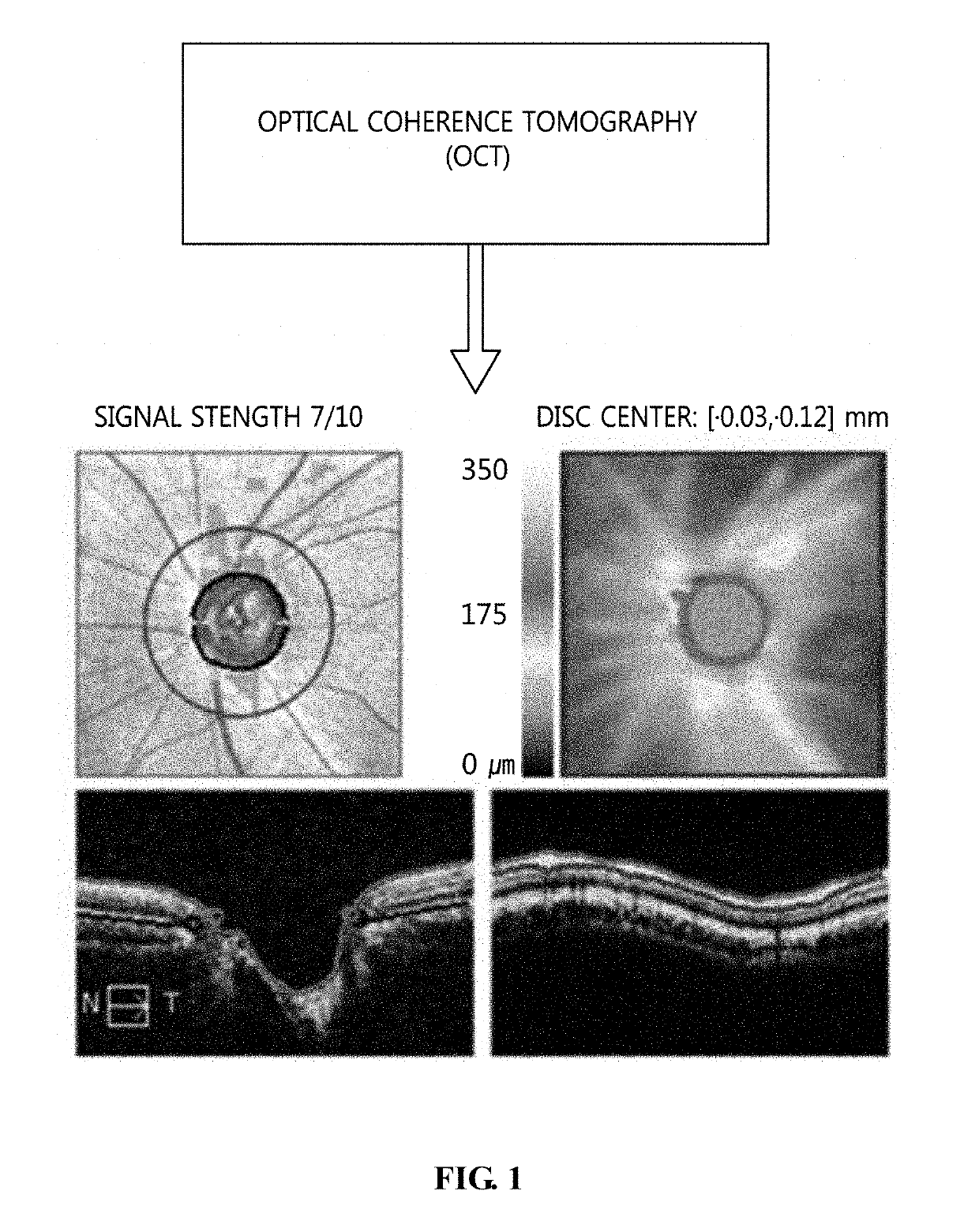
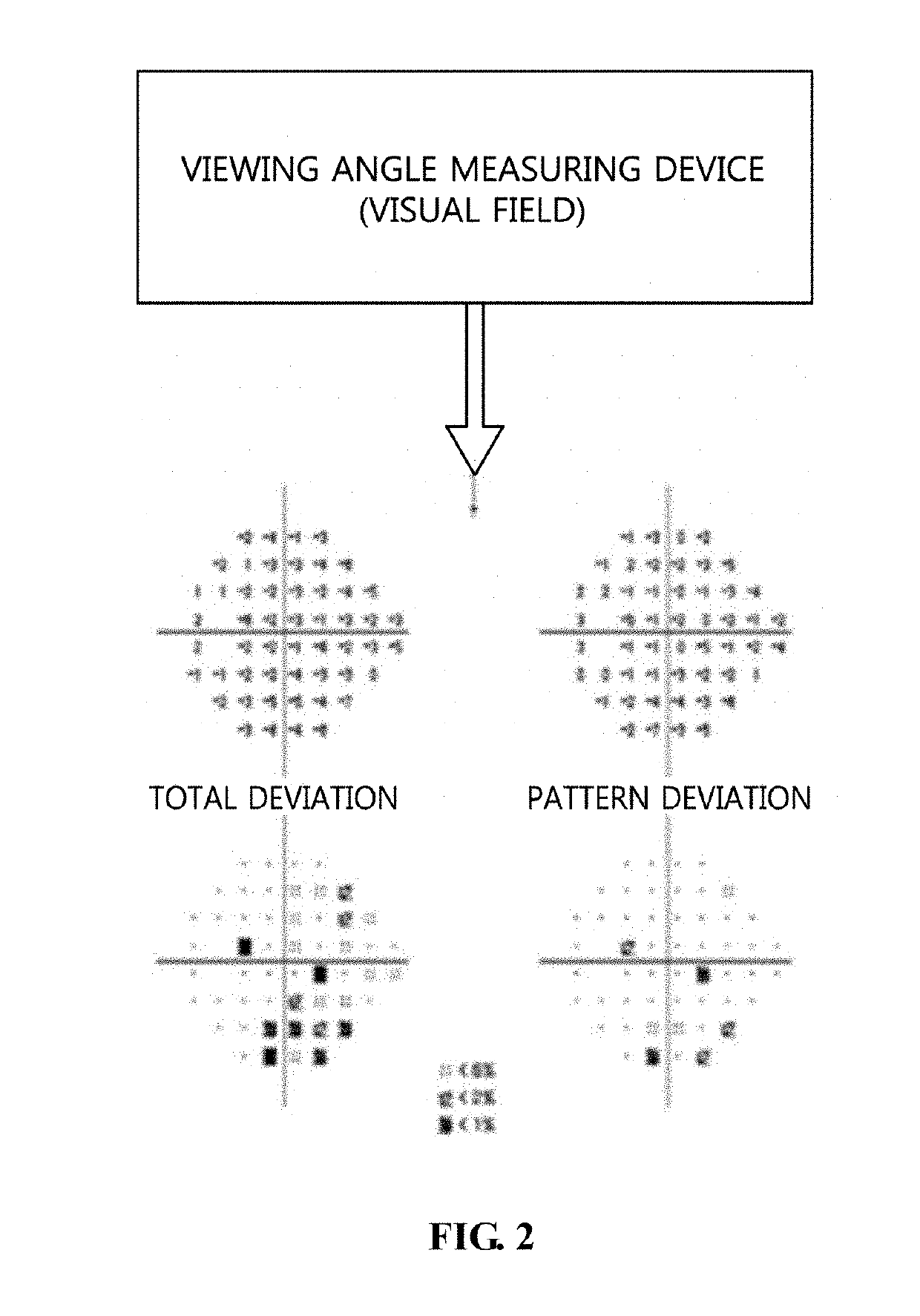
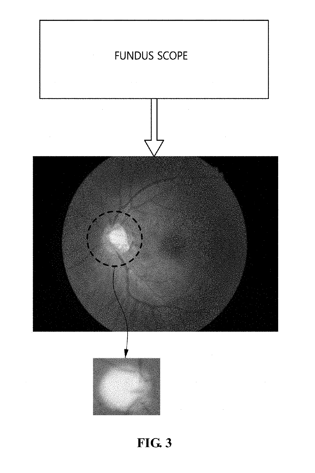
[0053]The present invention will be described in detail below with reference to the accompanying drawings. Repeated descriptions and descriptions of known functions and configurations which have been deemed to make the gist of the present invention unnecessarily obscure will be omitted below. The embodiments of the present invention are intended to fully describe the present invention to a person having ordinary knowledge in the art to which the present invention pertains. Accordingly, the shapes, sizes, etc. of components in the drawings may be exaggerated to make the description clearer.

[0054]Hereinafter, preferred embodiments of the present invention will be described in detail with reference to the attached drawings.

[0055]FIG. 4 is a diagram illustrating a glaucoma diagnosis system according to an embodiment of the present invention.

[0056]Referring to FIG. 4, the glaucoma diagnosis system according to the embodiment of the present invention includes a fundus scope 400, a glaucom...


PUM


Abstract

Disclosed herein are a glaucoma diagnosis method using a fundus image and an apparatus for the same. The glaucoma diagnosis method includes performing data amplification of generating multiple transformed images for an original fundus image based on a preprocessed image of the original fundus image, allowing multiple individual learning models of different types to be learned based on the multiple transformed images and generating a glaucoma determination model based on respective outputs of the learned multiple individual learning models, and diagnosing a class of glaucoma for the original fundus image based on the glaucoma determination model.

Description

CROSS REFERENCE TO RELATED APPLICATIONS[0001]This application claims the benefit of Korean Patent Application Nos. 10-2018-0005519, filed Jan. 16, 2018 and 10-2018-0134333, filed Nov. 5, 2018, which are hereby incorporated by reference in their entirety into this application.BACKGROUND OF THE INVENTION1. Technical Field[0002]The present invention relates generally to glaucoma diagnosis technology, and more particularly, to technology for diagnosing a glaucoma class for a fundus image based on a glaucoma determination model.2. Description of the Related Art[0003]Glaucoma is one of three ophthalmologic diseases causing blindness, together with cataracts and macular degeneration. According to epidemiological investigation of glaucoma conducted in 2014, about 3.54% (64,300,000) of adults worldwide aged 40 or older were glaucoma patients in 2013, and it is estimated that the number of glaucoma patients will increase to 7.6 million in 2020 (Tham et al., 2014). Also, an investigation indic...

Claims


Application Information

Patent Timeline
IPC IPC(8): G06T7/00G06T7/13G06T7/70G06T7/60G06K9/62G06T7/136A61B3/14G06V10/772
CPCG06T7/0012G06T7/13G06T7/70G06T7/60G06K9/6267G06T7/136A61B3/145G06T2207/30041G06T2207/20081G06T3/60G06T2207/20084G06K2209/05A61B3/102A61B3/12G06T7/66G06T2207/10024G06V10/507G06V2201/03G06V10/772G06F18/24
Inventor CHO, HYEON-SUNGJEONG, JAE-HOONKIM, JONG-YEUPPARK, JI-SANG
Owner ELECTRONICS & TELECOMM RES INST
Why Eureka
Try Eureka