
Bioprosthetic heart valve

Active Publication Date: 2019-10-03
UCL BUSINESS PLC
View PDF0 Cites 1 Cited by
  • Summary
  • Abstract
  • Description
  • Claims
  • Application Information

AI Technical Summary

Benefits of technology

The present invention relates to a new design for a heart valve that reduces the risk of regurgitation (leakage) and increases the orifice area of the valve. The design has fewer layers of biocompatible material and biological tissue located at the inner surface of the stent, which reduces the potential risk of regurgitation through a single layer of fabric covering the inner cuff end of the valve. The manufacturing process is simplified and cost-effective, with fewer potential points of failure on the valve. Additionally, a protective patch may be applied along the suture line to further reinforce the valve.

Problems solved by technology

Valvular disease, due to congenital defects or old age can lead to issues such as stenosis or regurgitation, placing a burden on the heart which can ultimately lead to heart failure.
However, such valves have an intricate layered structure with several layers of biocompatible textile and biological tissue sutured to each other.
Intricate suturing of multiple layers leads to an increase in potential failure points on the implant as well as an increase in manufacturing time and cost.
Furthermore, the need for multiple fabric layers and biological tissue layers leads to a decreased inner diameter, resulting in an increase in the transvalvular pressure drop.

Method used


Image

  • Bioprosthetic heart valve
  • Bioprosthetic heart valve
  • Bioprosthetic heart valve

Examples


Embodiment Construction

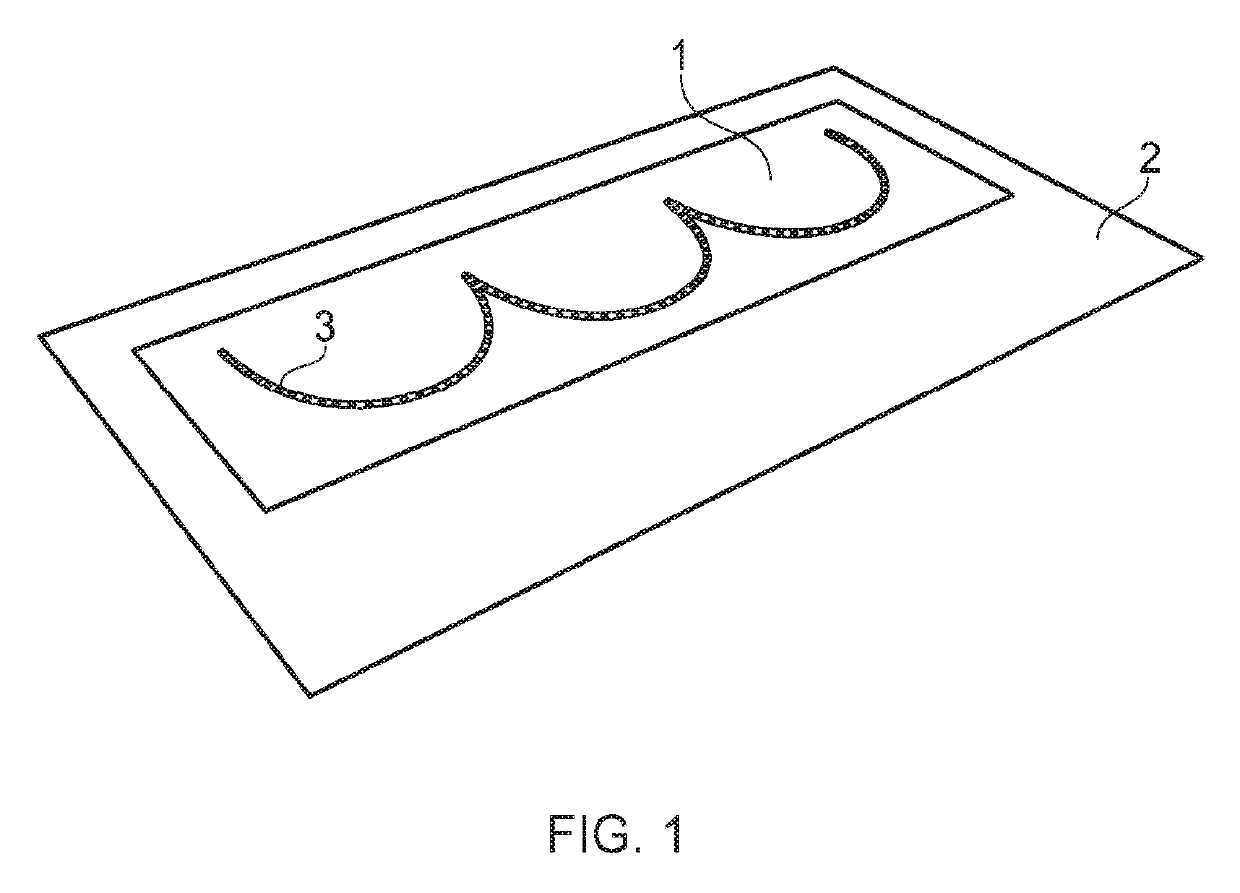
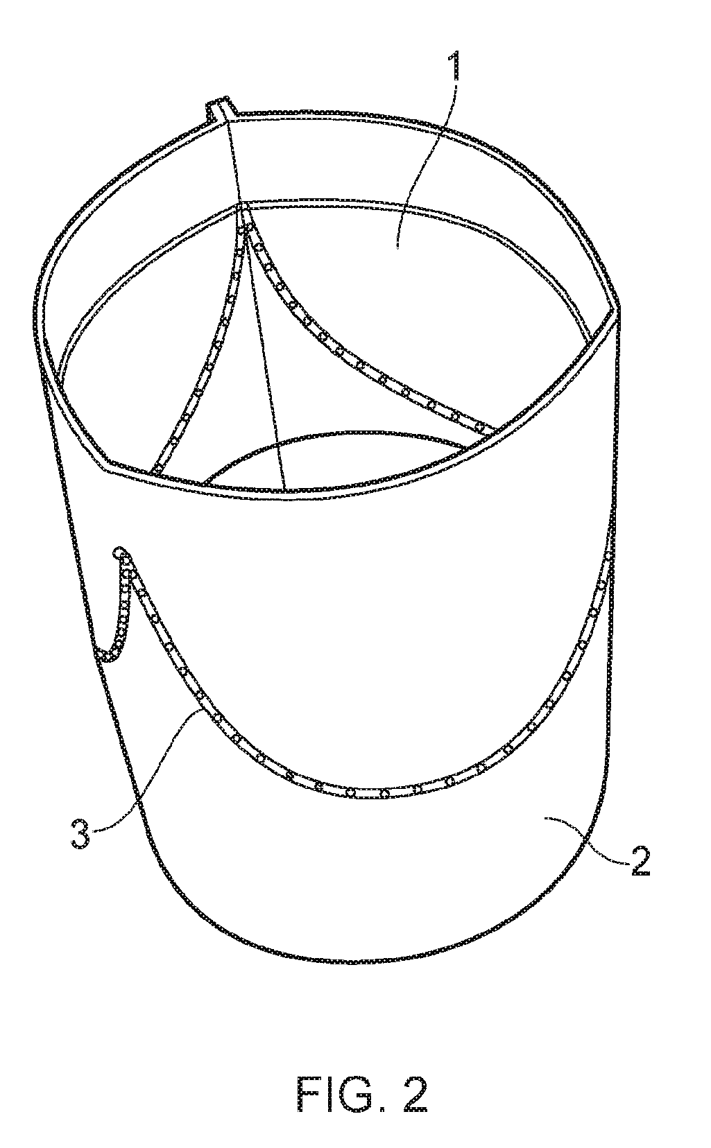
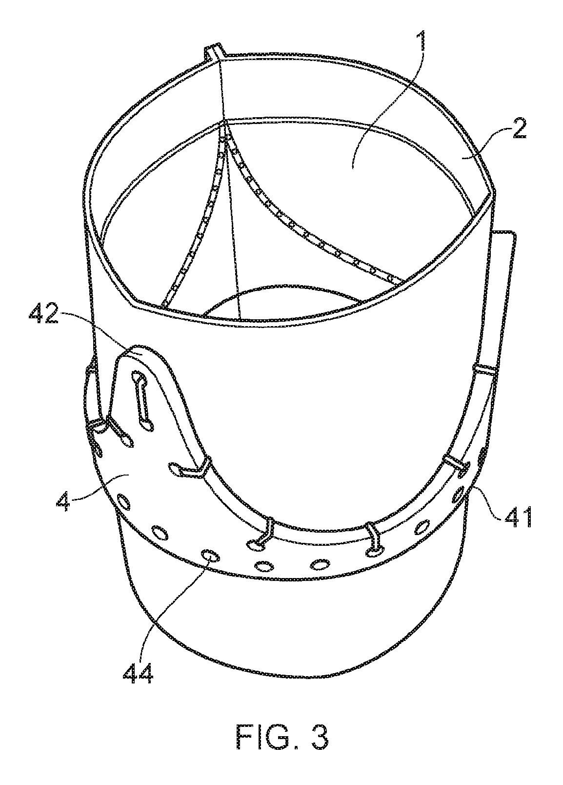
[0059]A first embodiment of a fabrication method according to the present invention is described below with reference to FIGS. 1 to 5.

[0060]In an initial step, a sheet of biological tissue 1 and a sheet of a biocompatible material 2 are provided. In the embodiment shown, each of the sheets has a rectangular shape. However, it is envisaged that alternative shapes could be provided, such as those shown in FIGS. 6a-6d.

[0061]The biological tissue typically takes the form of a sheet of human or animal pericardium, typically decellularised. Suitable types of animal pericardium include, but are not restricted to, porcine or bovine pericardium. In some embodiments, the biological tissue is tissue-engineered; that is to say it forms a scaffold material seeded with stem cells. This scaffold material may be formed from decellularised animal or human tissue, or from synthetic biomaterials. Such synthetic biomaterials may be bioresorbable or biostable.

[0062]The biocompatible material may take t...


PUM


Abstract

Bioprosthetic heart valves and methods for fabricating bioprosthetic heart valves are provided. Biological tissue is attached to biocompatible material. The biocompatible material is folded to form a tubular structure with the attached biological tissue located on the inner surface of the tubular structure, the biological tissue forming leaflets of the heart valve. A stent is secured around the outer surface of the tubular structure. A region of the biocompatible material is cut. The biocompatible material is folded at the cut region away from the tissue and around the downstream edge of the stent and is secured to the stent's outer surface. The biocompatible material is folded at the upstream end of the tubular structure around the upstream edge of the stent and attached to the stent's outer surface so that the stent's inner and outer surfaces are covered with no more than a single layer of the biocompatible material.

Description

FIELD OF THE INVENTION[0001]The present invention relates to heart valves, particularly to a bioprosthetic heart valve and to a method of fabrication for a heart valve with leaflets made from biological tissue.BACKGROUND OF THE INVENTION[0002]The four valves of the heart (tricuspid valve, pulmonic valve, mitral valve and aortic valve) are essential to maintain a unidirectional flow of blood around the chambers of the heart. Valvular disease, due to congenital defects or old age can lead to issues such as stenosis or regurgitation, placing a burden on the heart which can ultimately lead to heart failure.[0003]The concept of implanting replacement valves is well known with implantable heart valves being available since the 1950s. Nowadays there are a great number of implantable heart valves available.[0004]Important factors when choosing a heart valve to be implanted in a patient include: the ease of implantation; reliability / risk of failure; maximising resultant blood flow through th...

Claims


Application Information

Patent Timeline
IPC IPC(8): A61F2/24
CPCA61F2/2409A61F2/2418A61F2/2415A61L27/3604A61F2220/0075A61F2/2412B65B11/10A61F2250/0069A61F2240/001
Inventor BURRIESCI, GAETANORAHMANI, BENYAMINBYRNE, GUERARDMCGREGOR, CHRISTOPHER
Owner UCL BUSINESS PLC
Learn More