Systems and Methods for Infrared-Enhanced Ultrasound Visualization

a technology of infrared and ultrasound, applied in the field of systems and methods for infrared-enhanced ultrasound visualization, can solve the problems of multiple needles or catheters, difficulty in visualizing the needle crossing into the vessel, and difficulty in staying in the vessel under ultrasound guidan

Pending Publication Date: 2020-02-27
BARD ACCESS SYST
View PDF10 Cites 1 Cited by
  • Summary
  • Abstract
  • Description
  • Claims
  • Application Information

AI Technical Summary

Benefits of technology

[0006]Also disclosed herein is a method of accessing a vasculature of a patient, the method comprising, providing an ultrasound probe including a light detector disposed at a distal end thereof and a medical device for accessing the vasculature of the patient, placing an ultrasound probe adjacent a skin surface of the patient, the ultrasound probe including a light detector to detect a vessel, inserting a medical device through the skin surface of the patient, the medical device including an emitter at a distal tip thereof, illuminating subcutaneous tissue above the vessel with infrared light of the emitter to indicate a location of the vessel, advancing the tip of the medical device into the vessel and illuminating an inner portion of the vessel to indicate the distal tip of the medical device being in the vessel, and providing an enhanced ultrasound image, including light-based images, to indicate to a user a position of the tip of the medical device relative to the vessel.

Problems solved by technology

Accessing the patients vasculature is a common technique, but also one that continues to be problematic and require the use and loss of multiple needles or catheters in failed cannulation attempts.
However, visualizing the needle crossing into the vessel and staying in the vessel can still be difficult under ultrasound guidance.

Method used


Image

  • Systems and Methods for Infrared-Enhanced Ultrasound Visualization
  • Systems and Methods for Infrared-Enhanced Ultrasound Visualization
  • Systems and Methods for Infrared-Enhanced Ultrasound Visualization

Examples


Embodiment Construction

[0019]Reference will now be made to figures wherein like structures will be provided with like reference designations. It is understood that the drawings are diagrammatic and schematic representations of exemplary embodiments of the present invention, and are neither limiting nor necessarily drawn to scale.

[0020]For clarity it is to be understood that the word “proximal” refers to a direction relatively closer to a clinician using the device to be described herein, while the word “distal” refers to a direction relatively further from the clinician. For example, the end of a catheter placed within the body of a patient is considered a distal end of the catheter, while the catheter end remaining outside the body is a proximal end of the catheter. Also, the words “including,”“has,” and “having,” as used herein, including the claims, shall have the same meaning as the word “comprising.”

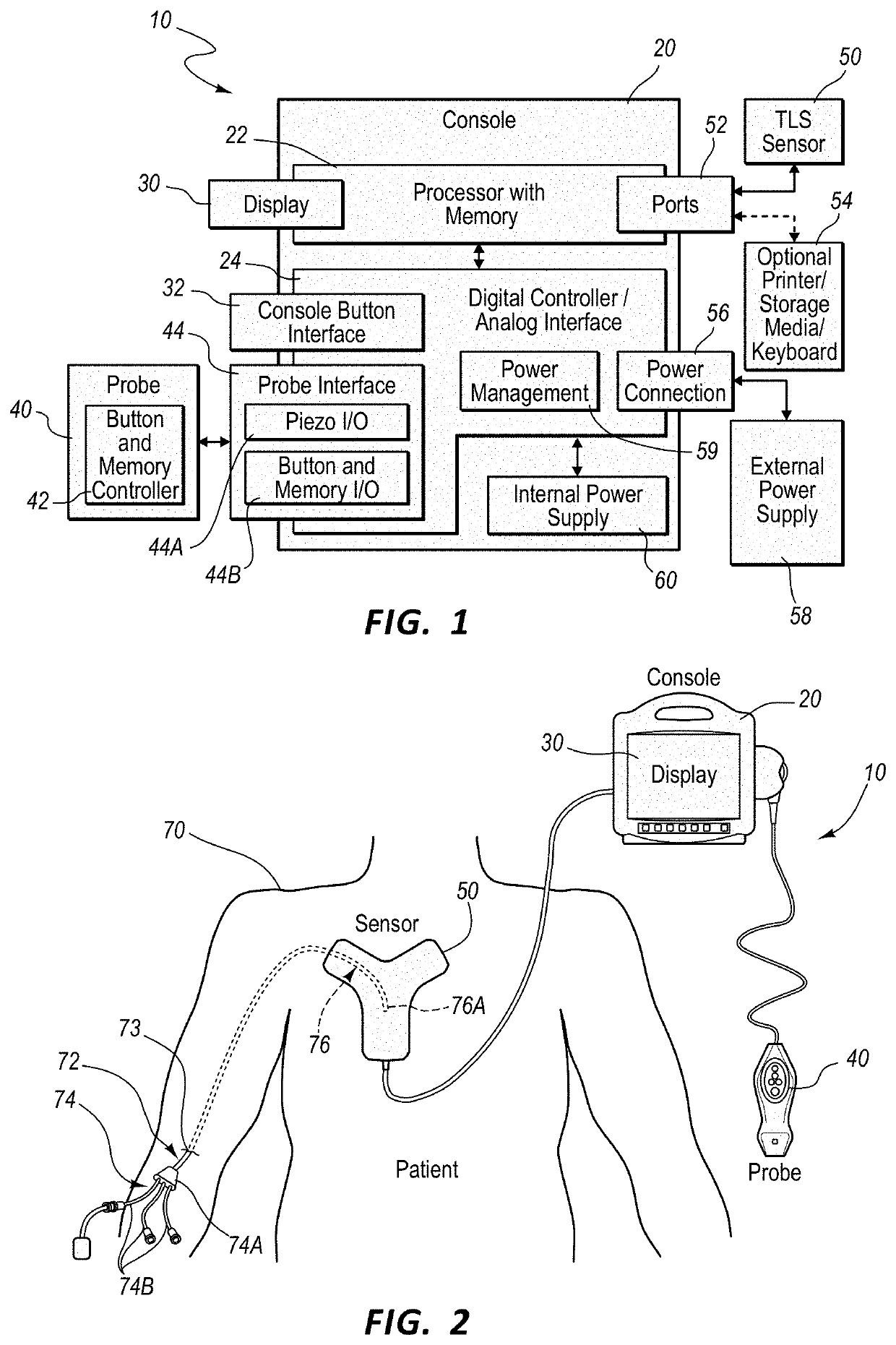
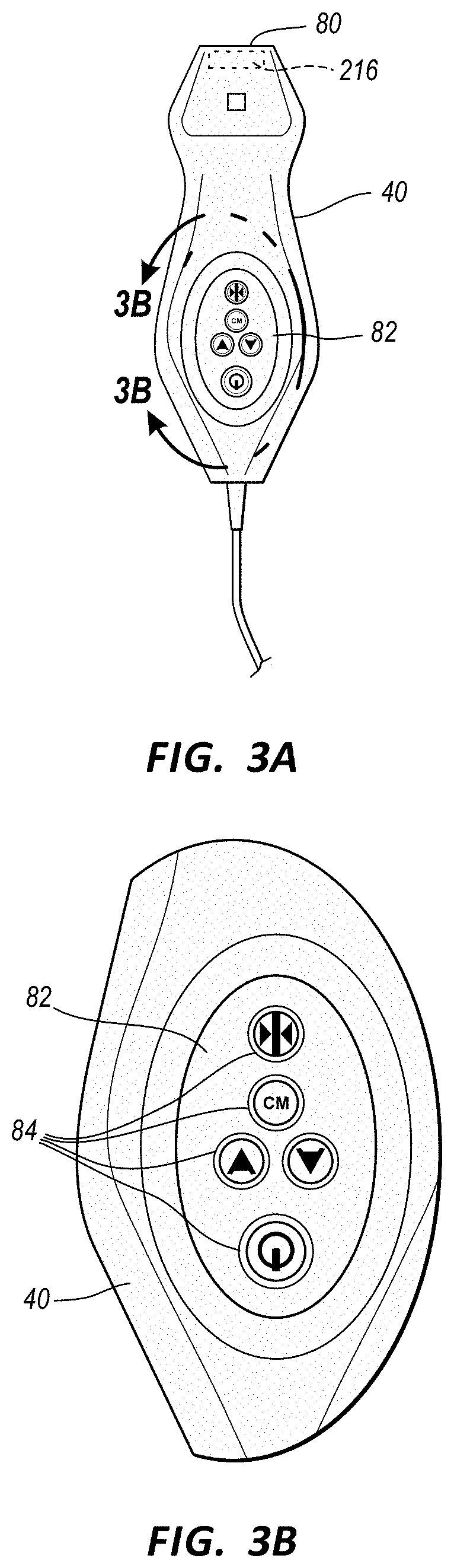
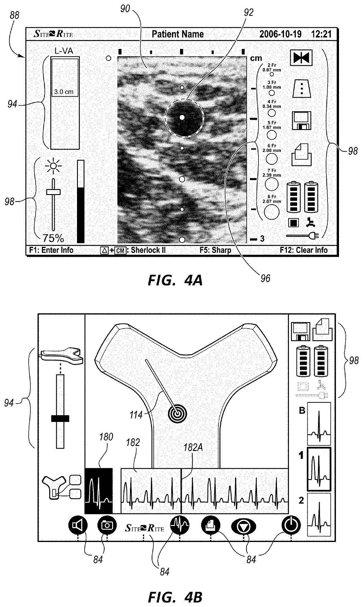
[0021]Embodiments of the present invention are generally directed to systems and methods for providing...


PUM


Abstract

A system and method for providing enhanced ultrasound images for accessing a vasculature of a patient is disclosed. The system includes an ultrasound imaging device including a probe with a light detector disposed at a distal end thereof. A medical device includes an infrared emitter disposed a distal tip thereof. The emitter provides infrared light that illuminates surrounding tissue. Reflected light is detected and interpreted based on the profile of absorbance spectra which can be used to identify different tissue structures. This light-based information is superimposed on an ultrasound image of the vessel to facilitate first-stick access of patient vasculature.

Description

PRIORITY[0001]This application claims the benefit of priority to U.S. Provisional Application No. 62 / 721,358, filed Aug. 22, 2018, titled, “Systems And Methods For Infrared-Enhanced Ultrasound Visualization,” which is incorporated by reference in its entirety into this application.BRIEF SUMMARY[0002]Accessing the patients vasculature is a common technique, but also one that continues to be problematic and require the use and loss of multiple needles or catheters in failed cannulation attempts. Ultrasound has become prevalent as a tool to improve first stick success through visualization of vessel size, depth and location. However, visualizing the needle crossing into the vessel and staying in the vessel can still be difficult under ultrasound guidance.[0003]Briefly summarized, a system and method for providing enhanced ultrasound images for accessing a vasculature of a patient is disclosed. The system includes an ultrasound imaging device including a probe with a light detector disp...

Claims


Application Information

Patent Timeline
Patent Type & Authority Applications(United States)
IPC IPC(8): A61B8/08A61B5/00A61B1/07
CPCA61B5/0035A61B8/0841A61B1/07A61B5/0075A61B8/4416A61B8/4444A61B8/467A61B8/0891A61B8/463A61B5/0086A61B1/000094A61B1/046
Inventor MISENER, ANTHONY K.
Owner BARD ACCESS SYST
Why Eureka
Try Eureka