

Method for detecting target molecule in which rolling circle amplification is used
- Summary
- Abstract
- Description
- Claims
- Application Information
AI Technical Summary
Benefits of technology
Problems solved by technology
Method used
Image

Examples
first embodiment
Detection Method: First Embodiment
[0043]The method of detecting a target molecule according to the first embodiment of the present invention is a method of detecting a target molecule, the method comprising the steps of:
[0044]forming a complex of a target molecule, a capture oligonucleotide, an oligonucleotide primer, and a single-stranded circular DNA;
[0045]performing a nucleic acid amplification reaction by rolling circle amplification based on the formation of the complex; and
[0046]detecting amplified nucleic acid.
[0047]The first embodiment is described below.
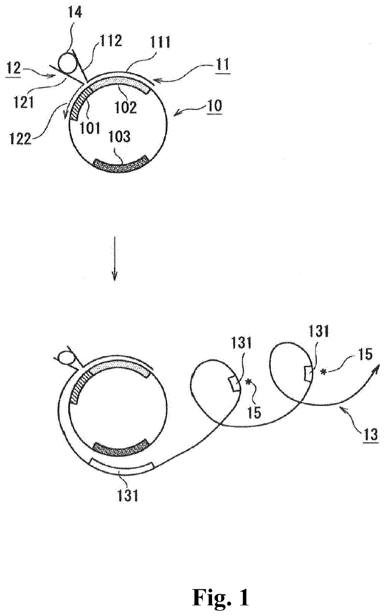
[0048]The single-stranded circular DNA contains: a first region, and a second region linked to the 3′-side of the first region; and preferably further contains a sequence complementary to a detection reagent-binding sequence such as a guanine quadruplex-forming sequence.
[0049]A description is given below showing an example with reference to FIG. 1. The single-stranded circular DNA is illustrated in the 5′→3′ clockwise direct...
second embodiment
Detection Method: Second Embodiment
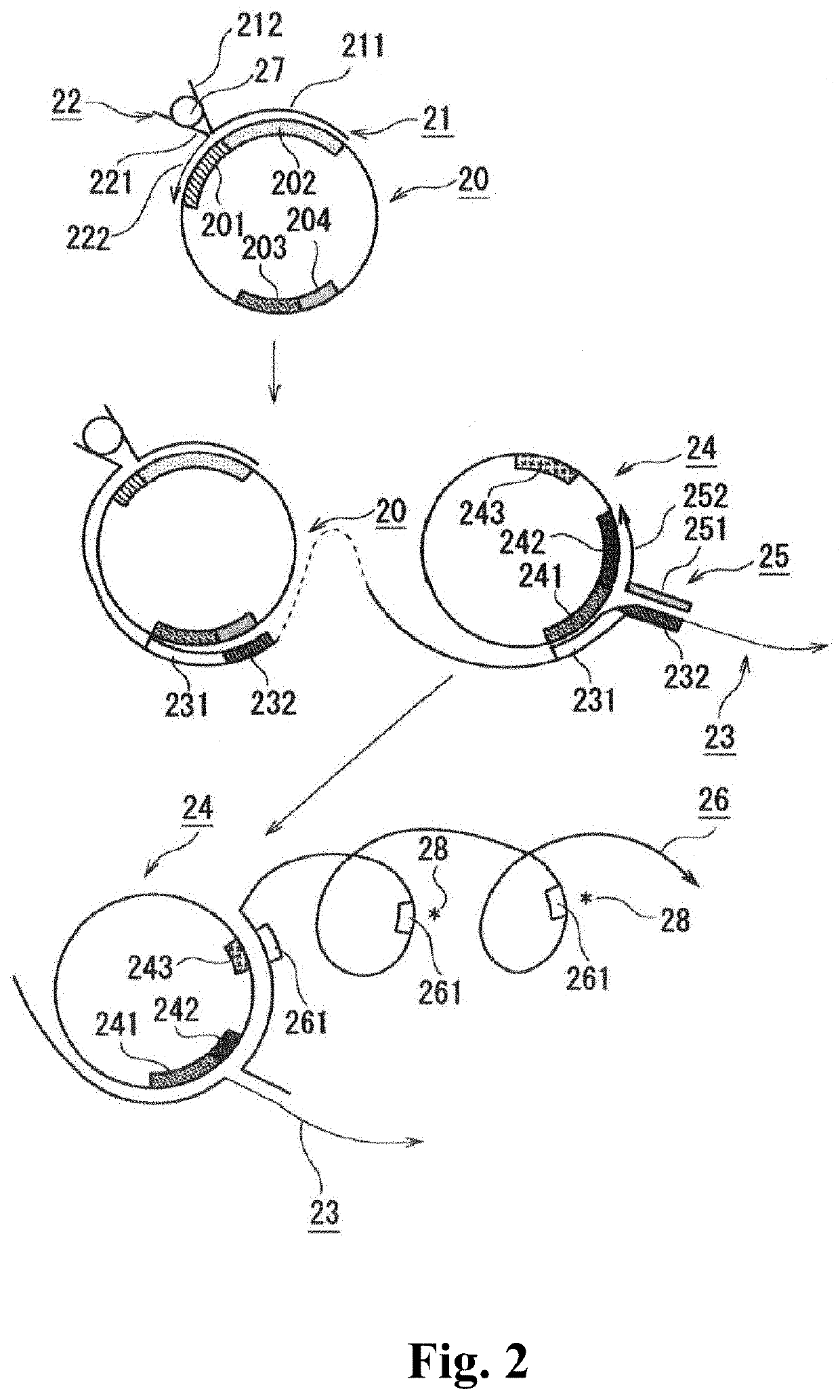
[0069]The detection method according to the second embodiment of the present invention comprises the steps of:
[0070]forming a first complex containing a target molecule, a capture oligonucleotide, a first oligonucleotide primer, and a first single-stranded circular DNA;
[0071]performing a nucleic acid amplification reaction by rolling circle amplification based on the formation of the first complex;
[0072]hybridizing a second single-stranded circular DNA and a second oligonucleotide primer with an elongated chain generated by the nucleic acid amplification reaction, to form a second complex containing the elongated chain, the second oligonucleotide primer, and the second single-stranded circular DNA;
[0073]performing a nucleic acid amplification reaction by rolling circle amplification based on the formation of the second complex; and
[0074]detecting amplified nucleic acid.
[0075]The second embodiment is described below.
[0076]The capture oligonucleotide...
example 1-1
Thrombin Detection Method Using Split Aptamer
Preparation of a1 to a5 of FIG. 4
[0117]A mixture was prepared with 2 μL of 100 nM DNA template [2] (final concentration, 10 nM), 2 μL of 400 nM DNA template [1] (final concentration, 40 nM), 2 μL of 120 nM Positive control Primer (final concentration, 12 nM), 2 μL of 480 nM DNA primer [1] (final concentration, 48 nM), 2 μL of 10×attached buffer, 1 μL of 20×attached BSA solution, 2 μL of 10 mM dNTPs (final concentration, 1 mM), 1 μL of 0 to 1000 mM KCl solution (final concentration, 0 to 50 mM), 2 μL of 1 U / μL Phi29 Polymerase (final concentration, 0.1 U / μL), and 4 μL of water (20 μL in total).
Preparation of a1 to b5 of FIG. 4
[0118]A mixture was prepared with 2 μL of 100 nM DNA template [2] (final concentration, 10 nM), 2 μL of 400 nM DNA template [1] (final concentration, 40 nM), 2 μL of 120 nM Capture Probe [1] (final concentration, 12 nM), 2 μL of 120 nM Detection Probe [1] (final concentration, 12 nM), 2 μL of 480 nM DNA primer [1] (fi...
PUM
| Property | Measurement | Unit |
|---|---|---|
| Molecular weight | aaaaa | aaaaa |
Abstract
Description
Claims
Application Information