Methods of preparing a re-usable single cell and methods for analyzing the epigenome, transcriptome, and genome of a single cell

a single cell and cell genome technology, applied in the field of preparing a reusable single cell and methods for analyzing the epigenome, transcriptome, and genome of a single cell, can solve the problems of many single cells not being re-used, high error rate of methods for amplifying and sequencing a cell genome, and inability to study dna-bound proteins, rna-bound proteins, and dna-bound proteins simultaneously

Active Publication Date: 2020-04-02
UNITED STATES OF AMERICA
View PDF0 Cites 3 Cited by
  • Summary
  • Abstract
  • Description
  • Claims
  • Application Information

AI Technical Summary

Benefits of technology

[0049]FIG. 9C shows reproduced pictures of gels run with products of the same 8 single cells used in FIGS. 9A and 9B produced according to embodiments of the invention. FIG. 9C shows production of DNA products and the DNA size distribution using antibodies for histone modifications, H3K27ac and H3K27me3 with the 8 re-usable single cells, which are used in experiments of FIGS. 9A and 9B. FIG. 9C is a result of agarose gel electrophoresis loaded with final DNA products of Round3 experiment derived from the 8 re-usable single cells produced according to embodiments of the invention. Area (A) of the gels includes products containing a genomic sequence and antibody barcodes. Area (B) of the gels includes byproducts. FIGS. 9A, 9B, and 9C support reusability of the re-usable single cells produced according to embodiments of the invention.

Problems solved by technology

The ability to study DNA, RNA, and DNA-bound proteins simultaneously, and determine the location of proteins with and without modifications in the genome of individual cells may be technically challenging.
Moreover, many single cells cannot be re-used after the cellular components of the single cell have been studied.
In addition, some methods of amplifying and sequencing a cell genome may have high error rates and may introduce mutations during the amplification process.

Method used


Image

  • Methods of preparing a re-usable single cell and methods for analyzing the epigenome, transcriptome, and genome of a single cell
  • Methods of preparing a re-usable single cell and methods for analyzing the epigenome, transcriptome, and genome of a single cell
  • Methods of preparing a re-usable single cell and methods for analyzing the epigenome, transcriptome, and genome of a single cell

Examples


example 1

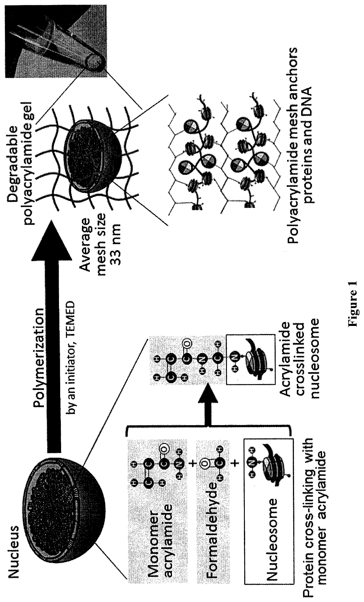
[0124]This example demonstrates a method of preparing a re-usable single cell by anchoring the cellular components inside the single cell with a polyacrylamide nano-scale scaffold.

[0125]A diagram illustrating an embodiment of the inventive method for preparing a polyacrylamide nano-scale scaffold of the invention is provided in FIG. 1. To prepare the polyacrylamide nano-scale scaffold, 1×10⁶ MS1, or K562 cells were dispersed in a phosphate-buffered saline solution comprising 4% formaldehyde (PFA) and either 1%, 4% 5%, 10%, 20% acrylamide, or 28% acrylamide. The dispersed cells were held for one hour on ice to allow the PFA and acrylamide to bind to the cellular proteins, followed by washing the dispersed cells with Tris buffered saline (TBS) solution to remove free PFA and free acrylamide. The cells were pelleted by centrifugation, the TBS was removed, and the cells were resuspended in a solution containing 1% sodium ammonium persulfate, and 4% acrylamide-bisacrylamide or 4% acrylam...

example 2

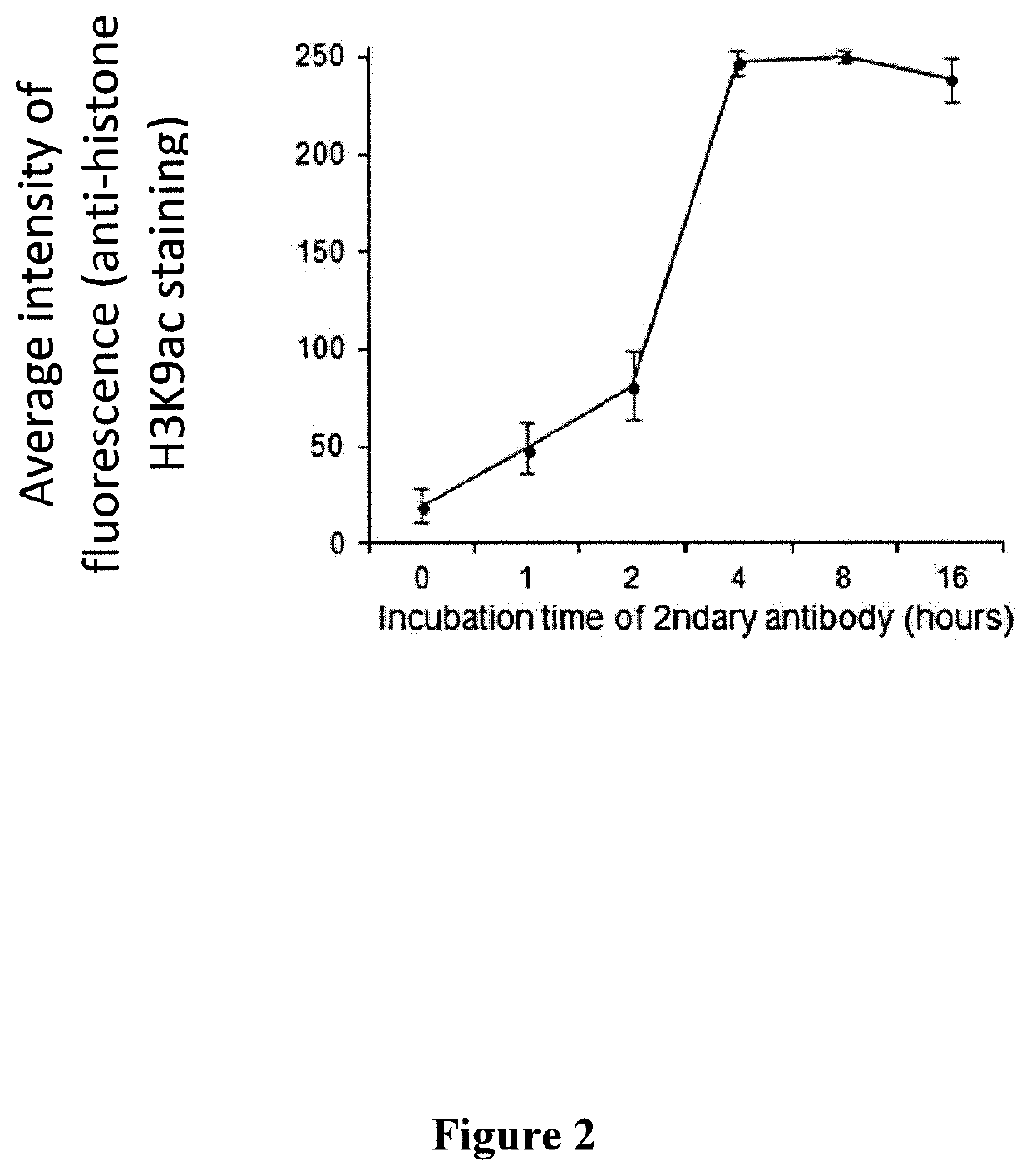
[0127]This example demonstrates that cellular components anchored by the polyacrylamide nano-scale scaffold are accessible to antibodies.

[0128]K562 cells were embedded in a polyacrylamide nano-scaffold prepared utilizing 20% acrylamide and 4% acrylamide-bisacrylamide, as described in Example 1. Once the polyacrylamide nano-scale scaffold was polymerized, the oil was removed, and the cells were incubated in 1% TRITON X-100 (“the surfactant” Dow Chemical Company, Midland, Mich.) for at least 15 minutes. Incubation in the presence of surfactant permeabilizes the cells and allows buffers and reagents to enter the cells. The cells were then immersed in annealing buffer (20 mM Tris-HCl, pH 8.8, 10 mM (NH4)2SO4, 10 mM KCl, 0.1% surfactant) and 0.2 to 20 μM of the first oligonucleotide primer. The cells were then heated at 94° C. for 3 minutes. To avoid non-specific binding of antibodies the cells were transferred to 12.5 mM EDTA containing 2.5% bovine serum albumin (BSA) and 1% surfactant....

example 3

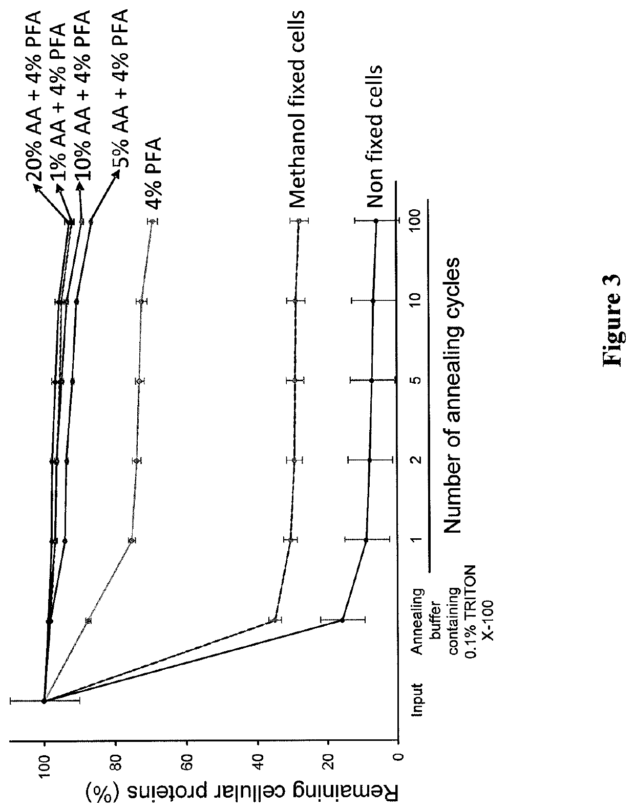
[0130]This example demonstrates that the polyacrylamide nano-scale scaffold retains proteins inside a single cell even after repeated heating and cooling cycles, thereby generating a “re-usable” single cell.

[0131]The experiments in this Example were performed to determine the effect of the polyacrylamide nano-scale scaffold on the retention of proteins in a single cell. A polyacrylamide nano-scaffold was prepared using 1×10⁶ K562 cells utilizing methanol, 4% formaldehyde (PFA), 4% PFA with 1% acrylamide, 4% PFA with 5% acrylamide, 4% PFA with 10% acrylamide, 4% PFA with 20% acrylamide, or no fixative, and 4% acrylamide-bisacrylamide as described in Example 1. The cells were washed once with hydrogel solution (3.88% acrylamide, 0.12% bis(acryloyl)cystamine, 1% ammonium persulfate, and 1× phosphate buffered saline (PBS)). Single cells were suspended in hydrogel solution at 3 μl per cell. Single cells (3 μl of cell suspension) were transferred into a PCR tube containing 0.2% TEMED in m...


PUM

PropertyMeasurementUnit
pHaaaaaaaaaa
pHaaaaaaaaaa
timeaaaaaaaaaa

Abstract

Methods and kits for preparing re-usable single cells are described. Cell components are anchored using a nano-scale scaffold to create a re-usable single cell. The nano-scale scaffold may be a polyacrylamide nano-scale scaffold. Methods to determine modifications of the genome, transcriptome, or epigenome are described.

Description

CROSS-REFERENCE TO RELATED APPLICATIONS[0001]This patent application claims the benefit of U.S. Provisional Patent Application No. 62 / 502,247, filed on May 5, 2017, which is incorporated by reference in its entirety herein.STATEMENT REGARDING FEDERALLY SPONSORED RESEARCH OR DEVELOPMENT[0002]This invention was made with Government support under contract number ZIA SC 010355, awarded by the National Institutes of Health, National Cancer Institute. The Government has certain rights in this invention.INCORPORATION-BY-REFERENCE OF MATERIAL SUBMITTED ELECTRONICALLY[0003]Incorporated by reference in its entirety herein is a computer-readable nucleotide / amino acid sequence listing submitted concurrently herewith and identified as follows: One 4,096 Byte ASCII (Text) file named “738705_ST25.TXT,” created on May 4, 2018.BACKGROUND OF THE INVENTION[0004]The ability to study DNA, RNA, and DNA-bound proteins simultaneously, and determine the location of proteins with and without modifications in...

Claims


Application Information

Patent Timeline
Patent Type & Authority Applications(United States)
IPC IPC(8): C12Q1/6834C12Q1/6806C12Q1/6827C12Q1/6874
CPCB82Y5/00C12Q1/6834C12Q1/6874C12Q2565/629C12Q1/6827C12Q1/6806C12Q2600/158C12Q2565/518C12Q2523/101C12Q2600/154C12Q2600/156C12N1/14C07K2317/77
Inventor TOSATO, GIOVANNAOHNUKI, HIDETAKA
Owner UNITED STATES OF AMERICA
Why Eureka
Try Eureka