Eureka AIR delivers breakthrough ideas for toughest innovation challenges, trusted by R&D personnel around the world.

Methods and systems for processing an ultrasound image

a processing system and ultrasound image technology, applied in the field of ultrasonic imaging, can solve the problems of sacrificing the performance of improving spatial resolution, affecting affecting the quality of ultrasound images, etc., to achieve the effect of improving the contrast of ultrasound images, reducing motion artifacts, and increasing the ultrasound image frame ra

Active Publication Date: 2020-04-23
KONINKLJIJKE PHILIPS NV
View PDF0 Cites 4 Cited by
  • Summary
  • Abstract
  • Description
  • Claims
  • Application Information

AI Technical Summary

Benefits of technology

The patent describes a method for improving ultrasound images by eliminating off-axis clutter signals. This is done by performing a spatial spectral estimation of the channel data and identifying the frequency content of the isolated channel data. The user can then selectively attenuate signals with high spatial frequencies to reduce or eliminate off-axis clutter in the final image. The method allows for improved image quality based on the user's subjective opinion.

Problems solved by technology

Off-axis clutter is a significant cause of image degradation in ultrasound.
Adaptive beamforming techniques, such as minimum variance (MV) beamforming, have been developed and applied to ultrasound imaging to achieve an improvement in image quality; however, MV beamforming is computationally intensive as an inversion of the spatial covariance matrix is required for each pixel of the image.
In addition, even though MV beamforming is developed primarily for an improvement in spatial resolution, and is not ideal for reducing off-axis clutter, its performance in terms of improving spatial resolution often needs to be sacrificed by reducing the subarray size.
Otherwise, image artifacts may occur in the speckle due to signal cancellation.
Further, these methods would only work for conventional imaging with focused transmit beams and are not suitable for plane wave imaging (PWI) or diverging wave imaging (DWI) involving only a few transmits.
The smaller the wavelength (or the higher center frequency), the better the lateral resolution will be; however, a smaller wavelength is achieved at the cost of penetration depth.
On the other hand, a larger aperture size D is needed to achieve a better lateral resolution; however, the aperture size is often limited by the human anatomy, hardware considerations, and system cost.
Since the spatial resolution in PWI / DWI is generally known to be only slightly worse than, or comparable to, conventional imaging with focused transmit beams, its main drawback is degradation in the image contrast, which is directly related to the number of transmit angles.
PWI / DWI also suffers from motion artifacts, particularly when imaging fast-moving organs such as the heart, as individual image pixels are constructed from signals from different transmit beams.
The effect of motion becomes more severe with increasing numbers of transmit angles; therefore, the dilemma in PWI / DWI systems is clear: more transmit angles are required to achieve high image contrast, but also result in more motion artifacts that degrade image quality.
Further, regardless of the number of transmit angles, PWI / DWI does not reduce reverberation clutter, which is one of the main sources of image quality degradation in fundamental B-mode ultrasound images.

Method used


Image

  • Methods and systems for processing an ultrasound image
  • Methods and systems for processing an ultrasound image
  • Methods and systems for processing an ultrasound image

Examples


Embodiment Construction

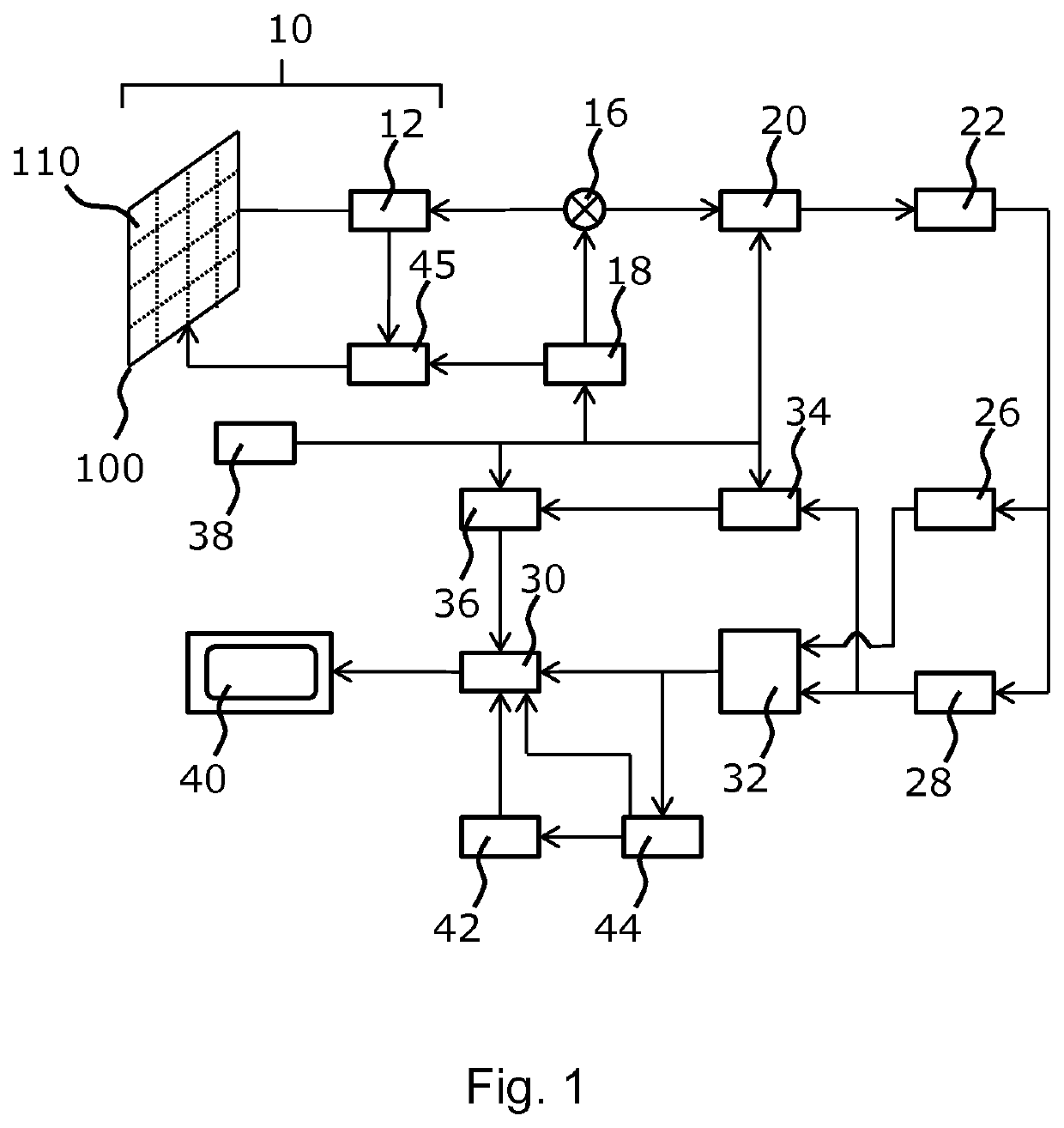
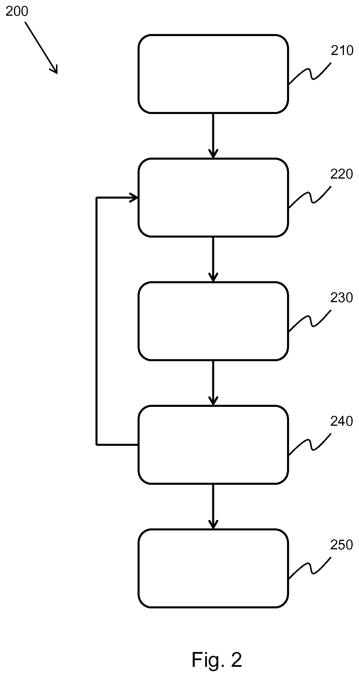
[0148]The invention provides methods and systems for generating an ultrasound image. In a method, the generation of an ultrasound image comprises: obtaining channel data, the channel data defining a set of imaged points; for each imaged point: isolating the channel data; performing a spatial spectral estimation on the isolated channel data; and selectively attenuating the spatial spectral estimation channel data, thereby generating filtered channel data; and summing the filtered channel data, thereby forming a filtered ultrasound image.

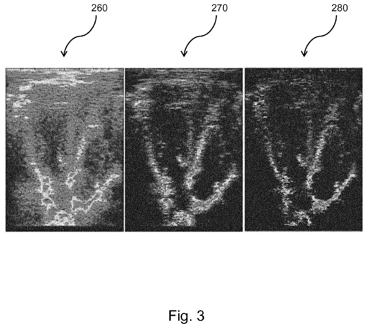
[0149]In some examples, the method comprises aperture extrapolation. The aperture extrapolation improves the lateral resolution of the ultrasound image. In other examples, the method comprises transmit extrapolation. The transmit extrapolation improves the contrast of the image. In addition, the transmit extrapolation improves the frame rate and reduces the motion artifacts in the ultrasound image. In further examples, the aperture and transmit extrap...


PUM


Abstract

The invention provides methods and systems for generating an ultrasound image. In a method, the generation of an ultrasound image comprises: obtaining channel data, the channel data defining a set of imaged points; for each imaged point: isolating the channel data; performing a spatial spectral estimation on the isolated channel data; and selectively attenuating the spatial spectral estimation channel data, thereby generating filtered channel data; and summing the filtered channel data, thereby forming a filtered ultrasound image. In some examples, the method comprises aperture extrapolation. The aperture extrapolation improves the lateral resolution of the ultrasound image. In other examples, the method comprises transmit extrapolation. The transmit extrapolation improves the contrast of the image. In addition, the transmit extrapolation improves the frame rate and reduces the motion artifacts in the ultrasound image. In further examples, the aperture and transmit extrapolations may be combined.

Description

FIELD OF THE INVENTION[0001]This invention relates to the field of ultrasound imaging, and more specifically to the field of ultrasound image filtering.BACKGROUND OF THE INVENTION[0002]Ultrasound imaging is increasingly being employed in a variety of different applications. It is important that the image produced by the ultrasound system is as clear and accurate as possible so as to give the user a realistic interpretation of the subject being scanned. This is especially the case when the subject in question is a patient undergoing a medical ultrasound scan. In this situation, the ability of a doctor to make an accurate diagnosis is dependent on the quality of the image produced by the ultrasound system.[0003]Off-axis clutter is a significant cause of image degradation in ultrasound. Adaptive beamforming techniques, such as minimum variance (MV) beamforming, have been developed and applied to ultrasound imaging to achieve an improvement in image quality; however, MV beamforming is c...

Claims


Application Information

Patent Timeline
IPC IPC(8): A61B8/08G06T7/00
CPCG06T2207/10132G06T2207/20024A61B8/5276G06T7/0012A61B8/5207G06T2207/20182G01S15/8977G01S15/8995G01S7/52047G01S15/8915G10K11/34
Inventor VIGNON, FRANCOIS GUY GERARD MARIESHIN, JUN SEOBHUANG, SHENG-WENROBERT, JEAN-LUC FRANCOIS-MARIE
Owner KONINKLJIJKE PHILIPS NV
Why Eureka
Eureka Blog
Learn More