Antibodies useful in cancer diagnosis
a technology for cancer and antibodies, applied in the field of antibodies useful in cancer diagnosis, can solve problems such as the difficulty of providing cldn6 antibodies with properties, and achieve the effect of reducing the risk of an onset of a cancer diseas
- Summary
- Abstract
- Description
- Claims
- Application Information
AI Technical Summary
Benefits of technology
Problems solved by technology
Method used
Image
Examples
example 1
Materials and Methods
[0345]Mapping of antibody binding site (FIG. 4A) Mapping of epitopes recognized by antibodies can be performed as described in detail in “Epitope Mapping Protocols (Methods in Molecular Biology) by Glenn E. Morris ISBN-089603-375-9 and in “Epitope Mapping: A Practical Approach” Practical Approach Series, 248 by Olwyn M. R. Westwood, Frank C. Hay.
[0346]Epitope mapping with biotinylated peptides (FIG. 4B)
[0347]A Peptide-ELISA was performed to identify the antibody-binding site of monoclonal murine lead antibodies and polyclonal rb-serum from IBL. Biotinylated overlapping peptides covering the C-terminal sequence of CLDN6 were coupled to SA coated plates. Purified murnAB (1 μg / mg) or IBL rb-serum (1 μg / ml) were applied to antigen coated ELISA-plates and unbound antibodies were washed off. Bound antibodies were detected with corresponding enzyme-labeled secondary antibody (alkaline phosphatase goat anti-mouse IgG(1+2a+2b+3) or alkaline phosphatase goat anti-rabbit I...
example 2
Generation of Monoclonal Antibodies
[0358]The aim of this project was to generate murine monoclonal CLDN6-specific antibodies capable of detecting CLDN6 expressing tumour cells in ovarian cancer or any other cancer tissue of any histology including primary peritoneal or fallopian tube tumours FFPE tissues.
[0359]To generate a highly specific, high affinity diagnostic CLDN6 antibody it was essential to start immunization protocols with a variety of different immunogens (Table 1) and adjuvants. During the project about 130 mice (C57BL / 6 and BALB / c) were subjected to various immunization strategies to trigger an a-CLDN6 immune response (Table 2).
[0360]To trigger the mouse immune system and to overcome the immune tolerance we used peptide-conjugates and recombinant proteins coding for different parts of human CLDN6 expressed as recombinant fusion proteins with different epitopes to facilitate affinity purification (Polyhistidine-(HIS)- and Glutathion-S-transferase-(GST)-tag) (Table 1).
[03...
example 3
Western Blot Screening of Monoclonal Antibodies
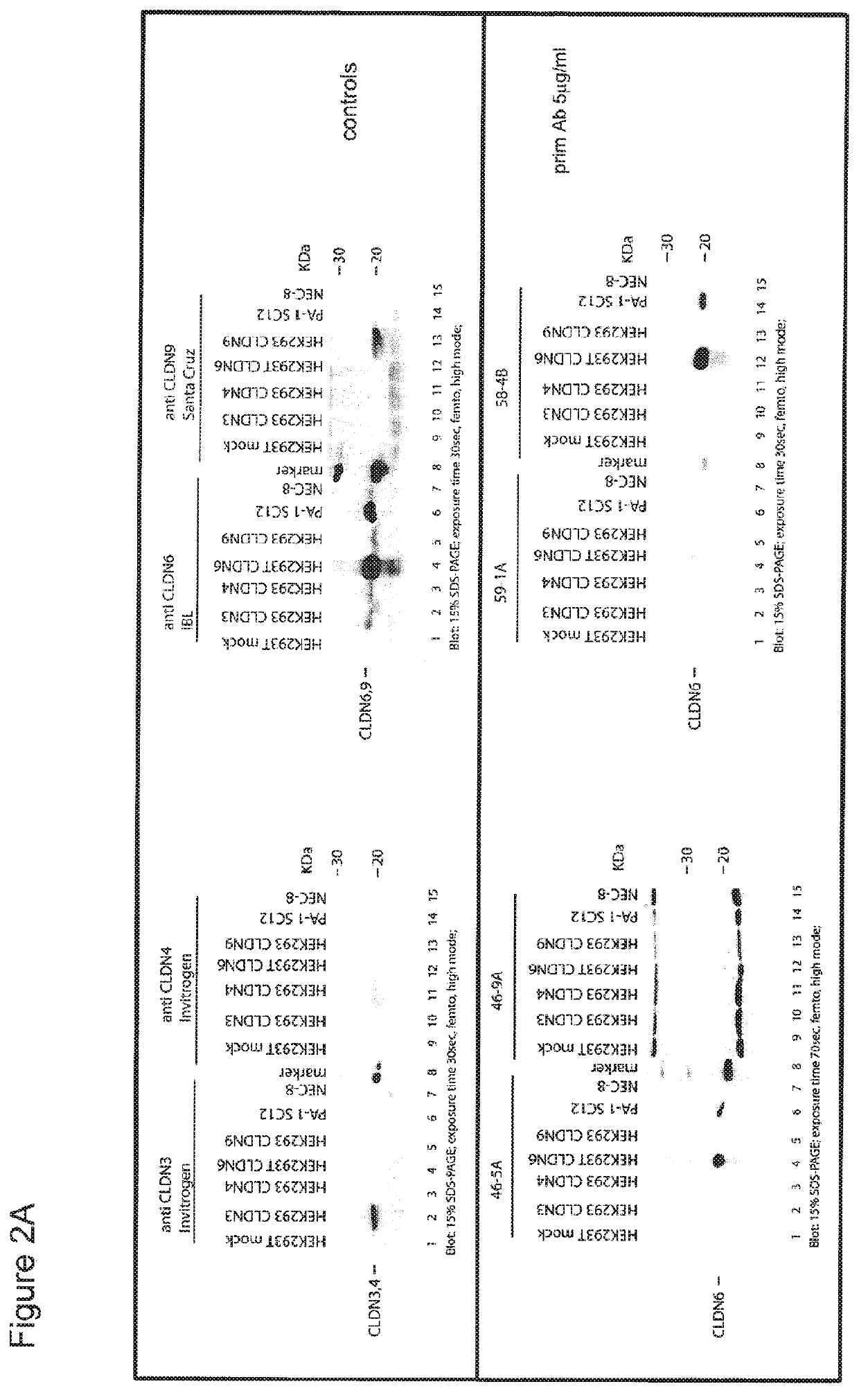
[0365]To test if ELISA-positive antibodies in the supernatants are able to bind specifically to recombinant claudin 6 they were analysed in Western Blot. Antibodies binding to CLDN6 but not to any other tagged-protein were purified and cells expanded and cryoconserved. Antibodies were purified via FPLC using MABselect Protein A affinity resin. The purified antibodies selected by the Western Blot screening were reanalyzed in Western Blot to assess the binding to CLDN6 positive tumour cell lines (PA-1, NEC-8) and to CLDN3, 4, 6 or 9 transfected HEK293 cells (FIG. 2). In addition, the antibodies were evaluated for their ability to bind their antigen in formalin fixed paraffin-embedded tissues (FFPE) by immunohistochemistry. Supernatants of 33 hybridoma specifically binding to Claudin 6 were purified and analysed further in Immunohistochemistry.
PUM
| Property | Measurement | Unit |
|---|---|---|
| Fraction | aaaaa | aaaaa |
| Volume | aaaaa | aaaaa |
Abstract
Description
Claims
Application Information
 - R&D
- Intellectual Property
- Life Sciences
- Materials
- Tech Scout
- Unparalleled Data Quality
- Higher Quality Content
- 60% Fewer Hallucinations
Browse by: Latest US Patents, China's latest patents, Technical Efficacy Thesaurus, Application Domain, Technology Topic, Popular Technical Reports.
© 2025 PatSnap. All rights reserved.Legal|Privacy policy|Modern Slavery Act Transparency Statement|Sitemap|About US| Contact US: help@patsnap.com


